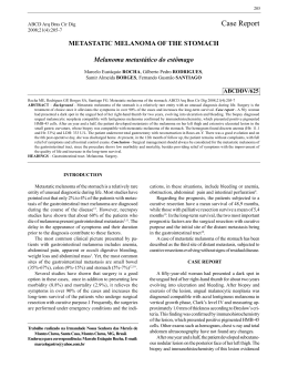
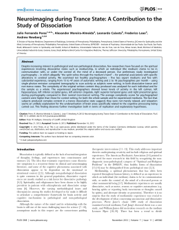
Materia 15
07.03.07
16:35
Page 104
Dementia & Neuropsychologia 2007;1:104-107
Visual agnosia and prosopagnosia
secondary to melanoma metastases
Case report
Norberto Anízio Ferreira Frota1, Lécio Figueira Pinto2, Claudia Sellitto Porto3,
Paulo Henrique Pires de Aguia4, Luiz Henrique Martins Castro5, Paulo Caramelli6
Abstract – The association of visual agnosia and prosopagnosia with cerebral metastasis is very rare. The
presence of symmetric and bilateral cerebral metastases of melanoma is also uncommon. We report the case of
a 34 year-old man who was admitted to hospital with seizures and a three-month history of headache, with
blurred vision during the past month. A previous history of melanoma resection was obtained. CT of the skull
showed bilateral heterogeneous hypodense lesions in the occipito-temporal regions, with a ring pattern of contrast enhancement. Surgical resection of both metastatic lesions was performed after which the patient developed visual agnosia and prosopagnosia. On follow-up, he showed partial recovery of visual agnosia, while
prosopagnosia was still evident. The relevance of this case is the rare presentation of metastatic malignant
melanoma affecting homologous occipito-temporal areas associated with prosopagnosia and associative visual
agnosia.
Key words: prosopagnosia, visual agnosia, metastasis, melanoma.
Agnosia visual e prosopagnosia secundárias a melanoma metastático: relato de caso
Resumo – A associação de agnosia visual e prosopagnosia com metástase cerebral são muito raras. A presença
de lesões metastáticas de melanoma, simétricas e bilaterais, também é incomum. Nós descrevemos o caso de
um homem de 34 anos que foi admitido no hospital com convulsões e uma história de cefaléia há três meses,
com dificuldade visual no último mês. História pregressa revelava ressecção de melanoma. TC de crânio evidenciou lesões hipodensas, heterogêneas, bilaterais nas regiões occipito-temporais, com captação de contraste
anelar. A ressecção cirúrgica de ambas as metástases cerebrais foi realizada e o paciente desenvolveu agnosia
visual e prosopagnosia após o procedimento. Na evolução houve melhora parcial da agnosia visual enquanto a
prosopagnosia ainda estava evidente. A relevância deste caso consiste na apresentação rara de melanoma
maligno metastático afetando as áreas occipito-temporais homólogas associada com prosopagnosia e agnosia
visual associativa.
Palavras-chave: prosopagnosia, agnosia visual, metastase, melanoma.
Visual agnosia is defined as a disorder of the visual
process leading to dysfunction in perception and/or recognition of objects, faces (prosopagnosia), letters (alexia)
or colors (achromatopsia), either in isolation or combination1-4.
Prosopagnosia was defined by Bodamer in 1947,
although it had been known since the XIX century, according to a description by Quaglino, Hughlings Jackson
and Charcot. It is associated with bilateral lesions in the
fusiform gyrus, although unilateral lesions involving the
right fusiform gyrus have also been described5.
Ischemic lesions are the most frequent causes, but
focal atrophy, congenital disorders and tumors can also
be associated with prosopagnosia1.
Brain metastases are the most frequently encountered
intracranial tumors. Malignant melanoma is the third
1
MD, Department of Neurology, University of São Paulo School of Medicine, Behavioral and Cognitive Neurology Unit Fellow. 2MD, Department of
Neurology, University of São Paulo School of Medicine, Electroencephalography-Epilepsy Fellow; 3MD, PhD, Behavioral and Cognitive Neurology Unit
Psychology. 4MD, PhD, Department of Neurosurgery, University of São Paulo School of Medicine; 5MD, PhD, Department of Neurology, University of
São Paulo School of Medicine; 6MD, PhD, Behavioral and Cognitive Neurology Unit Faculty of Medicine, Federal University of Minas Gerais.
Dr. Norberto Anízio Ferreira Frota – Rua Dr. Nicolau de Sousa Queiros, 406 / Ap. 94 - 04105001 São Paulo SP - Brazil. E-mail: [email protected].
104
Materia 15
07.03.07
16:35
Page 105
Dementia & Neuropsychologia 2007;1:104-107
most frequently encountered histologic type of brain
metastasis. Occipital lobe lesions represent 5.5% of brain
metastasis5,6. Association with prosopagnosia has previously been described in only one patient7.
Case report
A 34 year-old man, a professional cook, with an educational level of four years, presented a clinical history of
headache, nausea and vomiting in the past three months.
He also complained of a visual disorder described as a
“reading difficulty” in the last month.
The patient was admitted to the emergency room
because of a tonic-clonic seizure the day before. On examination, the patient was confused and somnolent, but
arousable, with reactive pupils and no motor deficits.
The magnetic resonance imaging (MRI) showed
bilateral occipito-temporal lesions suggestive of brain
metastasis. These lesions, involved the medial and inferior occipital gyrus bilaterally, the left fusiform and lingual
gyrus and part of the right fusiform gyrus (Figure 1).
Eight years before, the patient had undergone partial
resection of a malignant melanoma in the nose, without
further treatment.
Subsequently, he was submitted to a neurosurgical
procedure involving total metastasis resection through
occipital craniectomy. A ZEISS 588 microscope was used
to access both lesions. Following surgery, the patient was
admitted to the critical care unit.
After surgery, the patient had no motor, sensory or
coordination deficits, but presented associative visual
agnosia for colors, objects, faces and letters.
Upon hospital discharge, he underwent whole-head
Table 1. Neuropsychological testing: face and color discrimination.
Visual function
Test
Patient
Thematic figure
Severe difficulty in describing picture parts
Raven progressive matrices
Color series: 16/36 (10th percentile)
Face recognition
Familiar faces (12 stimuli): no recognition;
Famous faces (4 stimuli): recognition of one face (after prompt)
Table 2. Neuropsychological testing: attention, executive functions, memory, language and visuospatial skills.
Visuospatial skills
Attention
Planning
Block design (WAIS)
Severe impairment
Rey-Osterrieth complex figure
Severe impairment
Trail making test
Unable to perform test
Stroop test
Unable to recognize colors
Chapuis labyrinth
Mild impairment
Wisconsin card sorting test
Number of errors = 32/64
Number of categories = 01/03
Numbers of perseveration errors = 15
Memory
Wechsler memory scale–
review (WMS-R)
Logical memory (immediate recall) = normal
Visual reproduction (immediate recall) = 21/41 (4th percentile)
Delayed recall
Logical memory = normal
Visual reproduction =10/41 (1st percentile)
Language
Rey-Osterrieth complex figure
Delayed recall = 06/36 (moderate impairment )
Verbal fluency
Semantic category (animals): normal performance
Phonemic category (FA.S): normal performance
Boston naming test
10/60
105
Materia 15
07.03.07
16:35
Page 106
Dementia & Neuropsychologia 2007;1:104-107
Figure 1. Preoperative MRI: arrows showing bilateral occipito-temporal metastasis with contrast enhancement.
radiotherapy with 30 Gy, followed by chemotherapy
using dacarbazine. On follow-up the MRI was repeated,
showing no signs of residual brain metastasis (Figure 2).
The patient was re-evaluated at the outpatient unit 30
days after surgery. He complained of difficulties in recognizing faces.
On neurological examination, he scored 21 out of 30
points on the Mini Mental State Examination (MMSE).
No motor, sensory or coordination deficits were recorded. The examination of the cranial nerves was unremarkable. Visual recognition deficits were still evident, albeit
improved since surgery. Neuropsychological testing three
months after surgery confirmed visual recognition deficits, more markedly for faces and colors (Table 1).
Table 2 shows impaired performance in other neuropsychological tests.
The patient died nine months after surgery due to
multiple metastases (liver, spleen, skin and bones).
Figure 2. Postoperative MRI: To the right, white arrows showing
involvement of inferior and occipital gyrus, with black arrows
showing relative preservation of the right fusiform gyrus. To the
left the black arrows show involvement of inferior and medial
occipital gyrus and fusiform.
Discussion
The pattern of distribution of the lesions involving,
bilaterally and almost symmetrically, the medial and inferior occipital gyrus, the left fusiform and lingual gyrus
and part of the right fusiform gyrus is unusual, especially
for brain metastasis. These areas play a critical role in
visual recognition of objects, faces, colors and letters and
are known as the “what system” of the central visual processing1.
Deficits presented soon after surgery were more obvious, probably due to brain swelling, surgical manipulation and brain resection, characterizing a clinical picture
of associative visual agnosia. The patient underwent neuropsychological testing approximately three months postoperatively (Tables 1 and 2) which revealed below-aver-
106
age scores in almost all cognitive domains, except episodic memory and verbal fluency. Visual agnosia had become
more prominent and might have influenced performance
in the other tests. Recognition for faces and colors was
more affected than for objects.
Prosopagnosia was initially associated with bilateral
occipito-temporal lesions, most of them being ischemic1-3.
Right fusiform gyrus lesions are also described in association to prosopagnosia5,9. Degenerative, vascular, traumatic and congenital lesions are possible etiologies1, but a
metastatic lesion has been described in one case8.
Achromatopsy is also associated with occipito-temporal lesions, mainly on the right side. In the majority of
Materia 15
07.03.07
16:35
Page 107
Dementia & Neuropsychologia 2007;1:104-107
cases it has a favorable outcome, except when the lesion is
more posteriorly located, the case in our patient4.
In this patient, the right fusiform gyrus involvement
was partial, but the medial and inferior occipital gyri
were also involved. This pattern of anatomical distribution of the lesions probably disrupted the visual processing network for face recognition, as described by Rossion10.
The relevance of the present case is justified not only
because of the rarity of prosopagnosia due to brain
metastasis, but also to the relative preservation of the
right fusiform gyrus, indicating the importance of other
areas in the visual process related to face recognition.
4.
5.
6.
7.
8.
References
1. Damasio AR, Tranel D, Rizzo M. Disorders of complex visual processing. In: Mesulam MM, editor. Principles of behavioral and cognitive neurology. 2th ed. New York: Oxford
University; 2000:332-372.
2. Farah MJ, Feinberg TE. Visual object agnosia. Behavioral
Neurology and Neuropsychology. New York: Mc GrawHill;1997:239-244.
3. De Renzi E. Prosopagnosia. In: Farah MJ, Feinberg TE, edi-
9.
10.
tors. Behavioral Neurology and Neuropsychology. Bew
York: Mc Graw Hill; 1997:245-255.
De Renzi E. Disorders of visual recognition. Semin Neurol
2000;20:479-485.
De Renzi E, Perani D, Carlesimo GA, Silveri MC, Fazio F.
Prosopagnosia can be associated with damage confined to
the right hemisphere--an MRI and PET study and a review
of the literature. Neuropsychologia 1994;32:893-902.
Klos KJ, O’Neill BP. Brain metastases. Neurologist 2004;10:
31-46.
Delattre JY, Krol G, Thaler HT, et al. Distribution of brain
metastases. Arch Neurol 1988;45:741-744.
Seron X, Mataigne F, Coyette F, Rectem D, Bruyer R, Laterre
EC. A case of metamorphopsia restricted to faces and different familiar objects. Rev Neurol 1995:151:691-698.
Yamamot T, Wada Y. Selective impairment of facial recognition due to a haematoma restricted to the right fusiform
and lateral occipital region. J Neurol Neurosurg Psychiatr.
2001;71:254-257.
Rossion B, Caldara R, Seghier M, Schuller AM, Lazeyras F,
Mayer E. A network of occipito-temporal face-sensitive
areas besides the right middle fusiform gyrus is necessary
for normal face processing. Brain 2003;126:2381-2395.
107
Baixar