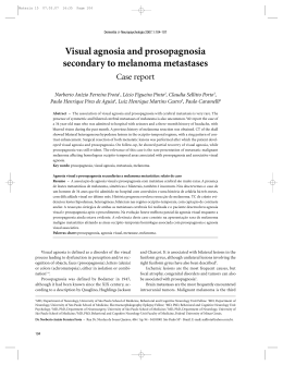
Revista do Instituto de Medicina Tropical de
São Paulo
versão impressa ISSN 0036-4665
Rev. Inst. Med. trop. S. Paulo v.41 n.1 São Paulo Jan./Fev. 1999
doi: 10.1590/S0036-46651999000100010
WIDESPREAD CUTANEOUS LESIONS DUE
TO Sporothrix schenckii IN A PATIENT
UNDER A LONG TERM STEROIDS THERAPY
Luiz Carlos SEVERO (1), Moira FESTUGATO (2), César
BERNARDI (3) & Alberto Thomaz LONDERO (4)
SUMMARY
A case of sporotrichosis in a woman presenting 63 cutaneous
lesions distributed all over the tegument is related. The patient had
both humoral (Immunoglobulins) and cellular (Lymphocytes
subpopulations) immunity within normal limits, but was under
treatment with steroid during a long time (Prednisone 10 mg daily
for 2 years), due to a sciatic pain. In addition a review of the
Brazilian literature on this type of lesions was carried out and
commented.
KEYWORDS: Sporotrichosis; Sporothrix schenckii; Corticosteroid;
Side effect.
INTRODUCTION
Sporothrix schenckii usually is introduced into the human body by
direct intracutaneous inoculation or, rarely, by the respiratory
route. Inoculated, it causes a subcutaneous mycosis; inhaled, it can
elicit manifestations similar to those caused, usually, by the other
thermal dimorphic pathogenic fungi, which are agents of systemic
mycoses. S. schenckii also may act as an opportunistic fungus6,8.
Widespread cutaneous lesions is a very uncommon occurrence in
subcutaneous sporotrichosis, but they are frequent in the systemic
and opportunistic presentation of the mycosis5,10,11.
A case of a patient under a long corticoid therapy presenting
widespread cutaneous lesions caused by S. schenckii will be herein
related. In addition the Brazilian literature will be reviewed and
commented.
CASE REPORT
The patient is a 65 year-old black housewife, living in Porto Alegre,
Rio Grande do Sul. One month ago, while bathing, she noted many
cutaneous nodules. She denied any symptoms. She was using
steroids since a long time (Prednisone 10 mg daily for 2 years), due
to a sciatic pain. On physical examination she presented with 63
non-painful, movable, nonattached subcutanoeus nodules of
irregular shape (averaging 3 cm in greatest dimension), distributed
all over her body (Fig. 1): 17 lesions on the lower right limb, 6 on
the lower left limb, 4 on the right limb, 14 on the upper left limb, 7
on the back, 6 on the buttocks, 6 on the thorax and 3 on the
abdominal wall (Fig. 2). None of these lesions had a lymphangitic
component. Laboratory findings were: hemogram, slight anemia;
fasting glycemia 104 mg/dL (normal 70-110); VDRL non reagent;
FTA-ABS negative; Mantoux non reactive. A chest X-ray showed no
pleuro-pulmonary abnormalities.
Fig. 1 - Cutaneous lesions widely distributed in breast, abdomen
and left upper limb.
Fig. 2 - Schematic distribution of patient's 63 cutaneous lesions.
Two biopsies were perfomed, one of which in a recent nodule. H&E
stained sections showed in both specimens epithelioid histiocytes in
concentric zones with central area of neutrophils with a dermal
necrotic focus. Homogenized fragments were seeded on Sabouraud
medium and incubated at 25°C; fast growing membranous white
colonies were obtained; microscopically the colonies were
composed of delicate septate branched hyaline hyphae and oval or
piriform conidia (2-3/4 µm). Subcultures on BHI (Difco), incubated
at 37°C, revealed the characteristic yeast-like forms of S. schenckii.
New sections of biopsied tissue were obtained and stained by
Gomori methenamine silver stain; several single and budding round
yeast cells compatible with the tissue forms of S. schenckii were,
then, detected.
Due to the atypical presentation of the mycosis, the patient
underwent immunological assessment: total lymphocytic count was
3162/mm3 (normal 1250-4500), from which 50% were T
lymphocyte and 9% B lymphocytes; subpopulations of lymphocytes
CD4 885/mm3, CD8 316/mm3, ratio CD4/CD8 = 2.8 (normal 0.93.0); Immunoglobulins: IgG 3.630 mg/dL (normal 690-1400), IgA
591 mg/dL (normal 70-370) and IgM 269 mg/dL (normal 40-240);
complement C3 163 mg/dL (normal 20-600) and C4 39 mg/dL
(normal 5-260).
Steroid therapy was discontinued and the patient received
potassium iodide in an initial dosage of 5 drops three times a day
followed by 120 drops per day. The lesions cleared in one month
but the treatment continued for two months. Seven months later
the patient remained very well.
DISCUSSION
Five cases of sporotrichosis presenting with widespread skin lesions
could be gathered in the Brazilian literature. Patient's data, clinical
manifestations and associated conditions and treatment are shown
in Table 1.
In four patients (cases 1, 3, 4, and 5) cutaneous lesions were
associated with osseous involvement, consequently a presentation
of systemic sporothricosis. Involvement of bones in the most
frequent extracutaneous lesion in this type of infection 5,11. One of
these four patients (case 5) presented also a very destructive
mucosal lesion, an unusual occurrence. In the remaining patient
(case 2) no extracutaneous lesions were detected, consequently a
presentation of subcutaneous sporothricosis.
In spite of no clinical evidence of extracutaneous lesions in our
patient, they can not be excluded because visceral lesions can run
an asymptomatic course, only detected by histological examination
of internal organs9. We may also presume that our patient acquired
the infection by traumatic route but spreading of skin lesions
resulted from hematogenous dissemination, which was facilitated
by steroid therapy3,5,6,11.
Treatment of iodides seems to be effective in patients with
widespread skin lesions without extracutaneous involvement, as it
occurred in case 2 and in our patient.
RESUMO
Lesões generalizadas causadas por Sporothrix schenckii em
paciente sob corticoterapia de longa duração.
É relatado caso de esporotricose em mulher apresentando 63
lesões cutâneas distribuídas pelo tegumento. A paciente tinha
imunidade humoral (Imunoglobulinas) e celular (Subpopulações
linfocitárias) dentro dos limites da normalidade, mas estava sob
corticoterapia de longa duração (Prednisona 10 mg/dia por 2 anos),
para ciática. Em acréscimo uma revisão da literatura brasileira,
deste tipo de lesões, é feita e comentada.
REFERENCES
1. CASTRO, R.M.; SABOGAL, M.F.; CUCE, L.C. & SALEBIAN, A. Disseminated sporotrichosis. Report of a clinical case with
mucocutaneous, osteo-articular and ocular lesion. Mykosen, 24:
92-96, 1981.
[ Links ]
2. CURBAN, G.V.; LACAZ, C. da S.; BELFORT, E.; DILLON, N. &
AUADA, J. - Esporotricose disseminada. Registro de um caso. Rev.
Hosp. Clin. Fac. Med. S. Paulo, 15: 369-371, 1960.
[ Links ]
3. FRIEDMAN, S.J. & DOYLE, J.A. - Extracutaneuos sporotrichosis.
Int. J. Derm., 22: 171-176, 1983.
[ Links ]
4. GOMES, J.M. - Um caso de esporotricose generalizada. An. paul.
Med. Cirurg., 11: 197-199, 1920.
[ Links ]
5. LYNCH, P.J.; VOORHEES, J.J. & HARREL, E.R. - Systemic
sporotrichosis. Ann. intern. Med., 73: 23-30, 1970.
[ Links ]
6. MORGAN, M.A.; COCKERILL, F.R.; CORTESE, D.A. & ROBERTS,
G.D. - Disseminated sporotrichosis with Sporothrix schenckii
fungemia. Diagn. Microbiol. infect. Dis., 2: 151-155, 1984.
[ Links ]
7. RIPPON, J.W. - Medical Mycology. The pathogenic fungi and
the pathogenic actinomycetes. 3. ed. Philadelphia, W. B.
Saunders, 1988. p. 373-380.
[ Links ]
8. SAMPAIO, S.A.P.; LACAZ, C. da S. & ALMEIDA, F. - Aspectos
clínicos da esporotricose em São Paulo. Análise de 235 casos. Rev.
Hosp. Clin. Fac. Med. S. Paulo, 9: 391-402, 1954.
[ Links ]
9. SCHAMROTH, J.M.; GRIEVE, T.P. & KELLER, P. - Disseminated
sporotrichosis. Int. J. Derm., 27: 28-30, 1988.
[ Links ]
10. SMITH, P.W.; LOOMIS, G.W.; LUCKASEN, J.L. & OSTERHOLM,
R.K. - Disseminated cutaneous sporotrichosis. Arch. Derm., 117:
143-144, 1981.
[ Links ]
11. WILSON, D.E.; MANN, J.J.; BENNETT, J.E. & UTZ, J.P. - Clinical
features of extracutaneous sporotrichosis. Medicine (Baltimore),
46: 265-279, 1967.
[ Links ]
(1) Pesquisador do CNPq; Faculdade de Medicina, Universidade
Federal do Rio Grande do Sul (UFRGS), RS, Brasil.
(2) Serviço de Dermatologia, UFRGS, Santa Casa, RS, Brasil.
(3) Chefe do Serviço de Dermatologia, UFRGS, Santa Casa, RS,
Brasil.
(4) Universidade Federal de Santa Maria, Santa Maria, RS, Brasil.
Correspondence to : L. C. Severo, M.D., Laboratório de
Microbiologia Clínica, IPD - Santa Casa, Annes Dias 285, 90020-090
Porto Alegre, RS, Brazil. Fax 00 55 51 214 8435 - E-mail:
[email protected]
Received: 02 June 1998
Accepted: 03 December 1998
Baixar