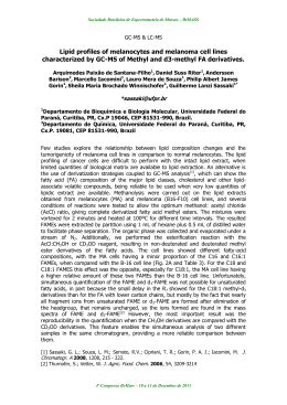
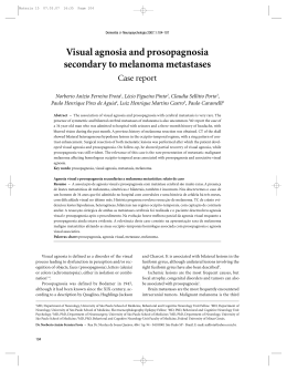
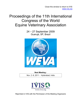
205
ABCD Arq Bras Cir Dig
2008;21(4):205-7
Case Report
Metastatic melanoma of the stomach
Melanoma metastático do estômago
Marcelo Eustáquio ROCHA, Gilberto Pedro RODRIGUES,
Samir Almeida BORGES, Fernando Gusmão SANTIAGO
ABCDDV/625
Rocha ME, Rodrigues GP, Borges SA, Santiago FG. Metastatic melanoma of the stomach. ABCD Arq Bras Cir Dig 2008;21(4):205-7
ABSTRACT – Background - Metastatic melanoma of the stomach is a relatively rare entity with an unusual diagnosis during life. Surgery is the
treatment of choice once it alleviates the symptoms in over 90% of the cases and increases the long-term survival. Case report - A 50y woman
had presented a dark spot in the ungual bed of her right-hand thumb for two years, evolving into ulceration and bleeding. The biopsy diagnosed
ungual malanocytic neoplasia compatible with lentiginous melanoma confirmed by immunohistochemistry, which presented positive pigmented
HMB-45 cells. After an year and a half, the patient developed metastasis of the melanoma on her left thigh and extensive ulcerated lesion in the
small gastric curvature, whose biopsy was compatible with metastatic melanoma of the stomach. The hemogram found discrete anemia (Hb: 11.1
and Ht: 33%) and LDH: 333 U/L. The patient underwent total gastrectomy with reconstruction in Roux-en-Y. There was a good evolution and on
the 6th post-operative day, she was discharged home. At present, in the 12th month of follow up, the patient remains without complaints, with full
relief of symptoms and all normal control exams. Conclusion - Surgical management should always be considered for the metastatic melanoma of
the gastrointestinal tract, since the procedure shows low morbidity and mortality, besides providing relief of symptoms with the improvement of
the quality of life and increase in the long-term survival.
HEADINGS – Gastrointestinal tract. Melanoma. Surgery.
INTRODUCTION
Metastatic melanoma of the stomach is a relatively rare
entity of unusual diagnostic during life. Most studies have
pointed out that only 2% to 4% of the patients with metastasis of the gastrointestinal tract melanoma are diagnosed
during the course of the disease12. However, necropsy
studies have shown that about 60% of the patients who
die of melanoma present gastrointestinal metastasis5,11. The
delay in the appearance of symptoms and their duration
prior to the diagnosis contribute to these factors.
The most common clinical picture presented by patients with gastrointestinal melanoma includes anemia,
abdominal pain, apparent or occult digestive bleeding,
weight loss and abdominal mass1. Yet, the most common
sites of the gastrointestinal metastasis are small bowel
(35%-67%), colon (9%-15%) and stomach (5%-7%)12,10.
Several studies have shown that surgery is a good
option in these cases, once in addition to presenting low
morbidity (8.8%) and mortality (2.9%), it relieves the
symptoms in over 90% of the cases and increases the
long-term survival of the patients who undergo surgical
resection with curative purpose.1 Frequently, the surgeries
are performed under emergency conditions and the indi-
Trabalho realizado na Irmandade Nossa Senhora das Mercês de
Montes Claros, Santa Casa, Montes Claros, MG, Brasil.
Endereço para correspondência: Marcelo Estáquio Rocha. E-mail:
[email protected]
cations, in these situations, include bleeding or anemia,
obstruction, abdominal pain and intestinal perfuration8.
Regarding the prognosis, the patients subjected to a
curative resection have a mean survival of 48,9 months,
while those with palliative resection survive a mean of 5,4
months10. In the long-term survival, the two most important
prognostic factors are the surgical resection with curative
purpose and the initial site of the distant metastasis being
in the gastrointestinal tract10.
A case of metastatic melanoma of the stomach has been
described as the third site of distant metastasis, subjected to
curative resections evolving without signs of residual disease.
CASE REPORT
A fifty-year-old woman had presented a dark spot in
the ungual bed of her right-hand thumb for about two years
evolving into ulceration and bleeding. After biopsy and
exeresis of the lesion, ungual malanocytic neoplasia was
diagnosed compatible with acral lentiginous melanoma in
vertical growth phase, Clark’s level IV and measuring approximately 1.0 mm of thickness according to Breslow’s criteria. This finding was confirmed by immunohistochemistry
of the lesion, which presented positive pigmented HMB-45
cells. Other exams such as hemogram, chest x-ray and total
abdomen ultrasonography have not found any changes.
After one year and a half, the patient developed subcutaneous nodular lesion on the posterior face of her left thigh. The
biopsy and immunohistochemistry of this lesion evidenced
206
Metastatic melanoma of the stomach
relapse of malignant melanoma. In the same period, the
patient reported epigastry, nausea, vomiting and weight loss.
The high digestive endoscopy presented gastric mucosa at the
small curvature level with extensive ulcerated lesion, covered
by dark fibrin of necrotic tissue with elevated margins and
occupying almost the entire small curvature (Figure 1). Endoscopic biopsy was performed revealing malignant neoplasia
suggestive of metastatic melanoma of the stomach. Hemogram
showed discrete anemia (Hb: 11.1 and Ht: 33%) and hepatic
and renal function without changes. LDH: 333 U/L.
The patient was referred to the surgical service and
underwent total gastrectomy with reconstruction in Rouxen-Y (Figure 2). The anatomopathologic presented lesion
compatible with metastatic melanoma of the stomach with
free resection margins. Moreover, metastatic melanoma
was found in two lymph nodes of the small curvature and in
one of the large curvature. There was good evolution and on
the 6th post-operative day, the patient was discharged home.
FIGURE 1 – Endoscopic image showing metastatic melanoma
in the small curvature of the stomach
FIGURE 2 – Gastrectomy specimen showing macroscopic lesion,
with free margins
In the 4th post-operative month, PET-SCAN was performed identifying no sign of the disease. Presently, in
the 12th month of follow up, the patient remains without
complaints, with full relief of the symptoms and with all
normal control exams.
DISCUSSION
Malignant melanoma which involves the gastrointestinal
tract can be classified as primary or metastatic15. The primary
melanoma of the gastrointestinal mucosa is very rare and it
can be clinically suggested if the patient does not have primary
cutaneous melanoma or if a gastrointestinal lesion is isolated
without another extraintestinal metastasis15. Regarding metastatic melanoma of the gastrointestinal tract, studies in cadavers
have shown that it is frequent in patients who die of melanoma,
however, due to the fact that these patients remain asymptomatic
for a long time, it is unusual during life, occurring only in 2%
to 4% of the patients with melanoma, and out of these only 5%
to 7% present the disease with metastasis to the stomach2,12.
The initial lesion thickness less than 0.75 mm is associated with excellent survival rates and low risk of metastasis14.
Some authors have stated that the main predictive factors of
survival after curative resections, include site of metastasis,
number of metastatic lesions and the disease-free interval
prior to the development of metastasis2,3. Hence, patients
without visceral metastasis have better prognosis than those
with visceral disease. Recurrence in multiple sites worsen the
prognosis, as well as the period of time without recurrence
shorter than one year3. Other papers have shown that the
presence of residual disease is what provokes greater impact
on these patients’ survival1,13. However, there is a consensus
in most studies that low LDH values (<200U/L) constitute
an independent factor with a positive predictive value in
the long-term survival1,6,7.In the case herein presented, the
disease was visceral, the pre-operation LDH value was over
200 U/L and the initial lesion thickness was > 0.75 mm, yet,
the patient has presented long-term survival and no relapse
so far, contradicting the factors advocated by the literature.
Surgical resection is the treatment of choice for metastatic
melanoma of the gastrointestinal tract. Chemotherapy can be
used, but the immunocompromised status it provokes in these
patients may cause serious complications15. Unfortunately, most
patients subjected to curative resection experience relapse of the
disease. There have been attempts to associate other adjuvant
therapies such as immunotherapy and anti-angiogenesis agents
in order to prevent the recurrence, but randomized studies have
shown no benefits in relation to the survival in the case of immunotherapy9.The approach with anti-angiogenesis agents is still
under study. In the case reported herein, despite the presence of
metastasis of more than one site (TGI and subcutaneous tissue)
the resection of the lesions was curative and even without the
administration of adjuvant therapy, the patient has remained
free of signs of relapse so far.
Due to the high recurrence rate of the metastatic melanoma, there is the need to perform a follow up with image
exams periodically. The exam of choice for this scanning is
the PET-SCAN4. Other exams such as high and low digestive endoscopy and CT can be carried out9.
CONCLUSION
Surgical treatment should always be considered for
metastatic melanoma of the gastrointestinal tract, once the
procedure presents low morbidity and mortality, besides
providing relief of the symptoms with the improvement of
the quality of life and increase in the long-term survival of
patients with curative resection even when some prognostic
factors are unfavorable as in the case presented here.
ABCD Arq Bras Cir Dig 2008;21(4):205-7
207
Rocha ME, Rodrigues GP, Borges SA, Santiago FG. Melanoma metastático do estômago. ABCD Arq Bras Cir Dig 2008;21(4):205-7
RESUMO – Introduction - O melanoma metastático do estômago é entidade relativamente rara e de diagnóstico incomum em vida. A cirurgia é o tratamento de escolha, pois alivia os sintomas em mais de 90% dos casos e aumenta a sobrevida a longo prazo. Objetivo - Relatar um caso de melanoma
metastático do estômago, submetida à ressecção curativa e que evoluiu sem sinais de doença residual. Relato do caso - Mulher de 50 anos apresentou
mancha escura em leito ungueal de dedo polegar de mão direita há dois anos, evoluindo com ulceração e sangramento. A biópsia diagnosticou neoplasia
melanocítica ungueal compatíveis com melanoma lentiginoso e confirmado pela imunohistoquímica que apresentou células pigmentadas HMB-45
positivas. Após um ano e meio a paciente evoluiu com metástase de melanoma em coxa esquerda e extensa lesão escavada em pequena curvatura
gástrica, cuja biopsia foi compatível com melanoma metastático do estômago. Hemograma com discreta anemia (Hb: 11,1 e Ht: 33%) e LDH: 333 U/L.
A paciente foi submetida à gastrectomia total com reconstrução em Y de Roux. Houve boa evolução e no 6º dia de pós-operatório teve alta hospitalar.
Atualmente, no 12º mês de seguimento, a paciente permanece sem queixas, com alívio completo dos sintomas e com todos os exames de controle normais. Conclusão - O tratamento cirúrgico deve ser sempre considerado no melanoma metastático do trato gastrointestinal, pois o procedimento possui
baixa morbidade e mortalidade, além de proporcionar alívio dos sintomas com melhora da qualidade de vida e aumento da sobrevida a longo prazo.
DESCRITORES – Trato gastrointestinal. Melanoma. Cirurgia.
REFERENCES
1.
2.
3.
4.
5.
6.
7.
8.
9.
Agrawal S, Yao TJ, Coit DG. Surgery for melanoma metastatic to the gastrointestinal tract. Ann Surg Oncol 1999; 6: 336–44.
Allen PJ, Coit DG. Thesurgical management of metastatic melanoma. Ann
Surg Oncol. 2002;9:762–70.
Balch CM, Soong SJ, Murad TM, Smith JW, Maddox WA, Durant JR. A
multifactorial analysis of melanoma. IV. Prognostic factors in 200 melanoma
patients with distant metastases (stage III). J Clin Oncol 1983; 1: 126–34.
Damian DL, Fulham MJ, Thompson E, Thompson JF. Positron emission tomography in the detection and management of metastatic melanoma. Melanoma
Res. 1996;6:325-9.
De la Monte SM, Moore GW, Hutchins GM, Patterned distribution of metastases
from malignant melanoma in humans. Cancer Res 1983; 43:3427-33.
Eton O, Legha SS, Moon TE. Prognostic factors for survival of patients treated
systemically for disseminated melanoma. J Clin Oncol 1998;16:1103–11.
Franzke A, Probst-Kepper M, Buer J. Elevated pretreatment serum levels of
soluble vascular cell adhesion molecule 1 and lactate dehydrogenase as predictors of survival in cutaneous metastatic malignant melanoma. Br J Cancer
1998; 78: 40–5.
Ihde JK, Coit DG. Melanoma metastatic to stomach, small bowel or colon. Am
J Surg 1991;162:208–11.
Liang KV, Sanderson SO, Nowakowski GS, Arora AS. Metastatic malignant
melanoma of the gastrointestinal tract. Mayo Clin Proc 2006; 81:511-6
10. Ollila DW, Essner R, Wanek LA, Morton DL. Surgical resection for melanoma
metastatic to the gastrointestinal tract. Arch Surg 1996; 131: 975–9.
11. Patel JK, Didolkar MS, Pickren JW, Moore RH. Metastatic pattern of malignant
melanoma. A study of 216 autopsy cases. Am J Surg 1978; 135:807-10.
12. Reintgen DS, Thompson W, Garbutt J, Seigler HF. Radiologic, endoscopic, and
surgical considerations of melanoma metastatic to the gastrointestinal tract.
Surgery 1984; 95: 635–9.
13. Ricaniadis N, Konstadoulakis MM, Walsh D, Karakousis CP. Gastrointestinal
metastases from malignant melanoma. Surg Oncol 1995; 4: 105–10.
14. Schmid-Wendtner MH, Baumert J, Eberle J, Plewig G, Volkenandt M, Sander
CA. Disease progression in patients with thin cutaneous melanomas (tumour
thickness ≤0.75 mm): clinical and epidemiological data from the Tumour Center
Munich 1977-98. Br J Dermatol. 2003;149:788-93.
15. Schuchter LM, Green R, Fraker D. Primary and metastatic diseases in malignant
melanoma of the gastrointestinal tract. Curr Opin Oncol. 2000;12:181-5.
Fonte de financiamento: não há
Conflito de interesse: não há
Recebido para publicação: 18/05/2008
Aceito para publicação: 22/08/2008
Baixar