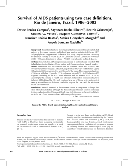
b r a z j i n f e c t d i s . 2 0 1 4;1 8(3):315–326 The Brazilian Journal of INFECTIOUS DISEASES www.elsevier.com/locate/bjid Review article Consensus of the Brazilian Society of Infectious Diseases and Brazilian Society of Clinical Oncology on the management and treatment of Kaposi’s sarcoma Érico Arruda a , Alexandre Andrade dos Anjos Jacome b , Ana Luiza de Castro Conde Toscano c,∗ , Anderson Arantes Silvestrini d , André Santa Bárbara Rêgo e , Evanius Garcia Wiermann f , Geraldo Felicio da Cunha Jr. g , Heloisa Ramos Lacerda de Melo h , Karen Mirna Loro Morejón i , Luciano Zubaran Goldani j , Luiz Carlos Pereira Jr. k , Mariliza Henrique Silva l , Mauro Sergio Treistman m , Mônica Cristina Toledo Pereira n , Patricia Maria Bezerra Xavier Romero o , Rafael Aron Schmerling p , Rodrigo Antonio Vieira Guedes q , Veridiana Pires de Camargo r a Sociedade Brasileira de Infectologia, Vila Mariana, SP, Brazil Hospital Mater Dei, Belo Horizonte, MG, Brazil c Instituto de Infectologia Emílio Ribas e Centro de Referência e Treinamento em DST/AIDS, São Paulo, SP, Brazil d Sociedade Brasileira de Oncologia Clínica e Grupo Acreditar, Brasília, DF, Brazil e Hospital Santa Lúcia, Brasília, DF, Brazil f Sociedade Brasileira de Oncologia Clínica, Brasília, DF, Brazil g Hospital da Baleia, Belo Horizonte, MG, Brazil h Hospital das Clínicas, Universidade Federal de Pernambuco (UFPE), Recife, PE, Brazil i Hospital das Clínicas da Faculdade de Medicina de Ribeirão Preto, Ribeirão Preto, SP, Brazil j Hospital de Clínicas de Porto Alegre, Porto Alegre, RS, Brazil k Hospital Dia, Instituto de Infectologia Emilio Ribas, São Paulo, SP, Brazil l Centro de Referência e Treinamento-DST-AIDS, São Paulo, SP, Brazil m Serviço de Infectologia de Rede Hospitalar Privada e Câmara Técnica de Doenças Infecciosas do CREMERJ n Fundação Hospitalar do Estado de Minas Gerais, Belo Horizonte, MG, Brazil o Hospital São Camilo, São Paulo, SP, Brazil p Centro de Oncologia Antonio Ermirio de Moraes, São Paulo, SP, Brazil q Centro de Oncologia do Hospital Sírio-Libanês, São Paulo, SP, Brazil r Instituto do Câncer do Estado de São Paulo e do Hospital Sírio Libanês, São Paulo, SP, Brazil b a r t i c l e i n f o a b s t r a c t Article history: Kaposi’s sarcoma is a multifocal vascular lesion of low-grade potential that is most often Received 27 September 2013 present in mucocutaneous sites and usually also affects lymph nodes and visceral organs. Accepted 23 January 2014 The condition may manifest through purplish lesions, flat or raised with an irregular shape, Available online 11 February 2014 gastrointestinal bleeding due to lesions located in the digestive system, and dyspnea and ∗ Corresponding author at: Av. Doutor Arnaldo, 165, São Paulo, SP, 01246-900, Brasil. E-mail address: [email protected] (A.L.C.C. Toscano). 1413-8670/$ – see front matter © 2014 Elsevier Editora Ltda. All rights reserved. http://dx.doi.org/10.1016/j.bjid.2014.01.002 316 Keywords: Kaposi’s sarcoma AIDS Consensus Cutaneous b r a z j i n f e c t d i s . 2 0 1 4;1 8(3):315–326 hemoptysis associated with pulmonary lesions. In the early 1980s, the appearance of several cases of Kaposi’s sarcoma in homosexual men was the first alarm about a newly identified epidemic, acquired immunodeficiency syndrome. In 1994, it was finally demonstrated that the presence of a herpes virus associated with Kaposi’s sarcoma called HHV-8 or Kaposi’s sarcoma herpes virus and its genetic sequence was rapidly deciphered. The prevalence of this virus is very high (about 50%) in some African populations, but stands between 2% and 8% for the entire world population. Kaposi’s sarcoma only develops when the immune system is depressed, as in acquired immunodeficiency syndrome, which appears to be associated with a specific variant of the Kaposi’s sarcoma herpes virus. There are no treatment guidelines for Kaposi’s sarcoma established in Brazil, and thus the Brazilian Society of Clinical Oncology and the Brazilian Society of Infectious Diseases developed the treatment consensus presented here. © 2014 Elsevier Editora Ltda. All rights reserved. Introduction General aspects of Kaposi’s sarcoma Kaposi’s sarcoma is a multifocal vascular lesion of low-grade potential that is most often present in mucocutaneous sites and usually also affects lymph nodes and visceral organs.1 Kaposi’s sarcoma was first described in 1872 by Hungarian dermatologist Moritz Kaposi. From that time to the identification of human immunodeficiency virus (HIV) associated with acquired immunodeficiency syndrome (AIDS), Kaposi’s sarcoma remained a rare tumor. While most of the cases identified in Europe and in North America occurred in elderly men of Italian descent or Eastern European Jews, the neoplasia also occurs in several other different populations: young black African men, children in pre-adolescence, receivers of allergenic renal transplant and other patients treated with immunosuppressive therapy. The disseminated and fulminant form of Kaposi’s sarcoma associated with AIDS is referred to as epidemic Kaposi’s sarcoma to distinguish it from the classical, African and transplant-related forms. In addition, Kaposi’s sarcoma was identified in homosexual men without HIV virus.2,3 Although the histopathology of different types of Kaposi’s tumors is essentially identical among the various affected groups, the clinical manifestations and course of the disease differ dramatically.2 A key to understanding the pathogenesis of Kaposi’s sarcoma was the discovery in 1994 of a gamma herpes virus, human herpes virus type 8 (HHV-8), also known as herpes virus of Kaposi’s sarcoma.4 HHV-8 has been identified in tissue biopsies of Kaposi’s sarcoma of virtually all patients with different forms of the disease (classical, African, transplant-related and AIDS-associated), but was absent in the tissue not involved by the neoplasia.2 Considered a rare disease, Kaposi’s sarcoma in its classical form occurs more often in males, with a ratio of about 10–15 men for every woman affected. Among Americans and Europeans, the usual age of onset is between 50 and 70 years of age.2 In the 1950s, Kaposi’s sarcoma was recognized as a relatively common endemic neoplasia in native populations of equatorial Africa, comprising about 9% of all cancers seen in males in Uganda. In Africa, indolent or locally more aggressive forms of Kaposi’s sarcoma occur at a man/woman ratio comparable to that observed for the classical tumor seen in North America and Europe. However, patients in Africa are significantly younger than European patients. A lymphadenopathic form is also seen in Africa, primarily in children in preadolescence, at a male/female ratio of 3 cases to 1,2,5 and mortality rate of nearly 100% in 3 years.5,6 In 1969, the first case of Kaposi’s sarcoma associated with immunosuppressive therapy in a patient with renal transplantation was described. Since then, it has been observed that several patients receiving renal transplants and other allergenic transplants who were treated with prednisone and azathioprine developed Kaposi’s sarcoma shortly after initiation of immunosuppressive therapy.2,7 Estimates of the incidence of Kaposi’s sarcoma among renal transplant recipients subjected to immunosuppressive therapy are between 150 and 200 times higher than the expected incidence of the tumor in the general population. The average time to develop Kaposi’s sarcoma after transplantation is 16 months.2 Epidemiological aspects of epidemic Kaposi’s sarcoma In 1981, a disseminated and fulminant form of Kaposi’s sarcoma was described in homosexual or bisexual men and was first reported as part of an epidemic now known as AIDS.8 The etiology of AIDS is a retrovirus with tropism for T lymphocytes known as HIV.9 The immune deficiency that characterizes AIDS is a profound disorder of cell-mediated immune functions. This immune dysfunction and deregulation of the immune system predispose patients to the development of a wide range of opportunistic infections and unusual neoplasm such as Kaposi’s sarcoma. HIV can play an indirect role in the development of Kaposi’s sarcoma.9 Approximately 95% of all cases of epidemic Kaposi’s sarcoma in the United States were diagnosed in homosexual or bisexual men. In the past, approximately 26% of all male homosexuals with HIV presented with or developed Kaposi’s sarcoma over the course of AIDS. As a comparison, less than 3% of all heterosexual injection drug users with HIV developed Kaposi’s sarcoma. The proportion of AIDS patients with Kaposi’s sarcoma has declined dramatically since the outbreak of the disease was identified in 1981.10 About 48% of patients diagnosed with AIDS in 1981 presented with Kaposi’s sarcoma at diagnosis. By August 1987, this proportion had declined to less than 20%. The introduction of highly active antiretroviral therapy (HAART) delayed or b r a z j i n f e c t d i s . 2 0 1 4;1 8(3):315–326 317 Número de casos 900 800 700 600 500 400 300 200 100 0 Feminino Masculino 1980 1981 1982 1983 1984 1985 1986 1987 1988 1989 1990 1991 1992 1993 1994 1995 1996 1997 1998 1999 2000 2001 2002 2003 2004 2005 2006 2007 2008 2009 2010 2011 2012 12 14 13 30 39 54 58 59 102 87 110 83 79 93 77 82 102 86 83 72 64 68 58 65 56 20 0 2 0 1 0 0 0 27 116 167 297 398 485 585 656 754 760 786 721 633 541 559 448 398 408 376 423 353 311 308 282 269 291 268 269 111 1 0 0 9 Source: SINAN-ministry of health [16] Fig. 1 – Incidence of Kaposi’s sarcoma – Brazil 1980 to June/2012. Source: SINAN – Ministry of Health.16 prevented the emergence of HIV strains resistant to treatment and profoundly decreased viral load, leading to increased survival and decreased incidence of opportunistic infections among AIDS patients.11,12 The use of HAART is associated with a substantial and sustained decline in the incidence of Kaposi’s sarcoma among patients with AIDS.13–15 In Brazil, cases of Kaposi’s sarcoma related to AIDS must be reported to the Information System for Notifiable Diseases (SINAN), which is fed mainly by the reporting and investigation of cases of diseases and conditions which appear on the national list of diseases subject to compulsory notification. Fig. 1 shows a graph with the number of new cases of Kaposi’s sarcoma since the beginning of the epidemic to 06.30.201216 : we observe a decline in disease incidence following the availability of HAART in 1996. Clinical manifestations of epidemic Kaposi’s sarcoma The epidemic or AIDS-related Kaposi’s sarcoma has a highly variable clinical course, and can appear as minimal mucocutaneous disease or as disseminated disease with involvement of other organs. The lesions can involve the skin, oral mucosa, lymph nodes and visceral organs. Most patients present with cutaneous disease, but occasionally the visceral disease may precede cutaneous manifestation. Skin lesions can occur at any location, but are typically concentrated in the lower extremities and the head and neck. The lesions can be macular, papular, nodular or look like plaques, and they are almost all palpable and non-pruritic. The size of the skin lesions can vary from a few millimeters to several centimeters in diameter, and can be brown, pink or violet. The lesions can be discrete or confluent and typically appear in a symmetrical linear distribution along tension skin lines. Mucous membrane involvement is common (e.g., palate, gum and conjunctiva), and ulcerated or bulky tumors can interfere with speech and chewing. The lymphedema associated with the tumor, typically manifested in the lower extremities or the face, seems to be caused by secondary obstruction of lymphatic vessels. Pain when walking can be present in the case of lesions involving the soles. Lesions can occur anywhere in the gastrointestinal tract, usually an indicator of more advanced HIV infection, manifesting itself through symptoms that include odynophagia, dysphagia, nausea, vomiting, abdominal pain, hematemesis, hematochezia, melena or intestinal obstruction. Pulmonary involvement can be difficult to distinguish from opportunistic infections and can be expressed by cough, dyspnea, hemoptysis, or chest pain. Pulmonary lesions can be an asymptomatic radiographic finding and pleural effusions are often exudative and hemorrhagic. Lymphadenopathy can be the only manifestation of the disease, which requires a lymph node biopsy and can lead to significant lymphedema.1,17–20 The introduction of HAART in order to control HIV has caused a major change in the behavior of Kaposi’s sarcoma related to AIDS: it was accompanied by a dramatic decrease in incidence of disease and its less aggressive presentation, but the disease remains a severe problem in the Western world, as in the case of its manifestation with pulmonary involvement.21,22 HAART can cause partial or complete regression of Kaposi’s sarcoma, with partial or complete disappearance of spindle-shaped cells.23 Exacerbation of Kaposi’s sarcoma (flare) can be seen after corticosteroid therapy or rituximab or as part of the immune reconstitution inflammatory syndrome which can occur upon initiation of HAART by patients. The immune reconstitution inflammatory syndrome is a pathological exaggerated inflammatory response that is due to an exuberant immune response to opportunistic infections either apparent or concealed, or cancers. The exacerbation mechanism of Kaposi’s sarcoma after treatment with corticosteroids appears to be linked to an upregulation of the expression of steroid receptors.24,25 Diagnosis of epidemic Kaposi’s sarcoma Although a presumptive diagnosis of Kaposi’s sarcoma can be often made based on the clinical history and appearance of skin lesions, this hypothesis should be confirmed by biopsy of the lesions, whenever possible. Biopsy is especially important for atypical lesions that are associated with systemic symptoms or progress rapidly toward discarding bacillary angiomatosis.26 There are three histological findings that are characteristic of Kaposi’s sarcoma, both in cutaneous and visceral forms: angiogenesis, inflammation and proliferation. The lesions usually present two main abnormalities, which are spindle-shaped cells, arranged in a snail-like form with leukocyte infiltration and neovascularization with abhorrent proliferation of small vessels. These tiny vessels lack a baseline membrane, which gives rise to microhemorrhages and 318 b r a z j i n f e c t d i s . 2 0 1 4;1 8(3):315–326 Table 1 – ACTG – classification of Kaposi’s sarcoma. High risk (1) Any of the following findings Low risk (0) Any of the following findings Tumor Confined to the skin and/or lymph node and/or minimum oral diseasea • Edema or ulceration associated with tumor • Extensive oral disease • Gastrointestinal disease • Visceral disease other than lymph node Immune system CD4 cells ≥200 L−1 CD4 cells <200 L−1 Systemic disease • Absence of history of opportunistic infections or canker sores • Absence of symptoms B • Performance status (PS) ≥70 • History of opportunistic infections or canker sores • Presence of symptoms B • Performance status <70 • Another HIV related disease (neurological, lymphoma) a Non-nodular disease confined to the palate; symptoms B = unexplained fever, night sweats, >10% weight loss or persistent diarrhea lasting more than 2 weeks; PS = Karnofsky scale; adapted from ACTG–AIDS Clinical Trials Group Oncology Committee. deposition of hemosiderin in tissue. With the progression of the disease, lesions evolve from stain to plaques and then to a nodular form. The standard histological characteristic does not differ among the epidemiological groups affected by the disease.27 Additional supplementary tests might be needed for patients with systemic symptoms which might mean visceral involvement of the disease.28 Staging and prognostic factors There is no universally accepted classification available for epidemic Kaposi’s sarcoma, and staging schemes that incorporate laboratory parameters and clinical findings have been proposed. The majority of patients with epidemic Kaposi’s sarcoma do not die due to the disease; factors other than the tumor load are apparently involved in the survival of patients. The conventions used to stage Kaposi’s sarcoma and the methods used to assess the benefits of treatment continue to evolve because of changes in the treatment of AIDS and the recognition of the shortcomings of the standard evaluation of the tumor. The clinical course of Kaposi’s sarcoma, treatment selection and response to treatment are strongly influenced by the subjacent degree of immune deficiency and the occurrence of opportunistic infections. The AIDS Clinical Trials Group [ACTG] Oncology Committee published criteria for the evaluation of epidemic Kaposi’s sarcoma. The staging system incorporates measures of disease extent, severity of immunodeficiency and presence of systemic symptoms. As can be seen in Table 1, ACTG criteria categorize the extent of the tumor as localized or disseminated, CD4 cell count as high or low, and systemic disease as absent or present.29 A subsequent prospective analysis of 294 patients who participated in clinical studies for Kaposi’s sarcoma of the ACTG group between 1989 and 1995 showed that each variable of Table 1 (tumor, immune system and systemic disease) was independently associated with patient survival.30 Multivariate analyses showed that worsening of the immune system was the most important individual predictor of survival. In patients with relatively high CD4 cell counts, tumor stage was predictive; a CD4 count of 150 cells/mm3 can be a better discriminating index than the limit of 200 cells initially adopted.31,32 None of the previous studies was conducted in a time when HAART was already readily available. The impact of this therapy on survival in cases of Kaposi’s sarcoma requires continued evaluation.31–34 In 2003, an Italian study group published a research in order to evaluate new prognostic factors and validate the ACTG staging system for Kaposi’s sarcoma related to AIDS in the HAART era. Clinical, epidemiological, and staging information, as well as survival data were collected from 211 patients with AIDS-related Kaposi’s sarcoma included in two Italian cohort studies on HIV as of 1996, the year in which HAART became available in Italy. In light of the results, researchers proposed to refine the application of the stage system in which the immune system should be eliminated with a determining prognosis and only the extent of the tumor (T) and systemic disease (S) should be regarded as predictive variables of survival. Two categories of risk for death were identified: (a) high risk (T1S1) and low risk (T0S0, T1S0 and T0S1). In addition, researchers showed that pulmonary involvement predicts survival better than the extent of the tumor and identifies the category with the highest risk regardless of the variable S (systemic disease). Noteworthy, survival analysis of the interaction between pulmonary disease and systemic disease appears to provide a better distribution of risk among groups of patients, with hazard ratios (HR) progressive toward death (Tp1S1 > Tp1S0 > Tp0S1 > Tp0S0) when compared to the interaction between the classical extent of the tumor and systemic disease.35 Therapeutic approach in epidemic Kaposi’s sarcoma Although the benefits of the use of HAART are indisputable, Kaposi’s sarcoma has not disappeared as a clinical problem. Due to the fact that HAART can induce tumor regression and that the appropriate treatment of HIV infection requires the administration of that therapy, distinguishing the antitumor effects of HAART from those induced by a chemotherapeutic agent for Kaposi’s sarcoma presents difficulties.21,22 Moreover, as described below, the scientific evidence suggests that tumor response to isolated use of HAART occurs mainly in patients who were not using this therapy. b r a z j i n f e c t d i s . 2 0 1 4;1 8(3):315–326 Major treatment goals are to relieve symptoms, prevent disease progression and decrease the size of the tumor to alleviate edema, involvement of a possible affected organ and psychological stress.36 Many low-risk patients in accordance with the ACTG (AIDS Clinical Trials Group Oncology Committee) classification present tumor regression with HAART alone.37 High-risk patients usually require the combination of HAART and chemotherapy, which is suspended after disappearance of skin lesions.37 HAART There are no randomized clinical trials comparing the treatment of Kaposi’s sarcoma with HAART versus the treatment without HAART, since it is virtually recommended for all patients with AIDS-related Kaposi’s sarcoma.61 The introduction of this therapy is associated with a substantial decrease in the incidence and severity of cases of Kaposi’s sarcoma recently diagnosed in patients with HIV infection. A French study that analyzed a database with 54,999 patients with over 180,000 patient-years of follow-up showed that the incidence rate of Kaposi’s sarcoma dropped from 32 per 1000 person-years in 1993–1994 down to 3 per 1000 person-years after 1999.38 Furthermore, the incidence of visceral involvement by Kaposi’s sarcoma upon diagnosis dropped from over 50% to less than 30%.38 A Swiss cohort study showed that the relative risk of development of Kaposi’s sarcoma between 1997 and 1998 (HAART era) compared to the time period between 1992 and 1994 (pre-HAART era) was 0.08 (95% CI, 0.03–0.22).39 The addition of HAART to systemic chemotherapy also increased the survival of patients with pulmonary involvement by Kaposi’s sarcoma.40 Observational studies indicate that the natural history of AIDS-related Kaposi’s sarcoma has changed since the introduction of HAART, along with decreased tumor incidence.41,31 A retrospective study that analyzed cases of Kaposi’s sarcoma in a database of 4439 people with HIV infection from pre-HAART therapy (1990–1996) and after the introduction of HAART therapy (1997–2002) showed that the mean count of CD4 cells and mean levels of HIV RNA were similar in the 366 patients pre-HAART and in the 40 patients in the HAART era. However, the overall risk of death was significantly lower in the HAART era (HR, 0.24).31 Due to the control of HIV infection, immune reconstitution is the most likely explanation for this changed prognosis, much more than a direct effect on the tumor. Although inhibitors of HIV protease have antiangiogenic properties and block the development and progression of lesions resembling Kaposi’s sarcoma in mice,42 there was no difference in the possibility of clinical response associated with the use of these agents.41,43 Furthermore, the decreased incidence of Kaposi’s sarcoma has been observed with the use of treatment regimens that do not contain protease inhibitors.38 Recent chemotherapy is associated with the improvement of Kaposi’s sarcoma; recent low viral load of HIV and HAART are associated with the improvement and resolution of Kaposi’s sarcoma. The response is not associated with the type of HAART regimen (inhibitor of non-nucleoside reverse transcriptase, protease inhibitor, or enhanced with protease inhibitor ritonavir).44 319 Although treatment with HAART promotes increased counts of CD4 cells to levels above those typically associated with increased susceptibility to infection, some patients develop AIDS-related Kaposi’s sarcoma despite the apparent correction of their immunodeficiency.45 In some patients with HIV who have Kaposi’s sarcoma disease (moderate to advanced) HAART used in isolation may not be sufficient to tumor treatment, making it necessary to use other adjuvant therapy such as systemic chemotherapy, which showed good efficacy and resulted in significant clinical improvements when combined with antiretroviral therapy, as demonstrated by phase III studies comparing HAART in isolation with HAART combined with systemic chemotherapy.46–49 A systematic review carried out in the post-HAART era with the aim of determining whether patients with advanced Kaposi’s sarcoma can respond to HAART in isolation was not explanatory. Of the available studies, there were only five cases in which patients with advanced Kaposi’s sarcoma (T1) responded to HAART in the absence of concomitant therapy for the disease.37 Immune reconstitution inflammatory syndrome The expression “immune reconstitution inflammatory syndrome” is used to describe a series of host responses that can occur soon after the start of HAART and has been linked to the progression of Kaposi’s sarcoma in three to six weeks after initiation of treatment. The relationship between immune reconstitution inflammatory syndrome and Kaposi’s sarcoma was shown in two studies: (1) a series of cases of 150 treatment-naïve patients who presented with Kaposi’s sarcoma, 10 (7%) developed tumor progression when HAART was initiated; the risk of the immune reconstitution inflammatory syndrome seemed enhanced in patients with higher counts of CD4 cells or with edema associated with Kaposi’s tumor; despite tumor progression, maintenance of HAART therapy was possible in those patients50 ; (2) in another series of nine patients in an institution, progression of Kaposi’s sarcoma occurred in an average of five weeks after initiation of HAART therapy and was associated with increases in CD4 cells and decrease in viral load; in all patients in whom systemic chemotherapy was used we observed tumor regression, and interruption of antiretroviral therapy was not necessary.51 Antiviral agents Although HHV-8 viremia is associated with an increased risk of developing Kaposi’s sarcoma,52 there is currently no consensus on the therapeutic benefits of the use of antiviral drugs in this group of patients. In vitro studies demonstrating sensitivity of HHV-8 to antiviral agents are in disagreement. One study demonstrated that HHV-8 is sensitive to antiviral agents such as cidofovir and gancyclovir, and weakly sensitive to acyclovir. However, these drugs do not act in the latent form of the virus and it is unlikely they are effective against established tumor lesions.53 Another study, however, found no sensitivity to acyclovir but showed activity to gancyclovir, foscarnet and cidofovir, also in the replicative phase.54 In a clinical study of patients with AIDS and cytomegalovirus retinitis, patients who were treated with oral or intravenous gancyclovir had reduced risk of developing KS.55 However, the drugs studied to 320 b r a z j i n f e c t d i s . 2 0 1 4;1 8(3):315–326 Table 2 – Epidemic Kaposi’s sarcoma – therapeutic approaches. Local treatment Retinoids Radiotherapy Intralesional chemotherapy Systemic treatment Chemotherapy • Liposomal doxorubicin • Liposomal daunorubicin • Bleomycin • Vincristine, vinblastine • Paclitaxel date have important systemic effects with intravenous administration, making their prophylactic use impractical in the long run. Local treatments Small localized lesions of Kaposi’s sarcoma were treated using electro dissection and curettage, cryotherapy, or through surgical excision in the pre-HAART era. Kaposi’s tumors are also generally very responsive to local radiotherapy, with excellent results being obtained with 20 Gy or slightly higher doses.56–58 Radiation therapy is generally reserved for treating localized skin areas and in the oral cavity. It is less used to control pulmonary lesions, lesions of the gastrointestinal tract or other sites of Kaposi’s sarcoma. Localized lesions also tend to be effectively treated with intralesional injections of vinblastine.59 Alitretinoin gel 0.1% was also effective in local control of the lesions of Kaposi’s sarcoma in a prospective multicenter randomized trial in which the substance was used two times a day for 12 weeks; overall response rate was 37%.60 Cytotoxic agents In epidemic Kaposi’s sarcoma, systemic chemotherapy is generally used in patients with advanced disease or when there is evidence of rapid disease progression.61 When treatment is indicated, the use of pegylated liposomal doxorubicin or liposomal daunorubicin is usually recommended as first line treatment.61 Other chemotherapeutic agents also used in the treatment of epidemic Kaposi’s sarcoma include bleomycin, vincristine, vinblastine, etoposide and paclitaxel as monotherapy or in combination therapy.61,62 Indications generally accepted for the use of systemic chemotherapy as adjuvant therapy to antiretroviral agents include: (a) extensive involvement of skin (e.g., more than 25 lesions), (b) extensive cutaneous Kaposi’s sarcoma that does not respond to local treatment, (c) extensive edema, (d) symptomatic visceral involvement and (e) immune reconstitution inflammatory syndrome.61 The mode of use of various drugs considered in chemotherapy for the treatment of Kaposi’s sarcoma is briefly described in Table 2. Liposomal anthracyclines, doxorubicin and daunorubicin constitute a considerable advance in the chemotherapy of Kaposi’s sarcoma. The advantages of liposomal formulation include increased drug uptake by the tumor, which leads to a more favorable pharmacokinetic profile. Clinical studies of liposomal anthracyclines in the treatment of Kaposi’s sarcoma associated with HIV/AIDS were conducted in pre-HAART era, but clinicians continue to regard them as first-line treatment agents of Kaposi’s sarcoma. Both liposomal daunorubicin (40 mg/m2 every two weeks) and pegylated liposomal doxorubicin (20 mg/m2 every two to three weeks) showed good antitumor activity. Toxicity profile of both agents is better than that of other anthracyclines, and there are no reports of either cardiotoxicity or significant alopecia, even with high cumulative doses. However, these agents still cause significant myelosuppression and occasional emesis. In addition, infusion-related hypotension and hand-foot syndrome are new adverse events seen with the use of such liposomal formulations.61 A phase III randomized study comparing liposomal daunorubicin and ABV regimen (doxorubicin, bleomycin and vincristine) revealed no difference in terms of overall response rates (partial response + complete response), time to treatment failure and survival duration.63 Two randomized phase III studies compared pegylated liposomal doxorubicin with conventional chemotherapy combinations (ABV in a study and BV [bleomycin + vincristine] in another) as first-line treatment for patients with Kaposi’s sarcoma who were not receiving HAART. The two studies showed that response rates were higher among groups of patients who received pegylated liposomal doxorubicin, but responses were not sustained over time.64,65 The three studies mentioned cannot be directly compared. A small randomized study included 79 patients with Kaposi’s sarcoma who were randomized to receive pegylated liposomal doxorubicin (20 mg/m2 ) or liposomal daunorubicin (40 mg/m2 ) every two weeks for up to six cycles. Differences have been shown favoring pegylated liposomal doxorubicin.66 Myelosuppression (e.g., neutropenia) is a major side effect of pegylated liposomal doxorubicin, representing a limiting factor in the therapeutic regimen for the treatment of neoplasia. Patients who develop neutropenic fever (defined by the presence of fever, oral temperature >38.3 ◦ C or ≥38.3 ◦ C for more than 1 h; whereas neutropenia is defined as neutrophil count <500 mm−3 or between 500 and 1000 mm−3 and tend to fall in the next 48 h) in the course of chemotherapy should be carefully evaluated for whether they should receive treatment with myeloid growth factors.67 In the case of using filgrastim, the daily dose is 5 g/kg/day, treatment should be continued until absolute neutrophil counts reach their normal value.67 Hand-foot syndrome is a set of signs and symptoms of acute nature affecting the palms of the hands and soles of the feet, being strongly associated with antineoplastic chemotherapy, occurring to a lesser extent in patients treated with liposomal anthracyclines compared with other chemotherapy agents such as 5-fluorouracil and derivatives. In treatment schemes where there is an expected rate of occurrence of this syndrome, it is important that patients be able to recognize early signs, so that therapy can be adjusted immediately. Changes in dose or systemic and local approaches can be used. Once symptoms have subsided, therapy can usually be restarted according to initial planning. Upon recurrence of symptoms, especially if more severe, dose adjustment is required.68 Pyridoxine (vitamin B6) has shown benefits as systemic therapy of hand-foot syndrome. There are reports of b r a z j i n f e c t d i s . 2 0 1 4;1 8(3):315–326 complete disappearance of hand-foot syndrome with doses of 50–150 mg daily. However, some patients may not respond to this drug. Although the exact mechanism of action is not yet fully understood, pyridoxine can also be used prophylactically. Anti-inflammatory inhibitors of cyclooxygenase-2 (COX-2) have also proven effective in the prophylaxis of handfoot syndrome associated with chemotherapy. High potency corticosteroids and disinfectant treatment of vesicles and erosions were effective when considering a topical therapy. Preventive use of glucocorticoids, by contrast, proved ineffective. In mild cases of the syndrome, avoid mechanical irritation of the skin on the palms of hands and soles of feet, and the use of emollient creams or soft gels is enough for control and relief of symptoms. Cooling of the affected areas using cold baths of hands and feet (without intensive washing) also helps in relief of symptoms. Depending on the severity of the syndrome, cure occurs in a matter of days or weeks.68 Although paclitaxel is potentially more toxic than liposomal anthracyclines, it has remarkable efficacy as second-line treatment of Kaposi’s sarcoma,69,70 and can be an alternative to initial therapy of patients with advanced symptomatic disease. The effectiveness of paclitaxel was originally demonstrated in a phase II study with 28 evaluable patients in which 20 (71%) had higher responses to the treatment regimen of 135 mg/m2 every 3 weeks (18 partial responses, one full clinical response and one full response). Responses were observed in all five patients with pulmonary lesions of Kaposi’s sarcoma and all four patients had received prior anthracycline-based chemotherapy. Treatment toxicity included grade 4 thrombocytopenia in 6 of the 29 enrolled patients and grade 4 neutropenia in 22 of the 29 patients treated without the use of hematopoietic growth factors.71 Treatment regimen of paclitaxel 100 mg/m2 every 2 weeks was compared with the use of pegylated liposomal doxorubicin in a dose of 20 mg/m2 every 3 weeks in a randomized clinical trial conducted after the introduction of routine treatment with HAART.46 In this study, 73 evaluable patients with Kaposi’s sarcoma associated with AIDS were included between 1998 and 2002 (the trial was prematurely terminated because of low patient inclusion). There were no statistically significant differences between the two treatment regimens in terms of response rate, progression-free survival or overall survival. The two treatment regimens propitiated considerable improvement of pain and of edema secondary to tumor.46 Myelosuppression (e.g., neutropenia) induced by paclitaxel is an important side effect, representing a limiting factor in the therapeutic regimen for the treatment of neoplasia. Patients who develop neutropenic fever (defined by the presence of fever, oral temperature >38.3 ◦ C or ≥38.3 ◦ C for more than 1 h; whereas neutropenia is defined as neutrophil counts <500 mm−3 or between 500 and 1000 mm−3 and tend to fall in the next 48 h) in the course of chemotherapy should be carefully evaluated for whether they should receive treatment with myeloid growth factors.67 In the case of the use of filgrastim, the daily dose is 5 g/kg/day, and treatment should be continued until absolute neutrophil count reaches normal value.67 Before the era of HAART, several other chemotherapeutic agents (e.g., bleomycin, doxorubicin, vinblastine, vincristine, 321 and etoposide) were active in the treatment of Kaposi’s sarcoma related to AIDS in case reports and in small phase II trials that evaluated different combinations and doses of these drugs.61 The percentage of patients achieving a decrease of ≥50% in number of lesions ranged from 58% to 90% under treatment with vinca alkaloids, from 74% to 76% with etoposide was 97% with the combination of vinblastine and bleomycin. However, clinical studies that evaluated these regimens presented low quality due to the lack of a standardized classification of disease activity and clinical outcomes analyzed. Therefore, evidence for the effectiveness of any of these treatment regimens is of low quality and does not support recommendation of any of these regimens in particular.72 For the duration of treatment with HAART, both pegylated liposomal doxorubicin and paclitaxel are isolated active agents in the treatment of epidemic Kaposi’s sarcoma, with response rates close to 50%.61 Immunotherapy The use of biological response modifier interferon-␣ was approved for the treatment of Kaposi’s sarcoma before the availability of HAART and liposomal anthracyclines. Interferons-␣ were extensively studied and showed objective response rate of 40% in patients with epidemic Kaposi’s sarcoma.73,74 In these studies, responses differed significantly according to the prognostic factors of disease extent, prior or coexistent opportunistic infections, previous treatment with chemotherapy, CD4 lymphocyte counts of less than 200 cells/mm3 , presence of circulating acid-labile interferon-␣ and increased 2-microglobulin. Response to interferon-␣ often requires continuous treatment for 6 months or more, since response time is typically longer than four months. Interferon-␣ should not be considered as a therapeutic option in case of progressive or visceral disease. Toxicity of high doses of interferon-␣ (e.g., fever, chills, neutropenia and depression) is common and low responses are observed in the presence of low counts of CD4 cells.61 Interferon-␣ is not very often used today due to its toxicity profile and because it does not work well in many patients with AIDS.28 Interleukin-12 has shown a response rate of 71% (95% CI, 48–89%) in the treatment of Kaposi’s sarcoma in 24 evaluable patients in a phase I study.75 Summary of therapeutic recommendations for epidemic Kaposi’s sarcoma Table 2 shows possible therapeutic approaches for patients with epidemic Kaposi’s sarcoma. Specifically, Table 3 presents main cytotoxic agents which can be used in the chemotherapy treatment of epidemic Kaposi’s sarcoma. Recommendations Based on the increased risk of mortality in the groups below, this committee makes the following recommendations related upon diagnosis and treatment of epidemic Kaposi’s sarcoma. 322 Table 3 – Cytotoxic agents used in the treatment of epidemic Kaposi’s sarcoma. Daunorubicin citrate liposome Dose schedule 20 mg/m2 every two or three weeks For doses <90 mg: dilute in 250 ml of glucose solution at 5% (50 mg/ml). For doses >90 mg: dilute in 500 ml of glucose solution at 5% (50 mg/ml) 40 mg/m2 15/15 days Should be diluted in glucose solution at 5–100 ml for 1 mg/ml concentration and infused within 60 min Guidelines and precautions The initial dose is administered at a rate not exceeding 1 mg/min. If no infusion reaction is observed, subsequent infusions can be administered over a period of 60 min In cases of palmar-plantar erythrodysesthesia, hematologic toxicity and stomatitis, the dose can be reduced or delayed Should not be administered with other chemotherapeutic drugs (no studies on interactions) Heart failure can occur with cumulative dose above 300 mg/m2 May cause myelosuppression (especially of granulocytic series) Doxorubicin hydrochloride Bleomycin sulfate Vincristine sulfate Vinblastine sulfate Paclitaxel 10–20 mg/m2 It should be dissolved in 0.9% sodium chloride or sterile water for injections Recommended concentration is 2 mg/ml 10 U/m2 (1 U = 1 mg) 15/15 days Dilute in 20 ml saline solution Apply in 10 min 0.01–0.03 mg per kg of bodyweight, as single dose every 7 days; or 0.4 to 1.4 mg/m2 of body surface, as single dose every 7 days Reconstitute in diluent provided with the product (benzyl alcohol) and only administer intravenously Weekly dose when isolated and 15/15 days in combination with other chemotherapeutic agents Initial dose of 3.7 mg/m2 of body surface per week Sequential increases should follow from 1.8 to 1.9 mg/m2 of body surface at weekly intervals Dilute with saline solution at the concentration of 1 mg/ml Administer bolus in 10–15 min in “Y” 135 mg/m2 every 3 weeks OR 100 mg/m2 every 2 weeks Premedicate with diphenhydramine, 50 mg EV, dexamethasone 20 mg EV and cimetidine, 300 mg (or ranitidine, 50 mg EV) before paclitaxel Leucopenia reaches its lowest point in 10–14 days, with Retrieval approximately on day 21 Cumulative dose above 550 mg/m2 : risk of heart failure. Significant finding on ECG: QRS voltage reduction Can be used with other chemotherapeutic c agents When in combination with vincristine, administer it previously as it increases sensitivity to bleomycin. Risk of pulmonary fibrosis increases with cumulative doses above 400 U Low myelotoxicity. Can be associated with other chemotherapeutic agents Common occurrence of neuropathy Little Myelosuppressi ve effect Can be used with other chemotherapeutic agents Do not administer with doxorubicin When administered with bleomycin, apply vinblastine beforehand Alopecia Dose-dependent leukopenia Some antiretroviral drugs are enzyme inducers and can interfere with the activity of paclitaxel by increasing metabolism Use G-CSF or peg-GCSF as primary prophylaxis when there is risk of neutropenic fever >20% or secondary to filgrastim 5 mcg/kg/day or pegfilgrastim a 6 mg subcutaneousl y per dose (only if the interval between cycles >2 weeks). Note: Always start 24 h after the end of chemotherapy b r a z j i n f e c t d i s . 2 0 1 4;1 8(3):315–326 Pegylated liposomal doxorubicin b r a z j i n f e c t d i s . 2 0 1 4;1 8(3):315–326 Diagnostic approach • Patients with confirmed KS and who fit in category S1 must be submitted to upper and lower endoscopy as well as bronchoscopy, regardless of the presence of clinical symptoms. • If there is no contraindication, investigation of pulmonary involvement by bronchoscopy should be performed in all patients with KS. • Investigation of visceral lesions using endoscopic examinations should be performed in symptomatic patients. • Investigation of visceral lesions using endoscopic examinations should be performed in patients unable to immediately start HAART or with virological failure to antiretroviral therapy. • Investigation of visceral lesions using endoscopic examinations should be performed in patients who developed KS in regular use of HAART. Therapeutic approach HAART Because it is an AIDS-defining disease, all patients should receive HAART, regardless of CD4 count.76 Chemotherapy • • • • • • • Systemic chemotherapy should be initiated in: S1T1 patients. Patients with pulmonary involvement. Patients with symptomatic visceral lesions. Patients with lymphedema secondary to Kaposi’s sarcoma. Patients with rapidly progressive skin disease. Patients with progression of clinical disease after introduction of HAART. • Patients with immune reconstitution inflammatory syndrome by Kaposi’s sarcoma. • Patients who developed Kaposi’s sarcoma in regular use of HAART, regardless of disease stage. 323 with intralesional chemotherapy and who do not have an indication of systemic chemotherapy, it is recommended to use radiotherapy (evidence level 2C). - Liposomal anthracyclines cannot be used in combination with other drugs. - Cytotoxic agents vincristine, bleomycin and doxorubicin (non-liposomal formulation) can be used in isolation or in combination. The sum of the adverse effects of the respective drugs should be monitored. Criteria of response to chemotherapy Definitions recommended for criteria of response to chemotherapy29 are described below. Full response (FR): absence of any detectable residual disease, including tumor-associated edema, persisting for at least 4 weeks. In patients in whom macular pigment (brown or beige) skin lesions persist after apparent FR, biopsy of at least one representative lesion is required to document the absence of malignant cells. In patients known to have had visceral disease, revaluation with appropriate endoscopic or radiological procedures should be made. If there are contraindications for such procedures the patient can be classified as having clinical FR. Partial response (PR): a decrease of 50% or more in the number and/or size of pre-existing lesions maintained for at least 4 weeks without the appearance of new skin lesions or oral lesions or visceral lesions or worsening of effusions/edema associated with the tumor, or an increase of 25% or more in the product of two-dimensional diameters of any indicative lesion. Stable disease: any response that does not meet criteria for partial response (PR) or progressive disease. Progressive disease: an increase greater than or equal to 25% in the number or size of pre-existing lesions and/or appearance of new lesions. In patients with an indication for systemic chemotherapy, this committee recommends that treatment be discontinued in the following situations: Systemic chemotherapy should be considered: • Patients unable to immediately start HAART or with current virological failure to antiretroviral therapy. • Patients with cosmetically disfiguring lesions that did not respond to local therapy. - For initial chemotherapy treatment, it is recommended to use a liposomal anthracycline (either pegylated liposomal doxorubicin or liposomal daunorubicin [evidence level 1b]). Other cytotoxic agents can be used in combination in locations where liposomal anthracyclines are not available. - For patients whose disease has progressed following treatment with liposomal anthracycline, it is recommended to use chemotherapy with paclitaxel (evidence level 3B). - For patients who present Kaposi’s sarcoma or limited disease causing symptoms or cosmetic disfigurement, it is recommended to use local treatment adjuvant to HAART (evidence level 2C). - For patients who present small lesions, it is recommended to use intralesional chemotherapy (evidence level 2C); for those patients with greater lesions that cannot be treated • after regression of cutaneous lesions; • after complete remission of lesions in the respiratory tract, if any; • after remission of symptoms in visceral lesions, if any; • if there is evidence of clinical benefit of continuing treatment. Final considerations Above recommendations have been prepared considering the prognosis of patients with Kaposi’s sarcoma in use of HAART. Where this therapy is not used for any reason, patients with visceral lesions, even asymptomatic, should be considered in accordance with the classification of ACTG (AIDS Clinical Trials Group Oncology Committee) and treatment should be optimized. Conflicts of interest The authors declare no conflicts of interest. 324 b r a z j i n f e c t d i s . 2 0 1 4;1 8(3):315–326 Appendix A. Levels of scientific evidence used in guidelines (according to the Oxford Centre for Evidence-Based Medicine) Level of recommendation Evidence level A 1A Systematic review (with homogeneity) of RCTs 1B RCT using narrow CI 1C Therapeutic results of the “all or nothing at all” type 2A Systematic review (with homogeneity) of cohort studies 2B Cohort study (including RCTs of lower quality) Observation of therapeutic results. Ecological study Systematic review (with homogeneity) of case-control studies Case-control study B 2C 3A 3B C 4 D 5 Treatment/prevention-etiology Case report (including cohort or case-control of lower quality) Opinion without critical evaluation or based on basic materials (physiological study or animal study) Diagnosis Systematic review (with homogeneity) of level 1 diagnostic studies, level 1B diagnostic criterion, in different clinical centers Validated cohort using good reference standard, diagnostic criterion tested in a single clinical center Sensitivity and specificity close to 100% Systematic review (with homogeneity) of diagnostic studies level >2. Exploratory cohort using good reference standard, diagnostic criterion derived or validated in fragmented samples or database Systematic review (with homogeneity) of diagnostic studies of level >3B Non-consecutive selection of cases or reference standard applied in a not very consistent manner Case-control or poor reference standard or not independent references 1. Pantanowitz L, Dezube BJ. Kaposi sarcoma in unusual locations. BMC Cancer. 2008;8:190, http://dx.doi.org/10.1186/1471-2407-8-190. 2. Antman K, Chang Y. Kaposi’s sarcoma. N Engl J Med. 2000;342:1027–38. 3. Friedman-Kien AE, Saltzman BR, Cao YZ, et al. Kaposi’s sarcoma in HIV-negative homosexual men. Lancet. 1990;335:168–9. 4. Chang Y, Cesarman E, Pessin MS, et al. Identification of herpes virus-like DNA sequences in AIDS associated Kaposi’s sarcoma. Science. 1994;266:1865–9. 5. Taylor JF, Templeton AC, Vogel CL, et al. Kaposi’s sarcoma in Uganda: a clinico-pathological study. Int J Cancer. 1971;8:122–35. 6. Templeton AC, Bhana D. Prognosis in Kaposi’s sarcoma. J Natl Cancer Inst. 1975;55:1301–4. 7. Penn I. Kaposi’s sarcoma in organ transplant recipients: report of 20 cases. Transplantation. 1979;27:8–11. 8. Kaposi’s sarcoma and pneumocystis pneumonia among homosexual men – New York City and California. MMWR Morb Mortal Wkly Rep. 1981;30:305–8. 9. Vogel J, Hinrichs SH, Reynolds RK, et al. The HIV tat gene induces dermal lesions resembling Kaposi’s sarcoma in transgenic mice. Nature. 1988;335:606–11. 10. Selik RM, Starcher ET, Curran JW. Opportunistic diseases reported in AIDS patients: frequencies, associations, and trends. AIDS. 1987;1:175–82. 11. lexner C. HIV-protease inhibitors. N Engl J Med. 1998;338:1281–92. 12. Palella Jr FJ, Delaney KM, Moorman AC, et al. Declining morbidity and mortality among patients with advanced human immunodeficiency virus infection. HIV Outpatient Study Investigators. N Engl J Med. 1998;338:853–60. 13. International Collaboration on HIV and Cancer. Highly active antiretroviral therapy and incidence of cancer in human immunodeficiency virus-infected adults. J Natl Cancer Inst. 2000;92:1823–30. 14. Portsmouth S, Stebbing J, Gill J, et al. A comparison of regimens based on non-nucleoside reverse transcriptase inhibitors or protease inhibitors in preventing Kaposi’s sarcoma. AIDS. 2003;17:F17–22. 15. Lodi S, Guiguet M, Costagliola D, et al. Kaposi sarcoma incidence and survival among HIV-infected homosexual men after HIV seroconversion. J Natl Cancer Inst. 2010;102:784–92. b r a z j i n f e c t d i s . 2 0 1 4;1 8(3):315–326 16. Ministério da Saúde. SINAN – Sistema de Informação de Agravos de Notificação; 2013. http://dtr2004.saude.gov.br/sinanweb/ [accessed 11.04.13]. 17. Krigel RL, Laubenstein LJ, Muggia FM. Kaposi’s sarcoma: a new staging classification. Cancer Treat Rep. 1983;67:531–4. 18. Gill PS, Akil B, Colletti P, et al. Pulmonary Kaposi’s sarcoma: clinical findings and results of therapy. Am J Med. 1989;87:57–61. 19. Dezube BJ. Clinical presentation and natural history of AIDS-related Kaposi’s sarcoma. Hematol Oncol Clin North Am. 1996;10:1023. 20. Tiussi RM, Caus ALO, Diniz LM, Lucas EA. Kaposi’s Sarcoma: clinical and pathological aspects in patients seen at the Hospital Universitário Cassiano Antônio Moraes – Vitória – Espírito Santo – Brazil. An Bras Dermatol. 2012;87:220–7. 21. Wang T, Liron Pantanowitz LD. Recent advances in Kaposi sarcoma. J Oncopathol. 2013;00:1–12. http://www. oncopathology.doctors.md/Vol1IssueI/tjop100002.pdf [accessed 10.07.2013]. 22. Cheung TW. AIDS-related cancer in the era of highly active antiretroviral therapy (HAART): a model of the interplay of the immune system, virus, and cancer. On the offensive – the Trojan Horse is being destroyed – Part A: Kaposi’s sarcoma. Cancer Invest. 2004;22:774–86. 23. Pantanowitz L, Dezube BJ, Pinkus GS, Tahan SR. Histological characterization of regression in acquired immunodeficiency syndrome-related Kaposi’s sarcoma. J Cutan Pathol. 2004;31:26–34. 24. Gill PS, Loureiro C, Bernstein-Singer M, Rarick MU, Sattler F, Levine AM. Clinical effect of glucocorticoids on Kaposi sarcoma related to the acquired immunodeficiency syndrome (AIDS). Ann Intern Med. 1989;110:937–40. 25. Guo WX, Antakly T. AIDS-related Kaposi’s sarcoma: evidence for direct stimulatory effect of glucocorticoid on cell proliferation. Am J Pathol. 1995;146:727–34. 26. Velho PENF, Souza EM, Cintra ML, Mariotto A, Moraes AM. Angiomatose bacilar: revisão de literatura e documentação iconográfica. An Bras Dermatol. 2003;78:601–9. 27. Mitsuyasu RT. Clinical variants and staging of Kaposi’s sarcoma. Semin Oncol. 1987;14 Suppl. 3: 13–8. 28. American Cancer Society, Last revised: 2/20/2013 Sarcoma de Kaposi; 2013. http://www.cancer.org/ acs/groups/cid/documents/webcontent/003106-pdf.pdf [accessed 11.07.13]. 29. Krown SE, Metroka C, Wernz JC. Kaposi’s sarcoma in the acquired immune deficiency syndrome: a proposal for uniform evaluation, response, and staging criteria. AIDS Clinical Trials Group Oncology Committee. J Clin Oncol. 1989;7:1201–7. 30. Krown SE, Testa MA, Huang J. AIDS-related Kaposi’s sarcoma: prospective validation of the AIDS Clinical Trials Group staging classification. AIDS Clinical Trials Group Oncology Committee. J Clin Oncol. 1997;15:3085–92. 31. Gallafent JH, Buskin SE, De Turk PB, Aboulafia DM. Profile of patients with Kaposi’s sarcoma in the era of highly active antiretroviral therapy. J Clin Oncol. 2005;23:1253–60. 32. Stebbing J, Sanitt A, Nelson M, Powles T, Gazzard B, Bower M. A prognostic index for AIDS-associated Kaposi’s sarcoma in the era of highly active antiretroviral therapy. Lancet. 2006;367:1495–502. 33. Cattelan AM, Calabro ML, Gasperini P, et al. Acquired immunodeficiency syndrome-related Kaposi’s sarcoma regression after highly active antiretroviral therapy: biologic correlates of clinical outcome. J Natl Cancer Inst Monogr. 2001;28:44–9. 34. Chan J, Kravcik S, Angel JB. Development of Kaposi’s sarcoma despite sustained suppression of HIV plasma viremia. J Acquir Immune Defic Syndr. 1999;22:209–10. 325 35. Nasti G, Talamini R, Antinori A, et al. AIDS Clinical Trial Group Staging System in the Haart Era – the Italian Cooperative Group on AIDS and Tumors and the Italian Cohort of Patients Naive from Antiretrovirals. AIDS-related Kaposi’s Sarcoma: evaluation of potential new prognostic factors and assessment of the AIDS Clinical Trial Group Staging System in the Haart Era – the Italian Cooperative Group on AIDS and Tumors and the Italian Cohort of Patients Naive From Antiretrovirals. J Clin Oncol. 2003;21:2876–82. 36. Dezube BJ, Pantanowitz L, Aboulafia DM. Management of AIDS-related Kaposi sarcoma: advances in target discovery and treatment. AIDS Read. 2004;14:236–8, 243–4, 251–3. 37. Krown SE. Highly active antiretroviral therapy in AIDS-associated Kaposi’s sarcoma: implications for the design of therapeutic trials in patients with advanced, symptomatic Kaposi’s sarcoma. J Clin Oncol. 2004;22:399–402. 38. Grabar S, Abraham B, Mahamat A, Del Giudice P, Rosenthal E, Costagliola D. Differential impact of combination antiretroviral therapy in preventing Kaposi’s sarcoma with and without visceral involvement. J Clin Oncol. 2006;24:3408–14. 39. Ledergerber B, Telenti A, Egger M. Risk of HIV-related Kaposi’s sarcoma and non-Hodgkin’s lymphoma with potent antiretroviral therapy: prospective cohort study. Swiss HIV Cohort Study. BMJ. 1999;319:23–4. 40. Holkova B, Takeshita K, Cheng DM, et al. Effect of highly active antiretroviral therapy on survival in patients with AIDS-associated pulmonary Kaposi’s sarcoma treated with chemotherapy. J Clin Oncol. 2001;19:3848–51. 41. Gill J, Bourboulia D, Wilkinson J, et al. Prospective study of the effects of antiretroviral therapy on Kaposi sarcoma-associated herpesvirus infection in patients with and without Kaposi sarcoma. J Acquir Immune Defic Syndr. 2002;31:384–90. 42. Sgadari C, Barillari G, Toschi E, et al. HIV protease inhibitors are potent anti-angiogenic molecules and promote regression of Kaposi sarcoma. Nat Med. 2002;8:225–32. 43. Martinez V, Caumes E, Gambotti L, et al. Remission from Kaposi’s sarcoma on HAART is associated with suppression of HIV replication and is independent of protease inhibitor therapy. Br J Cancer. 2006;94:1000–6. 44. Nguyen HQ, Magaret AS, Kitahata MM, Van Rompaey SE, Wald A, Casper C. Persistent Kaposi sarcoma in the era of highly active antiretroviral therapy: characterizing the predictors of clinical response. AIDS. 2008;22:937–45. 45. Krown SE, Lee JY, Dittmer DP. AIDS malignancy consortium. More on HIV-associated Kaposi’s sarcoma. N Engl J Med. 2008;358:535–6. 46. Cianfrocca M, Lee S, Von Roenn J, et al. Randomized trial of paclitaxel versus pegylated liposomal doxorubicin for advanced human immunodeficiency virus-associated Kaposi sarcoma: evidence of symptom palliation from chemotherapy. Cancer. 2010;116:3969–77. 47. Mosam A, Shaik F, Uldrick TS, et al. A randomized controlled trial of highly active antiretroviral therapy versus highly active antiretroviral therapy and chemotherapy in therapy-naive patients with HIV associated Kaposi sarcoma in South Africa. J Acquir Immune Defic Syndr. 2012;60:150–7. 48. Cooley T, Henry D, Tonda M, Sun S, O’Connell M, Rackoff W. A randomized, double-blind study of pegylated liposomal doxorubicin for the treatment of AIDS-related Kaposi’s sarcoma. Oncologist. 2007;12:114–23. 49. Martin-Carbonero L, Barrios A, Saballs P, et al., Caelyx/KS Spanish Group. Pegylated liposomal doxorubicin plus highly active antiretroviral therapy versus highly active antiretroviral therapy alone in HIV patients with Kaposi’s sarcoma. AIDS. 2004;18:1737–40. 50. Bower M, Nelson M, Young AM, et al. Immune reconstitution inflammatory syndrome associated with Kaposi’s sarcoma. J Clin Oncol. 2005;23:5224–8. 326 b r a z j i n f e c t d i s . 2 0 1 4;1 8(3):315–326 51. Leidner RS, Aboulafia DM. Recrudescent Kaposi’s sarcoma after initiation of HAART: a manifestation of immune reconstitution syndrome. AIDS Patient Care STDS. 2005;19:635–44. 52. Engels EA, Biggar RJ, Marshall VA, et al. Detection and quantification of Kaposi’ sarcoma-associated herpesvirus to predict AIDS-associated Kaposi’ sarcoma. AIDS. 2003;17:1847–51. 53. Medveczky MM, Horvath E, Lund T, Medveczky P. In vitro antiviral drug sensitivity of the Kaposi’s sarcoma associated herpesvirus. AIDS. 1997;11:1327–32. 54. Kedes DH, Ganem D. Sensitivity of Kaposi’s sarcoma-associated herpesvirus replication to antiviral drugs. J Clin Invest. 1997;99:2082–6. 55. Martin DF, Kuppermann BD, Wolitz RA, Palestine AG, Hong L, Robinson CA. Oral Ganciclovir for patients with cytomegalovirus retinitis treated with ganciclovir implant. N Engl J Med. 1999;340:1063–70. 56. Cooper JS, Steinfeld AD, Lerch I. Intentions and outcomes in the radiotherapeutic management of epidemic Kaposi’s sarcoma. Int J Radiat Oncol Biol Phys. 1991;20:419–22. 57. Nobler MP, Leddy ME, Huh SH. The impact of palliative irradiation on the management of patients with acquired immune deficiency syndrome. J Clin Oncol. 1987;5:107–12. 58. Singh NB, Lakier RH, Donde B. Hypofractionated radiation therapy in the treatment of epidemic Kaposi sarcoma – a prospective randomized trial. Radiother Oncol. 2008;88:211–6. 59. Epstein JB, Lozada-Nur F, McLeod WA, et al. Oral Kaposi’s sarcoma in acquired immunodeficiency syndrome. Review of management and report of the efficacy of intralesional vinblastine. Cancer. 1989;64:2424–30. 60. Bodsworth NJ, Bloch M, Bower M, et al. Phase III vehicle-controlled, multi-centered study of topical alitretinoin gel 0.1% in cutaneous AIDS-related Kaposi’s sarcoma. Am J Clin Dermatol. 2001;2:77–87. 61. Bower M, Collins S, Cottrill C, et al. AIDS malignancy subcommittee. British HIV association guidelines for HIV-associated malignancies 2008. HIV Med. 2008;9:336–88. 62. Lee FC, Mitsuyasu RT. Chemotherapy of AIDS-related Kaposi’s sarcoma. Hematol Oncol Clin North Am. 1996;10:1051–68. 63. Gill PS, Wernz J, Scadden DT, et al. Randomized phase III trial of liposomal daunorubicin versus doxorubicin, bleomycin, and vincristine in AIDS-related Kaposi’s sarcoma. J Clin Oncol. 1996;14:2353–64. 64. Northfelt DW, Dezube BJ, Thommes JA, et al. Pegylated-liposomal doxorubicin versus doxorubicin, bleomycin, and vincristine in the treatment of AIDS-related Kaposi’s sarcoma: results of a randomized phase III clinical trial. J Clin Oncol. 1998;16:2445–51. 65. Stewart S, Jablonowski H, Goebel FD, Arasteh K, Spittle M, Rios A, et al. Randomized comparative trial of pegylated liposomal doxorubicin versus bleomycin and vincristine in the treatment of AIDS-related Kaposi’s sarcoma. International Pegylated Liposomal Doxorubicin Study Group. J Clin Oncol. 1998;16:683–91. 66. Henry D, Cooley P, Volberding P. Final results of a phase III randomized trial of Doxil vs. DaunoXome in patients with AIDS-related Kaposi’s sarcoma (KS). Proc Am Soc Clin Oncol. 2002;21:411a [abstract 1640]. 67. National Comprehensive Cancer Network. NCCN clinical practice guideline in oncology. Myeloid growth factors v1.2013; 2013 [accessed 15.07.2013, restricted access to health professionals registered on the organization’s website] http://www.nccn.org/professionals/physician gls/pdf/ myeloid growth.pdf 68. Janusch M, Fischer M, Marsch WCh, Holzhausen HJ, Kegel T, Helmbold P. The hand-foot syndrome – a frequent secondary manifestation in antineoplastic chemotherapy. Eur J Dermatol. 2006;16, 494–9.v. 69. Gill PS, Tulpule A, Espina BM, et al. Paclitaxel is safe and effective in the treatment of advanced AIDS related Kaposi’s sarcoma. J Clin Oncol. 1999;17:1876–83. 70. Tulpule A, Groopman J, Saville MW, et al. Multicenter trial of low-dose paclitaxel in patients with advanced AIDS-related Kaposi sarcoma. Cancer. 2002;95:147–54. 71. Welles L, Saville MW, Lietzau J, et al. Phase II trial with dose titration of paclitaxel for the therapy of human immunodeficiency virus-associated Kaposi’s sarcoma. J Clin Oncol. 1998;16:1112–21. 72. Régnier-Rosencher E, Guillot B, Dupin N. Treatments for classic Kaposi sarcoma: a systematic review of the literature. J Am Acad Dermatol. 2013;68:313–31. 73. Real FX, Oettgen HF, Krown SE. Kaposi’s sarcoma and the acquired immunodeficiency syndrome: treatment with high and low doses of recombinant leukocyte A interferon. J Clin Oncol. 1986;4:544–51. 74. Groopman JE, Gottlieb MS, Goodman J, et al. Recombinant alpha-2 interferon therapy for Kaposi’s sarcoma associated with the acquired immunodeficiency syndrome. Ann Intern Med. 1984;100:671–6. 75. Little RF, Pluda JM, Wyvill KM, et al. Activity of subcutaneous interleukin-12 in AIDS-related Kaposi sarcoma. Blood. 2006;107:4650–7. 76. Minsitério da Saúde Protocolo Clínico e Diretrizes Terapêuticas para adultos vivendo com HIV/Aids 2013 – versão preliminar; 2013. http://www.aids.gov.br/sites/ default/files/anexos/publicacao/2013/52934/protocolo clinico e diretrizes terapeuticas para a 15126.pdf [accessed 23.07.13].
Baixar