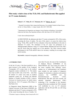
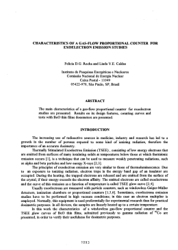
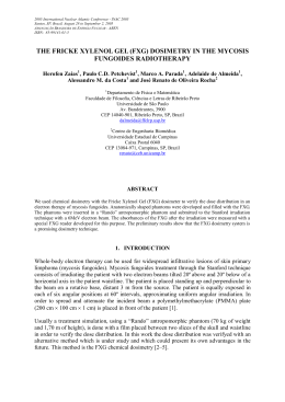
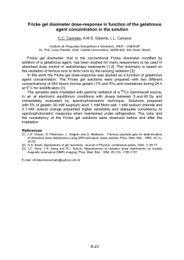
Artigo Original
Revista Brasileira de Física Médica. 2011;5(2):139-42.
The contribution of optically stimulated
luminescence dosimetry in quality control
in radiotherapy
A contribuição da dosimetria por luminescência opticamente
estimulada no controle de qualidade na radioterapia
Renato S. Fernandes1 and Yvone M. Mascarenhas2
Universidade de Mogi das Cruzes, Hospital das Clínicas da Faculdade de Medicina da Universidade de São Paulo – São Paulo (SP), Brazil.
2
Universidade de Mogi das Cruzes/Sapra Landauer – São Carlos (SP), Brazil.
1
Abstract
In 2011, Brazil will have more than 489,000 new cases of cancer. Of these patients, a considerable contingent will be submitted to
radiotherapy procedures. Thus, efficient systems that guarantee the quality of the beams used in radiotherapy procedures are extremely
important because collaborate with the overall success of treatment. This paper presents the use of OSL (optically stimulated luminescence)
dosimetry procedures in quality control in radiotherapy, making verification of the symmetry of treatment of radioactive fields emitted by
linear accelerators. The use of OSL dosimetry was compared to procedures performed daily, using ionization chambers. Dosimeters of
aluminum oxide doped with carbon (Al2O3: C) were distributed on a map, with the delimitation of a field 20x20 cm arranged as follows:
one in the center of the field; four equidistant distributed, forming a square 10x10 cm; and 4 remaining distributed equidistant, forming
a square of 20x20 cm. This arrangement is similar to equipment used for checking the symmetry of radioactive fields using ionization
chambers. The analysis of data obtained in the symmetry axis X made a variation of 0.2% in OSL dosimetry while using the equipment
provided with an ionization chamber, 0.1%. Noting the Y axis, the variation of data for OSL dosimetry was 0.04% and ionization chambers,
0.1%. The use of OSL dosimetry proved to be simple, with accessible instrumentation and making a possible reinterpretation of the data
obtained. However, the data suggest that the broader performance of procedures using OSL dosimetry, seeking greater familiarity with the
system, can reduce the variation in results.
Keywords: dosimetry, radiation therapy, quality control, cancer, ionization chambers.
Resumo
Em 2011, o Brasil terá mais de 489.000 novos casos de câncer. Desses pacientes, um notável grupo será submetido a procedimentos
de radioterapia. Desse modo, sistemas eficazes que garantam a qualidade dos feixes utilizados nos procedimentos de radioterapia são
extremamente importantes, pois colaboram com o sucesso total do tratamento. Este trabalho apresenta o uso dos procedimentos de
dosimetria de luminescência opticamente estimulada (OSL) no controle de qualidade na radioterapia, verificando a simetria dos campos
radioativos emitidos por aceleradores lineares. O uso de dosimetria OSL foi comparado aos procedimentos realizados diariamente, utilizando
câmaras de ionização. Dosímetros de óxido de alumínio dopados com carbono (Al2O3: C) foram distribuídos em um mapa, com a delimitação
de um campo de 20x20 cm organizado da seguinte maneira: um no centro do campo; quatro distribuídos equidistantes, formando um
quadrado de 10x10 cm; e quatro restantes, sendo distribuídos equidistantes, formando um quadrado de 20x20 cm. Essa organização é
similar ao equipamento utilizado para verificar a simetria dos campos radioativos que utilizam câmaras de ionização. A análise dos dados
obtidos no eixo X da simetria fez uma variação de 0,2% na dosimetria de OSL, enquanto ao utilizar o equipamento fornecido com uma câmara
de ionização fez 0,1%. Ao notar o eixo Y, a variação dos dados para a dosimetria de OSL foi de 0,04% e as câmaras de ionização, 0,1%. O uso
da dosimetria de OSL provou ser simples, com instrumentação acessível e tornou possível a reinterpretação dos dados obtidos. Entretanto, os
dados sugerem que a realização de procedimentos mais amplos utilizando dosimetria OSL, visando uma maior familiaridade com o sistema,
podem reduzir a variação dos resultados obtidos.
Palavras-chave: dosimetria, terapia de radiação, controle de qualidade, câncer, câmaras de ionização.
Corresponding author: Renato da Silva Fernandes – Hospital das Clínicas da FMUSP – Av. Dr. Enéas Carvalho de Aguiar, 255 – CEP: 05403-000 – São
Paulo (SP), Brazil – E-mail: [email protected]
Associação Brasileira de Física Médica®
139
Fernandes RS
Introduction
The Instituto Nacional do Cancer (INCA) showed, in
2009, an estimated new cancer cases in Brazil for the
year 2010. These data reported that more than 489,000
new patients were affected by some cancer in 2010. This
information will also be valid for the year 20111.
The use of radiotherapy, either alone or combined
with other methods (chemotherapy and surgery), has
emerged as an effective curative modality, especially in
malignant neoplasm in the initial stages2.
Radiotherapy is the use of ionizing radiation for therapeutic purposes. Radiation doses used are high and any
error during the treatment procedure can cause serious
patient injury, including death3.
The need to systematize actions for the quality control of radiotherapy treatments has been evident in recent
years4.
According to World Health Organization, the quality
control in radiotherapy is established based on the actions that ensure consistency between the clinical prescription and their administration to patients in relation to
the target volume, the lowest level in healthy tissue, minimal exposure of personal checks on the patient and
adequate patient monitoring for determining the outcome
of treatment5.
An efficient system of quality assurance in radiotherapy
minimizes errors in treatment planning and administration
of the dose to the patient, allows the intercomparison of results between different treatment centers and provides the
possibilities and functionalities of a processing equipment
to be used with a high level of consistency and accuracy4.
Several types of dosimeters can be used for quality control in radiotherapy beams: ionization chambers,
diodes, films, metal oxide semiconductor (MOSFET) and
TLD (thermoluminescent dosimeters)6-8.
New dosimeters and dosimetry procedures have
been developed. We can highlight the optically stimulated
luminescence dosimetry (OSL)9.
The arrival of use of technology in radiotherapy increases its effectiveness, as well as the errors incidence10.
It is critical that the verification procedures of the
beam are simple and reliable4.
Thus, the use of OSL dosimetry shows promise. It has
advantages such as high luminescence efficiency, stability, sensitivity, precision and accuracy, control of luminescence emitted, speed reading, rereading and possibility
of low power consumption, making possible the use of
handheld devices11.
The symmetry of the fields was the parameter chosen
for this analysis.
These dosimeters were arranged on a map, with the delimitation of a field 20x20 cm arranged as follows: one
in the center of the field; four equidistant distributed,
forming a square 10x10 cm; and the remaining four
equidistant distributed, forming a square of 20x20 cm
(Figure 1). The dosimeters were arranged in this way to
ensure intercomparison with data obtained during the
commissioning of the linear accelerator.
Dosimeters were placed on 1.5 cm of solid water
(acrylic) to ensure set build up (Figure 2).
The model was irradiated with 100 cGy at a distance
of 100 cm (isocenter), using a 6 MeV beam produced in
a linear accelerator from Varian Medical Clinac 600c sn
515. To read these dosimeters, it was used MicroStar
produced by Landauer Co. (Figure 3).
The data obtained were compared with readings taken every day from Double CheckTM equipment
(Figure 4), Victoreen®. This equipment consists of a
reader, equipped with nine ionization chambers which,
when irradiated by a radiation field of 20x20 cm, capture
signals in the center of the field, 5 and 10 cm. The readings were taken between 1 and 7 October 2010.
Figure 1. Placed in fields dosimeters 10x10 cm and 20x20 cm.
Materials and methods
OSL dosimeters were irradiated 9 (InLightTM with detectors of aluminum oxide doped carbon in the atmosphere).
140
Revista Brasileira de Física Médica. 2011;5(2):139-42.
Figure 2. Dosimeters arranged on the solid water. On the whole,
1.5 cm in the same field in order to ensure build up.
The contribution of optically stimulated luminescence dosimetry in quality control in radiotherapy
Discussion
The data obtained with the OSL dosimetry allow us to
observe a change in the symmetry of the field in the X
direction of approximately 0.2% (Figure 5A). With the
equipment use in the reading routine, this variation was
around 0.1%. Note that the fluctuations in the readings
are accompanied by two similar systems. In Y, we see
a variation around 0.04% when using the OSL dosimetry (Figure 5B). With the routinely use of the equipment,
we note a variation of 0.10%. Fluctuations in the readings are noted in a similar way in the two systems.
Conclusions
The OSL dosimetry was presented as an important tool
in the daily check of the symmetry of the radiation fields
emitted by linear accelerators.
Features, such as possibility of re-readings, stability,
sensitivity and ease of handling equipment, ensure wide
use OSL dosimetry in radiotherapy.
Expansion in the frequency and quantity measures can
provide more familiar to all, with steps leading to a smaller
range of variation.
More reading using the OSL dosimetry should be conducted to obtain more data for comparison with the system used routinely.
Figure 3. MicroStar Reader.
Acknowledgment
The authors thank the Department of Radiation
Oncology and Department of Medical Physics of Istituto
de Radiologia do Hospital das Clínicas da Faculdade de
Medicina da USP and Sapra Landauer for their support
in this work.
Figure 4. Double CheckTM model 7200, Victoreen®.
Results
-0,00%
-0,10% 0
-0,20%
-0,30%
-0,40%
-0,50%
-0,60%
-0,70%
-0,80%
-0,90%
1
2
3
4
5
0,10%
6
0,08%
Ionization
Chamber
OSL
Dosimetry
Readings
Variation of Simmetry Axis X - Ionization Chamber X OSL
Dosimetry
Variation
Variation
Variation of Simmetry Axis X - Ionization Chamber X OSL
Dosimetry
0,06%
Ionization
Chamber
0,04%
0,02%
-0,00%
-0,02%
OSL
Dosimetry
0
1
2
3
4
5
6
Readings
Figure 5. Comparison between data obtained with both ionization chamber and OSL dosimetry for variation of symmetry of the field
in the X (a) and Y (b) directions.
Revista Brasileira de Física Médica. 2011;5(2):139-42.
141
Fernandes RS
References
1. Brasil. Ministério da Saúde. Instituto Nacional de Câncer. Estimativa 2010:
incidência de câncer no Brasil/Instituto Nacional de Câncer. Rio de Janeiro:
INCA; 2009.
2. Perez CA, Brady LW. Principles and pratice of Radiation Oncology.
Philadelphia: JB Lippincott; 2004.
3. Berdaky MF, Caldas LVE. Implantação de um programa de controle de
qualidade de um acelerador linear de 6 MeV de fótons. Radiol Bras.
2001;34(5):281-4.
4. Brasil. Ministério da Saúde. Instituto Nacional de Câncer. TEC DOC - 1151:
aspectos físicos da garantia da qualidade em radioterapia. Rio de Janeiro:
INCA, 2000.
142
Revista Brasileira de Física Médica. 2011;5(2):139-42.
5. World Health Organization. Quality assurance in radiotherapy. Geneva:
WHO; 1988.
6. Bentel GC. Radiation therapy planning. New York: McGraw-Hill; 1996.
7. Hendee WR, Ibbott GS. Radiation therapy physics. St. Louis: Mosby; 1996.
8. Scaff LAM. Física da Radioterapia. São Paulo: Sarvier; 1997.
9. Yukihara, EG, Mckeever SWS. Optically stimulated luminescence (OSL)
dosimetry in medicine. Phys Med Biol. 2008;53(20):351-79.
10. Alvarez L. Tecnologia aumenta eficácia e incidência de erros da radioterapia.
O Estado de São Paulo [Internet] [cited 2011 Jan 16]. Available from: http://
www.estadao.com.br/estadaodehoje/20100208/not_imp507824,0.php.
11. McKeever SW, Moscovitch M. On the advantages and disadvantages of
optically stimulated luminescence dosimetry and thermoluminescence
dosimetry. Radiat Prot Dosimetry. 2003;104(3):263–70.
Baixar