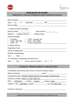
UNIVERSIDADE FEDERAL DE PERNAMBUCO CENTRO DE CIÊNCIAS BIOLÓGICAS PROGRAMA DE PÓS-GRADUAÇÃO EM CIÊNCIAS BIOLÓGICAS (PPGCB/CCB/UFPE) APLICAÇÃO DE HIDROLASES DE TILÁPIAS-DO-NILO (Oreochromis niloticus) COMO BIOMARCADORES DE EXPOSIÇÃO AO ALUMÍNIO VAGNE DE MELO OLIVEIRA RECIFE, 2011 VAGNE DE MELO OLIVEIRA APLICAÇÃO DE HIDROLASES DE TILÁPIAS-DO-NILO (Oreochromis niloticus) COMO BIOMARCADORES DE EXPOSIÇÃO AO ALUMÍNIO Dissertação apresentada ao Programa de PósGraduação em Ciências Biológicas da Universidade Federal de Pernambuco (PPGCB/UFPE) para o cumprimento parcial das exigências para obtenção do título de Mestre em Ciências Biológicas. Orientador: Prof. Dr. Ranilson de Souza Bezerra – Departamento de Bioquímica/CCB/UFPE Co-orientador: Prof. Dr. Luiz Bezerra de Carvalho Júnior – Departamento de Bioquímica/CCB/UFPE RECIFE, 2011 “Verás que um Filho Teu Não Foge à Luta” (trecho do hino nacional brasileiro) In memóriam de Maria das Dores, À minha vó com eterna gratidão. Vagne de Melo Oliveira Agradecimentos AGRADECIMENTOS Agradeço a Deus e a todos que acreditaram - e continuam acreditando - no meu potencial. Agradeço ao Programa de Pós-Graduação em Ciências Biológicas da Universidade Federal de Pernambuco (PPGCB/UFPE), nas figuras da professora Dra. Maria Tereza dos Santos Côrreia, e a Fundação de Amparo à Ciência e a Tecnologia do Estado de Pernambuco (FACEPE) por terem acreditado no meu projeto de Pós-Graduação. Agradeço aos meus orientadores, Prof. Dr. Ranilson de Souza Bezerra e ao Prof. Dr. Luiz Bezerra de Carvalho Junior, pela confiança, dignidade e paciência de terem me orientado. Além de um agradecimento especial a Helane Costa, Caio Rodrigues e Renata França, pessoas de caráter inigualáveis e das quais tenho imenso carinho e admiração, sem as quais, talvez, não tivesse obtido êxito em minha jornada. Agradeço aos colegas de trabalho, de descontração, de debates, de ensino, Anderson Henriques, Augusto Vasconcelos, Charlles, Danielli Freitas, Dário Alves, Danielli Matias, Douglas Holanda, Fábio Freitas, Fernanda, Gilmar Cezar, Janilson Félix, Juliette Silva, Karollina Lopes, Karina Ribeiro, Kelma Souza, Marina Marcuschi, Mirela Assunção, Raquel Pereira, Renata França, Robson Coelho, Thiago Cahú e Werlayne Mendes. Pessoas que fazem do LABENZ uma grande família. Agradeço a todos os companheiros de Pós-Graduação (UFPE), pelos momentos impagáveis das disciplinas, dentro e fora da sala de aula, em especial a Alessandra Paulino, pela amizade ofertada. Além dos colegas de departamento pelo apoio moral em todos os momentos, em especial a Meiriana Xavier. Agradeço ao Prof. Dr. Eduardo Isidoro Beltrão, pela atenção dedicada durante a realização deste projeto. Assim como a Carmelita, pela dedicação e generosidade. Agradeço aos amigos que durante todo o período estiveram ao meu lado sempre desejando o melhor, Alcidézio Junior, Onézia Lourdes, Flávio Batista, João Lamartine, Jackeline Souza, Paula Souza, Suely Almeida, Ana Elizabeth e Antônio Carlos. Por tudo, devo um agradecimento especial a Claudineide Vieira - por toda generosidade e afeto, a quem tenho como terceira mãe. E por fim, venho agradecer ao meu grande amigo e irmão Gian Carlo, pelo carinho, dedicação, incentivo e, sobretudo, inspiração; e ao meu pai e professor (in memoriam), Paulo Beijamin de Oliveira, por ter concebido a razão do meu viver. A todos, muito obrigado. i Vagne de Melo Oliveira Resumo RESUMO O alumínio é um elemento ubiquitário e sem papel biológico definido, aplicado em tratamento de água devido sua ação floculante. Seu acúmulo em ambientes aquáticos, mesmo em condições alcalinas, acarreta ação tóxica para a biota aquática, sobretudo quando a exposição se dá num período prolongado, causando doenças em especies de peixes podendo levar a morte. Organismosteste como a tilapia tem grande potencial em ferramentas no biomonitoramento de áreas impactadas. Objetivamos, avaliar enzimas hidrolases da tilápia-do-Nilo por indução a exposição de concentrações desse metal. Neste contexto, foram utilizadas sete enzimas de tilápias-do-Nilo O. niloticus, a saber: AChE cerebral, AChE muscular, BChE muscular, pepsina, tripsina, quimotripsina e amilase intestinal; como biomarcadores, através da indução a exposição ao alumínio em ensaios in vivo (análises dos grupos induzidos a exposição) e in vitro (indução por incubação a partir de amostras do grupo controle do ensaio in vivo). Os peixes foram cultivados e expostos a diferentes concentrações de alumínio, delineando três grupos experimentais: Tratamento Grupo Controle (TGC, sem exposição ao Al2(SO4)3); Tratamento Grupo 1 ppm de Al2(SO4)3 (TG1); Tratamento Grupo 3 ppm de Al2(SO4)3 (TG3). O cultivo foi realizado durante um período ininterrupto de 14 dias, com alimentação ad libitum, fotoperíodo 12:12, troca dinâmica da água (80%) e monitoramento diário dos parâmetros físico-químicos da qualidade de água. Em seguida, coletadas amostras de fígado e brânquias e imersas em CH2O (10%) para posterior análise, enquanto cérebro, músculo e intestino foram macerados e homogeneizados, obtendo o extrato bruto (E.B.) para as análises enzimáticas. Não houve alterações morfológicas nas avaliações das brânquias e do fígado da tilápia. As três enzimas colinesterásicas apresentaram aumento de atividade nos grupos expostos ao alumínio (TG1 e TG3), tanto nos ensaios in vivo quanto nos in vitro. A AChE cerebral teve atividade máxima de 126.93 ± 13.20% e 160.13 ± 10.92%, in vivo e in vitro, respectivamente, nos tratamentos expostos a concentração de 3 ppm (TG3), em relação ao tratamento grupo controle (TGC, 100%). Nas mesmas condições, também aumentou a da AChE muscular, com 163.81 ± 0.30% e 138.77 ± 4.07%. Entre as colinesterases, a BChE apresentou resultado mais expressivo no tratamento in vitro, 168.28 ± 5.49% e 196.17 ± 4.08%, para os grupos TG1 e TG3. Das quatro enzimas digestivas analisadas, a pepsina in vivo apresentou menor atividade, 65.01 ± 1.04% (TG1) e 61.06 ± 1.90% (TG3), embora todas as demais, tripsina, quimotripsina e amilase intestinal tenham demonstrado redução de sua atividade em ambos os ensaios, in vivo e in vitro. Nas análises experimentais in vivo a atividade da quimotripsina e da amilase intestinal demonstraram redução significativa entre os tratamentos, sobretudo em relação ao TG3, 73.0 ± 1.8% e de 60.20 ± 3.95%, respectivamente. A atividade in vitro da tripsina foi menor do que a in vivo, inclusive entre os tratamentos TG1 e TG3. Os resultados demonstram a relevância da aplicação de biomarcadores enzimáticos de O. niloticus. O aumento da atividade colinesterásica e a redução na atividade de principais enzimas do metabolismo digestivos, forneceram resultados que indicam a presença do alumínio. Sugerimos o procedimento como ferramenta útil e alternativa no monitoramento de áreas impactadas pelo metal. Palavras-chave: alumínio, biomarcador, colinesterases, enzimas digestivas, tilápias. ii Vagne de Melo Oliveira Abstract ABSTRACT Aluminum is a ubiquitous element and without defined biological role, applied in water treatment due to its action flocculant. Their accumulation in aquatic environments, even under alkaline conditions, causes toxic effects on aquatic biota, particularly when exposure occurs over an extended period, causing disease in fish species may lead to death. Test organisms such as tilapia has great potential tools for the biomonitoring of impacted areas. We aimed to evaluate the enzymes of Nile tilapia induction the exposure to concentrations of that metal. In this context, we used seven enzymes hydrolases of O. niloticus, namely, brain AChE, muscle AChE, muscle BChE, pepsin, trypsin, chymotrypsin and intestinal amylase as biomarkers, by inducing exposure to aluminum in vivo assays (analysis of the induced exposure groups) and in vitro (induction by incubation from the control group samples of the in vivo). The fish were cultured and exposed to different concentrations of aluminum, outlining three groups: Treatment Control (TGC, without exposure to Al2(SO4)3); Treatment Group 1 ppm of Al2(SO4)3 (TG1), Treatment Group 3 ppm of Al2(SO4)3 (TG3). The cultivation was conducted for a continuous period of 14 days, with feeding ad libitum, 12:12 photoperiod, dynamic exchange of water (80%) and daily monitoring of physico-chemical parameters of water quality. Then, samples of liver and gills and immersed in CH2O (10%) for further analysis, as brain, muscle and intestine were ground and mixed, obtaining a crude extract (CE) for enzyme analysis. There were no morphological changes in the assessments of the gills and liver of Tilapia. The three cholinesterase enzyme showed increased activity in the groups exposed to aluminum (TG1 and TG3) in both in vivo and in vitro. The brain AChE activity was maximum 126.93 ± 13.20% and 160.13 ± 10.92%, in vivo and in vitro, respectively, in treatments exposed to a concentration of 3 ppm (TG3), compared to treatment control group (TGC, 100%). Under the same conditions, also increased muscle AChE, with 163.81 ± 0.30% and 138.77 ± 4.07%. Among the cholinesterase, muscle BChE showed the most significant result in the in vitro treatment, 168.28 ± 5.49% and 196.17 ± 4.08% for groups TG1 and TG3. Of the four digestive enzymes analyzed, pepsin in vivo showed the lowest activity, 65.01 ± 1.04% (TG1) and 61.06 ± 1.90% (TG3), while all the others, trypsin, chymotrypsin and intestinal amylase have been shown to reduce intestinal activity in both studies, in vivo and in vitro. In the experimental analysis in vivo activity of chymotrypsin and intestinal amylase were also significantly reduced among the treatments, especially in relation to the TG3, 73.0 ± 1.8% and 60.20 ± 3.95%, respectively. The in vitro activity of trypsin was lower than in vivo, including among treatments TG1 and TG3. The results demonstrate the relevance of applying biomarkers enzymatic O. niloticus. The increased activity cholinesterase and decreases in activity of key enzymes in the digestive metabolism, provided results that indicate the presence of aluminum. We suggest the procedure as a useful alternative and in monitoring areas affected by metal. Key-words: aluminum, biomarker, cholinesterase, digestive enzymes, tilapia. iii Vagne de Melo Oliveira Lista de Figuras LISTA DE FIGURAS Capítulo I – Revisão de Literatura Figura 1 Exemplar de Tilápia-do-Nilo (Oreochromis niloticus), vista lateral 6 Figura 2 Ligação de um substrato ao sitio ativo de uma colinesterase 10 Figura 3 Classificação internacional das enzimas 11 Figura 4 Especificidade enzima-substrato 14 Figura 5 Estrutura tridimensional da acetlcolinesterase (AChE) 17 Figura 6 Esquematização dos processos de absorção de poluentes 23 Capítulo II: Artigo Científico Figure 1 Histology of liver and gills of tilapia 65 Figure 2 Cholinesterase activity (mU/mg) in vitro: effects of sulfate and chloride 66 Figure 3 Protein profile of alkaline 67 iv Vagne de Melo Oliveira Lista de Tabelas LISTA DE TABELAS Capítulo I – Revisão de Literatura Tabela 1 Evolução das publicações com bioindicadores ambientais 19 Capítulo II: Artigo Científico Table 1 Enzyme activity measured as biomarkers of exposure to aluminum: in vivo) and in vitro assay 62 Table 2 Inhibitory effect of aluminum on the activity of digestive proteases of Nile tilapia 63 v Vagne de Melo Oliveira Lista de Abreviações/ Siglas LISTA DE ABREVIAÇÕES AChE – Acetilcolinesterase BChE – Butirilcolinesterase BApNA - Benzoil arginina ρ-nitroanilida ChEs – Colinesterases CONAMA – Conselho Nacional do Meio Ambiente IBAMA – Instituto Brasileiro do Meio Ambiente e dos Recursos Naturais Renováveis IUBMB – International Union of Biochemistry and Molecular Biology kDa – Quilo Dalton PMSF– Fenil-metil-sulfonil fluoreto SBTI – Inibidores de tripsina de soja SDS - Sódio dodecil sulfato SDS-PAGE - Eletroforese em gel de poliacrilamida utilizando SDS SAPNA – succinil- alanina-fenilalanina-p-nitroanilina SUCPHEPNAN - Succinil fenilalanina ρ-nitroanilida TAME – Tosil-Arginina-Metil-Éster TCA - Ácido tricloroacético TGC – Tratamento grupo controle (sem exposição) TG1 – Tratamento grupo exposição (exposição: 1 ppm de alumínio) TG3– Tratamento grupo exposição (exposição: 3 ppm de alumínio) t – Toneladas TPCK – tosil-lisina clorometil cetona ZPCK – carbobenzoxi-fenil-clorometilcetona vi Vagne de Melo Oliveira Índice ÍNDICE AGRADECIMENTOS i RESUMO ii ABSTRACT iii LISTA DE FIGURAS iv LISTA DE TABELAS v LISTA DE ABREVIAÇÕES vi 1 INTRODUÇÃO 3 2 REVISÃO DE LITERATURA 5 2.1 Pescado: consumo e produção 5 2.2 Tilápia-do-Nilo Oreochromis niloticus (LINNAEUS, 1758) 6 2.2.1 Parâmetros físico-químicos para cultivo de tilápias 7 2.3 Características estruturais das vísceras de tilápias 2.4 Bioquímica enzimática 10 2.4.1 Pepsina (EC, 3.4.23.1) 13 2.4.2 Tripsina (EC, 3.4.21.4) 13 2.4.3 Quimotripsina (EC, 3.4.21.1) 14 2.4.4 Amilase intestinal (EC, 3.2.1.1) 15 2.4.5 Enzimas colinesterásicas (ChEs) 16 2.4.5.1 Acetilcolinesterase (EC, 3.1.1.7) 16 2.4.5.2 Butirilcolinesterase (EC, 3.1.1.8) 17 2.5 Metais pesados 2.5.1 Alumínio (III) 2.6 Bioindicadores e biomarcadores ambientais 3 8 OBJETIVOS 18 20 22 23 3.1 Objetivo geral 23 3.2 Objetivos específicos 23 4 REFERÊNCIAS 26 5 ARTIGO - Hydrolases from Nile tilapia as biomarkers of aluminum exposure: in vivo 37 and in vitro assay Abstract 39 5.1 Introduction 40 5.2 Materials and Methods 43 5.2.1 Chemicals and Equipment 43 5.2.2 Experimental design 43 5.2.2.1 In vivo assay 44 1 Vagne de Melo Oliveira Índice 5.2.2.2 In vitro assay 44 5.2.3 Cholinesterase enzyme extraction 44 5.2.4 Digestive Enzyme Extraction 44 5.2.5 Protein Determination 45 5.2.6 Assay for Non-specific Digestive enzymes 45 5.2.7 Enzyme Activity Assay 45 5.2.7.1 Acetylcholinesterase and butyrylcholinesterase 45 5.2.7.2 Chymotrypsin and Trypsin 45 5.2.7.3 Pepsin 46 5.2.7.4 Intestinal amylase 46 5.2.8 Polyacrylamide gel electrophoresis (PAGE) and zymogram 46 5.2.9 Histological examination 47 5.2.10 Statistical analysis 47 5.3 Results and discussion 47 5.3.1 Biometric parameters and physico-chemical quality of water 47 5.3.2 Cholinesterase activity 48 5.3.3 Digestive activities 48 5.3.4 Histology 48 5.4 Discussion Acknowledgement 5.5 References 48 53 53 6 CONCLUSÕES 68 7 ANEXOS 69 7.1 Normas da revista Aquatic Toxicology 69 7.2 Parecer do Comitê de bioética CCB/UFPE 77 2 Vagne de Melo Oliveira Introdução 1 INTRODUÇÃO A qualidade de vida humana é, direta ou indiretamente, dependente da saúde, vitalidade e vigor do ambiente que nos rodeia. Os componentes abióticos e bióticos, incluindo ar, água, solo, alimentos, plantas e animais constituem nosso meio ambiente, e através de uma série de interações, a vida é sustentada, entretanto, qualquer alteração num destes componentes promoverá graves repercussões sobre a continuidade do ciclo biológico (JHA, 2004). O ambiente aquático recobre dois terços do planeta e é habitado pela maioria das espécies existentes nos diferentes nichos ecológicos, além disso, muitas delas são importantes fontes de alimentação (TUZAN e SOYLAK, 2007). Como conseqüência do crescimento da população e o desenvolvimento industrial, a produção, o consumo e o descarte de produtos químicos corroboram para o aumento da produção de resíduos antropogênicos, acarretando em danos ecológicos (TURKMEN et al., 2005; ONER et al., 2009), pois muitos deles são, em última instância, depositados nos ecossistemas fluviais, sendo a maioria substancialmente tóxicos (FERNANDES et al., 2007; FIRAT e KARGIN, 2010). A exposição a metais pesados pode levar a vários efeitos tóxicos em animais aquáticos, incluindo dano tecidual, alterações respiratórias, bioquímicas, fisiológicas e, finalmente, mortalidade (DURAL et al., 2007). Portanto, os parâmetros enzimáticos e não enzimáticos ganham importância como ferramentas sensíveis para estimar os efeitos das exposições aos metais pesados antes da ocorrência dos efeitos nocivos nos organismos (ONER et al., 2009). Devido a essa ação deletéria, é necessário controlar sua bioacumulação em espécies-chave, viabilizando a observação da extensão temporal e espacial da contaminação, bem como uma avaliação do impacto potencial sobre a saúde do organismo e do ecossistema como um todo (FERNANDES et al., 2007). Em sistemas de água doce, o peixe é um produto que os seres humanos mais consomem por ser uma fonte potencial de proteínas e por ser de baixo preço comercial, beneficiando famílias de baixa renda (MENDIL et al., 2005). Além disso, são freqüentemente utilizados como biomonitores por oferecer vantagens específicas na descrição das características naturais dos ecossistemas aquáticos e na avaliação das mudanças nos habitats. Primeiro por serem espécies de uma vida longa e integrarem as flutuações dos poluentes ao longo do tempo, além disso, os compostos de maior concentração ao longo do tempo serão abundantes nos tecidos dos organismos mais antigos; segundo por estarem sempre presentes na água, permitindo o monitoramento (LAMAS et al., 2007). Entre as espécies aquáticas, as tilápias desfrutam de destaque mundial na aquicultura voltada para alimentação, além do seu potencial uso nos cultivos experimentais em laboratório (TENGJAROENKUL et al., 2000) e, dentre elas, a tilápia-do-Nilo (Oreochromis niloticus) tem sido utilizada como bioindicadora na produção de respostas aos efeitos que ocorrem em níveis mais 3 Vagne de Melo Oliveira Introdução elevados da organização biológica (FERNANDES et al., 2007), através de medidas em nível individual (medidas biométricas), populacional (redução reprodutiva, mortalidade, tamanho e biomassa) e de comunidade (perturbação do ciclo de nutrientes). Estes organismos são amplamente utilizados para obtenção de respostas a agentes estressores, e dessa forma, sua utilização contribui para avaliação de risco ecológico e na definição de estratégias adequadas para conservação das espécies mais afetadas por poluentes (TEJEDA-VERA et al. 2007). Do ponto de vista toxicológico, níveis de diferentes metais pesados estão sendo amplamente difundidos pela literatura, entretanto, dados acerca do alumínio em peixes é bastante limitado, apesar de sua crescente utilização em indústrias metalúrgicas, tratamento de água (devido sua ação floculante) e para fins domésticos. Tendo em vista a problemática abordada, faz-se necessário a realização de pesquisas que possam determinar o efeito deste metal em diferentes enzimas, tais como as colinesterásicas e as digestivas, tanto no âmbito in vivo - buscando se aproximar da realidade ambiental -, quanto in vitro, como forma de mensurar a ação direta do poluente, até então pouco estudado; alertando para prováveis danos ambientais e possíveis prejuízos à saúde pública. A utilização de parâmetros físicos, químicos e biológicos para aferir a qualidade da água de um sistema supostamente impactado como, por exemplo, de natureza bioquímica, baseiam-se nas respostas dos organismos em relação ao meio onde vivem. Avaliar a atividade enzimática de organismos supostamente expostos à ação de metais pesados constitui uma ferramenta relevante para fins de monitoramento e gestão, até pelo crescente parque industrial brasileiro. Com esse intuito, nossa pesquisa utiliza a tilápia nilótica - por ser bastante sensível à alteração de seu ambiente natural e apresentar relações com o metabolismo de outros vertebrados - como organismo-teste, ferramenta bioindicadora na exposição ao alumínio. . 4 Vagne de Melo Oliveira Capítulo I: Revisão de Literatura 2 REVISÃO DE LITERATURA 2.1 Pescado: consumo e produção nacional O pescado é uma importante parte na dieta diária de muitos países, por apresentar alta qualidade, proteínas de fácil absorção e uma grande variedade de vitaminas (tiamina, riboflavina, niacina) e minerais (cálcio, ferro, fósforo, iodo), contribuindo com um quarto da oferta mundial de proteína de origem animal. Possui altos níveis de ácidos graxos poliinsaturados, especialmente ômega-3, que diminui as taxas de colesterol no sangue. Na maioria dos países, peixes, moluscos, crustáceos e bivalves são fontes relevantes de emprego, lucro e moeda externa (DELGADO et al. 2003). O consumo per capita de pescado aumentou de 11,6 kg em 1971 para 16,4 kg em 2005, principalmente devido aos países em desenvolvimento. O rápido crescimento da população, juntamente com o aumento do valor médio de peixe consumido por pessoas (o consumo individual brasileiro individual em 2009 foi de aproximadamente 9 Kg/ano), acarretou num maior consumo global de peixes. Nesse cenário, a China é a responsável por grande parte desse crescimento, uma vez que sua quota de produção cresceu de 21% em 2004 para 35% em 2005, tendo como abastecimento per capita o valor de 26,1 kg (FAO, 2009). O Brasil apresenta grande potencialidade para a produção pesqueira, sobretudo, por ser um país de dimensões continentais. É o quinto maior país do mundo, possuindo 1,7% do território do globo terrestre e representando 47% da América do Sul, ocupando uma área de 8.514.876,599 km², 7.367 km2 de costa oceânica, 3,5 milhões de km2 de Zona Econômica Exclusiva (faixa marítima sobre o qual o país detém os direitos de exploração, conservação e administração de todos os bens nela existentes). Sua população está estimada em 190.732.694 milhões de habitantes, segundo o senso estatístico realizado em 2010 (IBGE, 2010), além de ser detentor da maior quantidade de água com potencial para a aqüicultura continental no mundo. É um país onde o consumo de pescado ainda é variado e vem apresentando um significativo crescimento ao longo dos anos (FAO, 2009). A produção nacional de pescado possui um índice superior a outras atividades, como a agricultura e a pecuária, por exemplo, sobretudo, por possibilitar boa lucratividade e um retorno financeiro rápido. Em 2009, essa produção atingiu 1.240.813,1 t (Pernambuco contribuiu com aproximadamente 23.4771 t), divididas entre todas as suas atividades - pesca marinha, continental, piscicultura e carcinocultura -, apresentando crescimento de 25,2% em relação a 2003. Na comparação do crescimento da produção das atividades zootécnicas no período de 2007 a 2009, observa-se que a aquicultura registrou crescimento relativo de 43,8%, ao passo que as principais criações da pecuária nacional, aves e suínos, registraram apenas 12,9 e 9,2%, respectivamente, e de bovinos uma retração na ordem de 8,6% (MPA, 2010). 5 Vagne de Melo Oliveira Capítulo I: Revisão de Literatura O valor da produção nacional de pescado compreendido no período entre 2007 e 2009, em reais, correspondeu a R$ 1.240.813,4. Neste cenário, a piscicultura teve um incremento de 90% da produção. Houve um crescimento de 34% no ano de 2008 em relação a 2007, crescendo de 210.640,5 t para 282.008,4 t. No ano 2009 a piscicultura apresentou a produção total de 337.353 t, representando um aumento de 20% em relação a 2008. Em relação à tilapicultura, a produção nacional vem apresentando um padrão de crescimento desde 1994, atingindo em 2009 um total de 39% do pescado proveniente da piscicultura nacional. A produção de tilápia entre 2003 e 2009 cresceu cerca de 105%, saindo de 64.857,5 t para 132.957,7 t, respectivamente. Ressalta-se que do ano de 2007 para 2008 aumentando de 17% saindo de 95.0910 t para 111.145,3 t e deste para 2009 elevando este valor para 132.957,8 t, fixando um crescimento de 20%, tornando-se o carro chefe da produção aquícola do país (MPA, 2010). 2.2 Tilápia-do-Nilo Oreochromis niloticus (LINNAEUS, 1758) A tilápia foi um dos primeiros peixes criados em aquicultura pelos antigos egípcios, há aproximadamente 4.000 anos e, embora a distribuição mundial significativa de tilápias – principalmente a de Moçambique Oreochromis mossambicus - tenha ocorrido entre as décadas de 1940 e 1950, a difusão maior deste peixe ocorreu a partir da década de 1960 até a de 1980. A tilápia-do-Nilo, Oreochromis niloticus (Fig. 1), foi introduzida do Japão para a Tailândia, em 1965, deste último, enviados para outros países asiáticos e africanos (KUBITZA, 2006). Da Costa do Marfim, foi introduzida no Brasil em 1971, sendo enviados na mesma década para os Estados Unidos e a China, que hoje é líder mundial na produção de tilápia e produziu mais da metade da produção global nos anos de 1992 a 2003 (FAO, 2010). Figura 1: Exemplar de tilápia-do-Nilo (Oreochromis niloticus) em vista lateral. Fonte: Acervo pessoal. 6 Vagne de Melo Oliveira Capítulo I: Revisão de Literatura Segundo Kubitza (2000), mais de setenta espécies de tilápias já foram identificadas e catalogadas, dentre as quais, quatro conquistaram o mercado mundial: a tilápia-do-Nilo ou tilapia cinza (O. niloticus), a tilápia azul ou áurea (O. aureus), a tilápia-de-Moçambique ou vermelha (O. mossambicus) e a tilápia Zanzinbar (O. urolepis hornorum). No Brasil são cultivadas diversas linhagens de tilápias, dentre as quais a de cor cinza e a de cor vermelha merecem destaque pela produção e consumo nacional (KUBITZA, 2006). A O. niloticus é uma espécie tropical e subtropical, que prefere viver em águas doce e rasa (IGARASHI, 2008), alimentando-se de fitoplâncton, plantas aquáticas, pequenos invertebrados, fauna bentônica e detritos (ROTTA, 2003). Sistematicamente, são peixes teleósteos (Teleostei), possuindo uma das três infraclasses da classe Actinopterygii (do grego aktis, raio + pteryx, nadadeira; asa) de peixes ósseos (RODRIGUES, 2009). A característica biológica da O. niloticus inclui: corpo compacto; espessura do pedúnculo caudal igual ao comprimento, escamas ciclóides, protuberância similar a um botão, ausente na superfície superior do focinho. O comprimento da mandíbula superior não apresenta dimorfismo sexual (não diferindo entre os sexos) e o primeiro arco branquial apresenta de 27 a 33 trilhas/rastros. Peixes desta espécie apresentam ainda linha lateral interrompida, partes espinhosas e partes moles de raios da nadadeira dorsal contínuas, nadadeira dorsal com 16 a 17 espinhos, sendo a anal com 3 e a caudal é truncada. Atinge maturidade sexual em torno de 6 meses, além de serem altamente prolíferas (podem gerar 4 desovas/ano) (KUBITZA, 2005; FAO, 2010). A maioria das espécies protege sua cria na boca, onde são chocados. Isso ajuda os ovos a ficarem oxigenados e os previne de serem atacados por bactérias, fungos e demais predadores (KUBITZA, 2000). A tilápia é a espécie mais utilizada, entre os peixes de água doce, para cultivo comercial, por apresentar melhor desempenho entre os peixes, sobretudo os machos, além de possuir boas características zootécnicas, organolépticas, ser de boa aceitação pelo público; de seu valor nutricional em termos de proteínas (IGARASHI, 2008). Com relação ao filé, o mesmo é desprovido de espinhas intramusculares, e possui bom rendimento (KUBITZA et al., 2007). Somado a isso, está o amplo conhecimento técnico – tais como cultivo, alimentação, manejo -, e biológico acerca da espécie, o que viabiliza sua utilização econômica, potencializando-a para industrialização (ISHIKAWA et al., 2008). 2.2.1 Parâmetros físico-químicos para cultivo de tilápias As tilápias crescem e até conseguem se reproduzir em água salgada ou salobra, sem apresentar problema com odores desagradáveis, pois são bastante tolerantes à salinidade (KUBITZA, 2005). São peixes de águas quentes, preferindo ambientes entre 21º e 35ºC, ainda que 7 Vagne de Melo Oliveira Capítulo I: Revisão de Literatura tolerem até 15ºC ou acima de 35ºC, quando bem nutridas. Temperaturas abaixo de 20° tendem a reduzir o consumo de alimentos, retardar o crescimento e tornar o peixe mais susceptível as doenças (KUBITZA, 2000). O aumento da temperatura da água é proporcional a atividade metabólica, ao consumo de alimento e, por conseguinte, ao crescimento. A faixa de temperatura mais adequada para juvenis de O. niloticus é entre 28 e 32ºC, com melhoria no desempenho produtivo e na composição corporal. Além disso, o conforto térmico é um fator externo que pode influenciar na atividade enzimática digestiva de peixes, uma vez que a quantidade de pepsinogênio é influenciada por ela, sendo proporcional a esta e reduzida à medida que a temperatura diminui (ROTTA, 2003). A tilápia pode sobreviver numa larga faixa de pH, embora se desenvolva melhor entre 6 e 9. Quando exposta a baixo pH, apresenta sinais de asfixia (movimentos operculares acelerados e boquejamento na superfície), enquanto o corpo e as brânquias apresentam excesso de muco. Já quando a acidez é excessiva pode acarretar num aumento da secreção mucóide, irritação e inchaço nas brânquias, culminando com a destruição do tecido branquial, afetando o dinamismo do peixe (KUBITZA, 2000). Esta é uma espécie que resiste a baixas concentrações de oxigênio dissolvido (OD), suportando valores inferiores a 0.3 mg/L (POPMA e MASSER, 1999). Para Kubitza (2000) os alevinos de O. niloticus de 10 a 25 g, podendo sobreviver à exposição ao oxigênio dissolvido entre 0.4 a 0.7 mg/L, por 3 a 5 horas, até quatro manhãs consecutivas, sem registro de significativo de mortalidade. Ainda segundo o autor, quando a concentração de oxigênio dissolvido atinge um percentual entre 45 a 50% da saturação (cerca 3 a 3,5 mg/L, a 28 e 30°C), adultos desta espécie começam a reduzir sua atividade e, por conseguinte, seu consumo de oxigênio, numa espécie de mecanismo compensador pela redução do oxigênio da água de cultivo. As tilápias ainda resistem a altas concentrações de amônia tóxica e a baixa qualidade da água, comparadas a maioria dos peixes cultivados (DELONG et al., 2009). 2.3 Características estruturais das vísceras de tilápias As brânquias são órgãos de vital importância para os peixes, ocupando o primeiro lugar entre os órgãos envolvidos na manutenção da saúde e metabolização de nutrientes, servindo de parâmetros para o bom desenvolvimento destes animais. Sua multifuncionalidade, a vasta área de superfície que ocupa e a sua localização relativamente ao meio externo faz das brânquias um órgãochave para a ação dos poluentes existentes no meio aquático, assim qualquer alteração nesta estrutura causará um desequilíbrio orgânico comprometendo a sobrevivência e desempenho dos peixes. Dessa forma, alterações histológicas das brânquias são reconhecidas como um método rápido e válido para determinar os danos causados pela exposição a diferentes irritantes nos peixes. O epitélio branquial apresenta-se estratificado, sendo constituído por diversos tipos celulares, em 8 Vagne de Melo Oliveira Capítulo I: Revisão de Literatura particular, por três: as pavimentosas que aparecem em maior frequência e possuem função de trocas gasosas; as células cloreto responsáveis pela regulação iônica, equilíbrio ácido-base e excreção de metabólitos nitrogenados, e as células mucosas, responsáveis pela produção de muco, o qual possui função de lubrificação e bactericida (CARVICHIOLO, 2009). A mucosa estomacal de teleósteos é revestida por epitélio simples cilíndrico composto de células com núcleo basal e glândulas gástricas tubulares na lâmina própria. De acordo com o hábito alimentar, as glândulas gástricas podem sofrer alterações morfológicas (SANTOS et al., 2007). O intestino apresenta glândulas digestivas e um suprimento abundante de vasos de sangue e de linfa, além de ser o local onde ocorre a maior parte da absorção dos nutrientes, íons e água provenientes da dieta. Os cecos pilóricos estão presentes em algumas espécies, como nos salmonídeos, e ausentes em outras, como nas tilápias. Nesse caso, ocorre um maior desenvolvimento da mucosa e/ou maior comprimento do intestino médio para compensar a escassez ou ausência dessas estruturas. Os teleósteos apresentam como característica básica a presença de pelo menos dois segmentos intestinais. No primeiro ocorre a absorção de nutrientes em suas formas menores (monossacarídeos, aminoácidos e ácidos graxos), enquanto que, no segundo, ocorre à entrada de macromoléculas pelo processo de pinocitose (ROTTA, 2003; CAVICHIOLO, 2009). O fígado é o maior e mais versátil dos órgãos metabólicos, ocupante de uma posição central no metabolismo do organismo (SANTOS et al., 2004; JUNQUEIRA e CARNEIRO, 2008; RODRIGUES, 2009). Este órgão é um dos locais mais ativos na metabolização, desempenhando funções anabólicas, como a síntese de proteínas; e catabólicas, como metabolização do glicogênio e do nitrogênio, detoxicação, além de processos biotransformativos e tentativas de neutralização dos efeitos dos poluentes químicos ou metabólicos (WILLIAMS e IATROPOULOS, 2002; CAVICHIOLO, 2009). Os hepatócitos podem ser considerados o primeiro alvo da toxicidade de uma substância, o que caracteriza o fígado como um órgão biomarcador da poluição ambiental (SANTOS et al., 2004). A literatura relata que, no fígado, o metabolismo de biotransformação e detoxicação de compostos químicos, dentre eles dos xenobióticos, está intimamente relacionado a variações individuais e fatores externos, como a temperatura, o pH, turbidez do meio, salinidade e alimentação (DER OOST et al., 2003; RODRIGUES, 2009). As alterações histopatológicas são excelentes ferramentas de marcação biológica de exposição a agentes químicos por serem compatíveis às respostas bioquímicas, principalmente alterações nas cinéticas enzimáticas. Neste sentido, análises de vísceras como brânquias, fígado, estômago e/ou intestino podem subsidiar num processo de investigação biológica. A análise morfológica de tecidos também fornece informações importantes a respeito da fisiopatologia celular. Assim, padrões bioquímicos, teciduais e anatômicos podem ser analisados como 9 Vagne de Melo Oliveira Capítulo I: Revisão de Literatura biomarcadores de exposição de contaminantes em peixes e outros organismos aquáticos (VAN DER OOST et al., 2003). 2.4 Bioquímica enzimática As enzimas são proteínas especializadas na catálise de reações biológicas, além de eficientes dispositivos moleculares que determinam os padrões de transformações químicas, tornando-se essenciais para a manutenção da vida (KOOLMAN e ROEHM, 2005). Atuam de forma altamente específica com seus substratos. A reação enzimática ocorre no interior de uma cavidade na enzima chamada sítio ativo (Fig. 2), onde a molécula que se liga a essa região é contornada com resíduos de aminoácidos cujos grupos substituintes se ligam ao substrato e catalisam a sua transformação (NELSON e COX, 2004). Trp286 Substrato Tyr72 Figura 2: Ligação de um substrato ao sitio ativo de uma colinesterase. Fonte: Adaptado de Silman e Sussman, (2005). Para evitar ambigüidade, a International Union of Biochemistry and Molecular Biology (IUBMB) classificou as enzimas em seis classes, de acordo com o tipo de reação especifica que catalisam (NELSON e COX, 2004). As enzimas com especificidades de efeito semelhantes são reunidas em uma das seis classes principais, como ilustrado na figura 3. As enzimas óxido-redutases catalisam a transferência de equivalentes de redução entre dois sistemas de óxido-redução; as transferases catalisam a transferência de outros grupos de uma molécula para outra; as hidrolases - objeto de nosso estudo (classe 3, grupo das hidrolases) - são uma classe que catalisa reações de hidrólise de ligação covalente. Enzimas desta classe que promovem a cisão de um material orgânico por adição de água. Elas catalisam a hidrólise de ligações éster, éter, peptídico, glicosila, anidrido-ácido, C-C, C-haleto ou P-N. Como, por exemplo, as enzimas que agem nas ligações ésteres como a acil-colina acil-hidrolase (pseudocolinesterase), as 10 Vagne de Melo Oliveira Capítulo I: Revisão de Literatura que atuam sobre compostos glicosila como o β-D-galactosídase e as que atuam sobre ligações peptídicas. As liases catalisam a quebra ou a formação de uma ligação química, com a qual ocorre a formação ou o desaparecimento de ligações duplas; já as isomerases modificam grupos dentro de uma molécula, sem alterar a fórmula molecular do substrato; e, por fim, as ligases, que catalisam reações dependentes de energia e, por isso, encontram-se acopladas à hidrólise fosfatos de nucleosídeos (geralmente ATP) (KOOLMAN e ROEHM, 2005). CLASSE TIPO DE REAÇÃO Equivalente a redução SUBCLASSES Oxirredutases Oxidases Peroxidases Redutases Transferases C- transferases Aminotransferases Fosfotransferases Hidrolases Esterases Glicosidases Peptidases s Liases C-C-Liases C-O-Liases C-N-Liases Isomerases Epimerases Cis trans isomerases Transferases Ligases C-C-Ligases C-O-Ligases C-N-Ligases Figura 3: Classificação internacional das enzimas. C- carbono; O- oxigênio; N- nitrogênio; S- sintases. red – redução; oxi – oxidação. Fonte: Adaptado de Koolman e Roehm, (2005). A diversidade biológica das espécies de peixes fornece uma ampla variedade de enzimas com propriedades únicas. As proteases são enzimas que quebram ligações peptídicas entre os aminoácidos das proteínas, cujo mecanismo é denominado de clivagem protéica, catalisando a hidrólise total das proteínas, comum em processos de ativação ou inativação de enzimas, envolvido principalmente na digestão (RAO et al., 1998). Elas são enzimas que desempenham um papel essencial no crescimento e sobrevivência de todos os organismos vivos. Para os animais marinhos, proteases são produzidas predominantemente pelas glândulas digestivas dentre elas, pepsina, 11 Vagne de Melo Oliveira Capítulo I: Revisão de Literatura gastricsina, tripsina, quimotripsina, colagenase, elastase, carboxipeptidase e esterase carboxila (KLOMKLAO, 2008). Genericamente, as proteases são divididas em dois grupos principais, dependendo do seu local de ação: endopeptidases e exopeptidases. As endopeptidases são caracterizadas por sua ação preferencial nas regiões internas da cadeia polipeptídica, entre as regiões N- (α-amino) e C- (αcarboxila) terminal. As endopeptidases são divididas em quatro subgrupos com base no seu mecanismo catalítico, (i) proteases de serina, (ii) aspártico proteases, (iii) proteases de cisteína, e (iv) metaloproteases. As exopeptidases atuam apenas nos finais das cadeias polipeptídicas na região N ou C terminal. Aquelas que atuam na região amino terminal livre liberam um único resíduo de aminoácido (aminopeptidases), um dipeptídeo (dipeptidil-peptidases) ou um tripeptídeo (tripeptidilpeptidases). As exopeptidases que atuam na região carboxi terminal livre liberam um único aminoácido (carboxipeptidases) ou um dipeptídeo (peptidil-dipeptidases) (RAO et al., 1998). As proteases ainda são classificadas segundo o valor do pH no qual apresentam atividade máxima, diferenciando-se dessa forma em: proteases ácidas, neutras ou alcalinas (RAO et al., 1998). Tanto as ácidas quanto as básicas estão, geralmente, presentes em peixes que apresentam estômago bem delimitado e funcional, como por exemplo, na O. niloticus, enquanto espécies agástricas, como o Utah-sugekarpe Catostomus ardens, geralmente possuem apenas a básica, onde o esôfago está diretamente relacionado com o intestino, sendo ao nível deste que ocorrerem os processos bioquímicos essenciais da digestão, consequência da atividade enzimática oriunda do pâncreas ou da sua própria mucosa (SEIXAS FILHO, 2003). De acordo com a IUBMB, as proteases estão inseridas no subgrupo 4 do grupo 3 (hidrolases), pois clivam a proteína adicionando uma molécula de água à ligação peptídica (BERG et al., 2004). Em tilápias, proteases como pepsina, tripsina e quimotripsina estão diretamente envolvidas com o processo de digestão, atuando no processo de degradação de peptídeos (TENGJAROENKUL et al., 2000; EL-BELTAGY et al., 2004). Estas enzimas (pepsina, tripsina e quimotripsina) já foram extraídas, purificadas e caracterizadas a partir de vísceras de peixes de diferentes espécies, como no caso da pepsina com o peixe Luciano-vermelho Lutjanus purpureos (ZHOU et al., 2007), mandarim Siniperca chuatsi (ZHOU et al., 2008) e no albacora branca Thunnus alalunga (NALINANON et al., 2010); no caso da tripsina com o aruanã-dourado Scleropages formosus (NATALIA et al., 2004), tilápia-do-Nilo Oreochromis niloticus (BEZERRA et al., 2005) e do celacanto de Moçambique Latimeria chalumnae (TANJI et al., 2007); e, por fim, da quimotripsina como o acará-disco Symphysodon aequifasciata (CHONG et al., 2002) e a sardinopa-chilena Sardinops sagax caeruleus (CASTILLO-YÁÑEZ et al., 2006). 12 Vagne de Melo Oliveira Capítulo I: Revisão de Literatura 2.4.1 Pepsina (EC, 3.4.23.1) A pepsina (EC, 3.4.23.1) é a principal enzima digestiva estomacal de peixes, comumente utilizada para aplicação industrial (KAGEYAMA, 2002; KLOMKLAO et al., 2007), incluída na categoria das endopeptidases que clivam as ligações peptídicas das proteínas pelo seu lado amino terminal dos resíduos de aminoácidos cíclicos aromáticos, como a tirosina e a triptotirosina (WHITAKER, 1994), rompendo as longas cadeias polipeptídicas em peptídeos menores e em alguns aminoácidos livres (KOOLMAN e ROEHM, 2005). A enzima ativa é lançada a partir do seu zimogênio - o pepsinogênio - por autocatálise na presença de ácido clorídrico (HCl). É uma aspartil protease (DE LUCA et al., 2009), por depender de resíduos do ácido aspártico para sua atividade catalítica. Para a maioria das espécies de teleósteos, está enzima apresenta pH ótimo em torno de 2 (RAO et al., 1998; ROTTA, 2003), enquanto especificamente para tilápia é de 2,5, além de temperatura ótima de 35°C (EL-BELTAGY et al., 2004), tendo na hemoglobina seu principal e mais utilizado substrato (KLOMKLAO, 2008). O peso molecular da pepsina em tilápias nilóticas é de 31,00 kDa. A atividade enzimática é inibida quase que completamente pela pepstatina A (especifico para aspartato proteases), um inibidor competitivo, que se liga fortemente aos resíduos do sítio ativo da enzima e parcialmente pelo EDTA (especifico para metaloproteases). Em contrapartida, a atividade aumenta quando da presença de cátions bivalentes, como o CaCl2, MgCl2 e o CoCl2 (KLOMKLAO et al., 2007). 2.4.2 Tripsina (EC, 3.4.21.4) A tripsina (EC, 3.4.21.4) é a principal enzima digestiva intestinal, responsável pela hidrólise de proteínas da dieta (BARKIA et al., 2009). A enzima tripsínica é uma serinoprotease que hidrolisa ligações peptídicas em que as carboxilas são constituídas por resíduos de lisina e arginina (RAO et al., 1998; KLOMKLAO, 2008). Os zimogênios secretados pelo pâncreas são ativados por clivagem proteolítica (KOOLMAN e ROEHM, 2005) no intestino pela enteroquinase, convertendo o zimogênio pancreático tripsinogênico em tripsina (KOLKOVSKI, 2001; KANNO et al., 2009) pela remoção de um hexapeptídio N-terminal. A tripsina, subsequentemente, converte outras moléculas de tripsinogênio em tripsina. De tal modo, a enteroquinase desencadeia uma cascata de atividade proteolítica, pois a tripsina é o ativador comum de todos os zimogênios pancreáticos, como por exemplo, o quimotripsinogênio (KOLKOVSKI, 2001; ROTTA, 2003; KLOMKLAO et al., 2008). As tripsinas de animais marinhos lembram as de mamíferos no seu peso molecular, entre 21 a 30,00 kDa (WANG et al., 2010), possuindo a tilápia nilótica aproximadamente 23,5 kDa (BEZERRA et al. 2005); composição de aminoácidos e sensibilidade a inibidores. Apresenta 13 Vagne de Melo Oliveira Capítulo I: Revisão de Literatura inibição ou instabilidade em m pH abaixo de 5,0 e acima de 11,0 e inibição por diisopropildiisopropi fluorfosfato (DFP) e pelo fluoreto fenilmetilsulfonil fenilmetil (PMSF,, inibidor de serinoprotease) serinoprotease (KISHIMURA et al., 2008; MARCUSCHI et al., 2010), 2010) inibidor de tripsina de soja (SBTI) e aprotonina. Esta enzima hidrolisa hidro substratos sintéticos como N-α-benzoil benzoil-DL-arginina-pnitroanilida (BApNA) e tosil-arginina arginina-metil-éster (TAME) (WHITAKER, 1994). 1994) São enzimas sensíveis a diversos íons metálicos, tais como Ca2+ e Mg2+, sendo inativadas pelos íons de Fe2+, Mn2+, Cu2+, Al3+, Ba2+, Co2+ e Zn2, apresentando do temperatura ótima em torno de 35-50°C 35 (ALENCAR ALENCAR et al. 2003; BEZERRA et al. 2005; SOUZA et al., 2006). 2006 2.4.3 Quimotripsina (EC, 3.4.21.1) A quimotripsina (EC, 3.4.21.1) é uma endopeptidase membro da família de serinoproteases, armazenada no pâncreas na forma de um precursor, o quimotripsinogênio, ativada através de um efeito cascata a partir da ativação da tripsina (RAO et al., 1998). Ao sofrer clivagem com a tripsina, é quebrada em duas partes, que ainda ficam atreladas por uma ligação dissulfeto. Quando Quan o quimotripsinogênio quebrado perde dois peptídeos pequenos, numa etapa chamada de transproteólise, obtém-se se a quimotripsina. É uma enzima específica para a hidrólise de ligações peptídicas em que grupos carboxila são fornecidos por um dos três aminoácidos aminoácidos aromáticos, ou seja, fenilalanina, tirosina e triptofano (APPLEBAUM ( e HOLT, 2003; KUZ’MINA e GOLOVANOVA, 2004). A principal ação da quimotripsina é denominada de tríade-catalítica, tríade catalítica, uma rede de ligações de hidrogênio entre a Ser195, His57 e a Asp102. Tanto anto ela quanto a tripsina contém a mesma "tríade" de resíduos cataliticamente ativos,, como ilustrado na figura 4 (EISENMENGER e REYES-DEREYES CORCUERA, 2009). Substrato –X-Ag-YAsp -189 Substrato –X-Phe-YSer -189 Ser -195 Ser -195 His -57 Asp -102 His -57 Asp -102 Tripsina –X–Y–Arg Arg (Lys)–Z– (Lys) Quimotripsina –X–Y–Tyr (Trp, Phe, Leu)–Z– Figura 4: Especificidade enzima-substrato. substrato. 1 – Tripsina (foto esquerda); 2- Quimotripsina (foto direita); Asp: aspartato; His: histidina; Ser: serina. – local de clivagem. Fonte: Adaptado de Koolman K e Roehm, (2005 2005). 14 Vagne de Melo Oliveira Capítulo I: Revisão de Literatura Quimotripsinas de peixes apresentam peso molecular variando entre 22 a 30,00 kDa, temperatura ótima oscilando de 45 a 55°C, e pH ótimo entre 7 a 9 (ALENCAR et al., 2003; CASTILLO-YAÑEZ et al., 2009; YANG et al., 2009). Esta enzima apresenta sensibilidade a determinados inibidores específicos, tais como tosil fenilalanina clorometil cetona (TPCK, inibidor de serinoproteases), N-carbobenzoyi-Lphenylalanine clorometil cetona (ZPCK, inibidor de quimotripsina-like) e inibidor de tripsina de soja (SBTI). São sensíveis a diversos íons metálicos, tais como Ca2+ e Mg2+, enquanto que inativados pelos íons de Fe2+, Mn2+, Cu2+ e Zn2 (YANG et al., 2009). São instáveis a temperatuas acima de 55°C e em condições ácidas (DE VECCHI e COPPES, 1996), além de hidrolisar substratos sintéticos como o N-succinyl-Ala-Ala-Pro-Phe-pNa (SApNA) e o Succinil fenilalanina ρ-nitroanilida (Suc-Phe-p-Nan) (CASTILLO-YAÑEZ et al., 2009). 2.4.4 Amilase intestinal (EC, 3.2.1.1) Os peixes não possuem atividade de amilase salivar, diferindo dos mamíferos, sendo a produção desta enzima restrita ao pâncreas e intestino, sobretudo em animais onívoros e herbívoros (ALARCÓN et al., 2001), como é o caso do tambaqui (Colossoma macropomum) e da tilápia nilótica (MOREAU et al., 2001), espécies onívoras, apresentando maior atividade enzimática na região dos cecos pilóricos e intestino proximal. Peixes carnívoros e os de águas frias apresentam limitada secreção e atividade de amilase no trato intestinal, o que é suficiente apenas para digerir uma pequena quantidade de carboidratos, atacando as ligações glicosídicas α-1-4, produzindo a partir do amido uma variedade de oligossacarídeos. O aumento da produção de amilase pode ocorrer em resposta à presença de carboidratos ou de produtos da sua hidrólise no lúmen do trato gastrintestinal (MOREAU et al., 2001; ROTTA, 2003). As amilases são divididas em dois grupos: as endoamilases e as exoamilases. As endoamilases catalisam hidrólises de forma aleatória no interior da molécula do amido. Essa ação causa a formação de ramos lineares de oligossacarídeos de cadeias de vários comprimentos e dessa forma quebram as ligações glicosídicas α-1,4 presentes na parte interna (endo) das cadeias de amilose ou amilopectina. A α-amilase é a endoamilase mais conhecida. As exoamilases hidrolisam exclusivamente ligações glicosídicas α-1, 4, como a β-amilase ou ambas as ligações α-1,4 e α-1, 6, como amiloglicosidase e glicosidase (GUPTA et al., 2003). A atividade amilolítica intestinal pode ser inibida pela ação de inibidores específicos de αamilase tipo I. O pH ótimo desta enzima varia de acordo com a espécie, entre 6 e 9, enquanto a temperatura ótima situa-se entre 25 e 55°C - na tilápia nilótica é de aproximadamente 32°C -, embora espécies como pargo (Pagrus pagrus) e sargo-alcorraz (Diplodus annularis) permaneçam estáveis a temperatura superiores a 60°C (HIDALGO et al., 1999; ALARCÓN et al., 2001; 15 Vagne de Melo Oliveira Capítulo I: Revisão de Literatura FERNÁNDEZ et al., 2001). O peso molecular desta enzima vai variar de acordo com a origem em questão, entre 10 a 210,00 kDa. As α-amilases microbianas apresentam peso molecular entre 50 e 60,00 kDa, sendo que as α-amilases bacterianas apresentam variação de 28 a 78,00 kDa e as αamilases fúngicas de 41 a 69,00 kDa. Alguns peixes apresentam peso molecular maior que 100,00 kDa, como é o caso do dourada Sparus aurata (ALARCÓN et al., 2001). Íons de Al3+, Ag2+, Hg2+, Cu2+, Mn2+, Zn2+, Fe3+, Cd2+, Pb2+ e de Co2+ são inibidores em potencial para está enzima, enquanto Ca2+, K+ e Mg2+ ativam ou pelo menos conferem estabilidade para sua atividade (GUPTA et al., 2003). 2.4.5 Enzimas colinesterásicas (ChEs) Há dois tipos de colinesterases em mamíferos: a acetilcolinesterase (AChE, EC 3.1.1.7) e a butirilcolinesterase (BChE; EC 3.1.1.8), que também é chamado de pseudo-colinesterase ou colinesterase não-específica. Colinesterases são distinguidas principalmente pela sua especificidade de substrato: AChE hidrolisa a acetilcolina, a colina mais eficientemente do que outros ésteres, enquanto BChE hidrolisa principalmente a butirilcolina e outras colinas (SALLES et al., 2006; RODRÍGUEZ-FUENTES et al., 2008; SOLÉ et al., 2010). A principal função da AChE é a hidrólise de acetilcolina, um neurotransmissor nas sinapses colinérgicas do sistema nervoso, mas o papel da BChE continua a ser elucidado (ADRESI, 2003; MONTEIRO et al., 2005). 2.4.5.1 Acetilcolinesterase (EC, 3.1.1.7) A AChE é freqüentemente descrita como uma enzima perfeita porque suas propriedades catalíticas se conjugam para aproximar sua atividade do limite máximo de velocidade permitido pela própria difusão do substrato no meio circundante (TÕUGU, 2001; SILMAN e SUSSMAN, 2005; DVIR et al., 2010). Ela contém dois sítios catalíticos, um sítio esterásico e um sítio aniônico. O sítio esterásico da acetilcolinesterase situa-se dentro de uma cavidade estreita e é constituído de uma tríade catalítica formada pelos resíduos dos aminoácidos serina 200, histidina 440 e glutamato 327 (podendo variar ligeiramente suas posições). Na catálise, o sítio aniônico, situado às bordas da cavidade, atrai fortemente o nitrogênio quaternário, carregado positivamente, da acetilcolina. Uma vez dentro da fenda catalítica, a acetilcolina sofre o ataque nucleofílico da serina (desprotonada pelo resíduo de histidina) ao seu carbono carbonílico, criando um intermediário tetraédrico estabilizado por pontes de hidrogênio o qual, num primeiro momento, forma serina acetilada e libera colina. Ao final do processo de clivagem da ligação éster, o grupo acetila é liberado pela adição de água, formando ácido acético e regenerando o sítio catalítico (VIEGAS JUNIOR et al., 2004). 16 Vagne de Melo Oliveira Capítulo I: Revisão de Literatura O peso molecular da acetilcolinesterase, assim como nas demais enzimas, vai oscilar de acordo com a espécie em questão, embora exista registro de que cada monômero de unidade desta enzima possua cerca de 70 a 80,00 80 kDa. Dessa forma, em peixes, por apresentar estrutura tetraédrica, no cérebro e no músculo, o peso molecular da enzima pode se situar entre 280 a 320,00 kDa (QUIN et al., 1987). Figura 5 – Estrutura tridimensional da acetlcolinesterase (AChE). (AChE À direita, ilustração do tetrâmero da AChE, acompanhada de quarto subunidades catalisadoras (sitio ativo: mancha amarela). Fonte: Adaptado de Dvir et al. (2010) e de Silman e Sussman (2005). Bioquimicamente, são potenciais inibidores da AChE o BW284C51,, eserine e o iso-OMPA (STURM et al., 1999; RODRÍGUEZ-FUENTES RODRÍGUEZ et al., 2004; MONTEIRO et al., 2005; 2005 SOLÉ et al., 2010). Esta enzima possui temperatura ótima de atuação entre 20 e 45°C, em um pH oscilante de 6,5 a 8,5 (BOCQUENÉ BOCQUENÉ et al., 1990; ASSIS et al., 2010). A AChE pode ter sua atividade interrompida quando da interação com íons de Cu2+, Cd2+, Ag+, Ni2+, Hg2+ e Sn2+, ou ter sua atividade aumentada quando em m contato com o Ca2+, K+ e o Mg2+ (OLSON e CHRISTENSEN, 1980). 2.4.5.2 .2 Butirilcolinesterase (EC, 3.1.1.8) A BChE envolve três diferentes atividades enzimáticas em sua estrutura: estrutura esterase, arilacilamidase e peptidase. Ambas as colinesterases possuem as mesmas formas moleculares com distribuição em tecidos diferentes. diferentes A atividade esterásica da BChE se torna rna mais importante na eliminação de inibidores de xenobióticos (ex. pesticidas, organofosforados, metais pesados) antes de chegarem a AChE, na regulação da transmissão colinérgica na ausência da AChE e na inativação de algumas drogas. A atividade aril-acilamidase aril possui função fisiológica pouco conhecida. conhecida A ligação acil-amida amida rompida pela aril-acilamidase aril amino-sensível sensível (AAA) é semelhante à encontrada 17 Vagne de Melo Oliveira Capítulo I: Revisão de Literatura em drogas analgésicas, sugerindo a participação da BChE no mecanismo da dor. A atividade proteolítica tem uma função no desenvolvimento de doenças neurodegenerativas por causa da produção da proteína β-amilóide, produzindo efeitos tóxicos e processo inflamatório na célula nervosa (ADRESI, 2003). O peso molecular da BChE é de aproximadamente 85,00 kDa, para cada monômero em mamíferos (BRIMIJOIN, 1983). A BChE é fortemente ativada por íons de Mn2+. Outros inibidores já catalogados da butiril são o eserine e iso-OMPA (STURM et al., 1999; SALLES et al., 2006). 2.5 Metais pesados Os metais pesados são substâncias quimicamente reativas e bioacumuláveis, ou seja, os organismos não são capazes de eliminá-las; que possuem número atômico superior a 22, situados entre o cobre (Cu) e o chumbo (Pb) na tabela periódica, tendo peso atômico entre 63,546 e 200,590 e densidade superior a 4,0 g/cm3 (BAIRD et al., 2002). Esses elementos ocorrem naturalmente em pequenas concentrações, na ordem de partes por bilhão (ppb) a partes por trilhão (ppt), no meio ambiente e na matéria viva (FARIAS et al., 2007). Alguns metais tomam parte do metabolismo fisiológico, sendo considerados essenciais por atuarem como componentes funcionais, estruturas, e regulatório de numerosas biomoléculas no metabolismo, podendo ser classificados como potencialmente tóxicos (p. ex.: arsênio, cádmio, mercúrio, chumbo), provavelmente essenciais (p. ex.: níquel, vanádio, cobalto) e essenciais (p. ex.: cobre, zinco, ferro, manganês). Estes elementos tóxicos podem ser muito prejudiciais, mesmo em baixas concentrações, quando ingeridos durante um longo período de tempo (TUZEN et al., 2003; ULUOZLU et al. 2007). É crescente o número de pesquisas acerca dos metais e suas consequências para a biota aquática. De 2000 até 2010 foram registrados vários estudos na tentativa de elencar os principais elementos metálicos envolvidos na poluição marinha - os quais os principais trabalhos são listados na tabela 1 -, e seus efeitos toxicológicos na biota em geral. Dentre estes elementos, cádmio (Cd), prata (Ag), cobre (Cu), cobalto (Co), cromo (Cr), mercúrio (Hg), manganês (Mn), chumbo (Pb) e zinco (Zn) merecem destaque por já terem sido citados pela sua potencialidade tóxica, sobretudo ao atingir os sistemas nervoso, gastrointestinal e respiratório das espécies acometidas, além de ocasionar danos carcinogênicos quando do consumo de pescado contaminado por estes metais. Para os organismos aquáticos, a forma metálica livre é a mais tóxica, podendo acarretar efeitos crônicos quando expostos por períodos prolongados de tempo a concentrações subletais (BISINOTI et al., 2004). Este problema é agravado devido à tendência dos metais pesados como, por exemplo, o Cu, acumular-se no sedimento, onde poderão ser translocados através dos diversos nichos da cadeia trófica, ocasionando processo de bioconcentração, que consiste no aumento imediato da densidade 18 Vagne de Melo Oliveira Capítulo I: Revisão de Literatura de um poluente assim que passa da água para um organismo aquático (PERAZA et al., 1998; BOOCK e MACHADO NETO, 2000). Tabela 1 – Evolução das publicações com bioindicadores ambientais Referências Espécies aquáticas Elementos metálicos Ag Wong et al. (2000) Torre et al. (2000) Barwick e Maher (2003) Tuzen (2003) Scott e Sloman (2004) Atli et al. (2006) Chatterjee et al. (2006) Wu et al. (2007) Fernandes et al. (2007) Lamas et al. (2007) Quiao-quiao et al. (2007) Uluozlu et al. (2007) Dural et al. (2007) Onner et al. (2009) Al-Kahtani (2009) Firat e Kargin (2010) Onsanit et al. (2010) E-Quiñones et al. (2010) Oreochromis mossambicusa Cd Metapenaeus bennettaed Sarda sardae * * * * * * * * * * * * * * Salmo salarf Oreochromis mossambicusa Oreochromis niloticusb Liza saliensg Salmo truttah Hypophtalmichthy molitrixm Engraulis encrasicholusi Sparus aurataj Oreochromis niloticusb Oreochromis niloticusb Oreochromis niloticusb Lateolabrax japonicusl Astyanax altiparanaen Co * * * * * * Cr Hg Mn Pb Zn * * * * * * * * * * * * Cyprinus carpioc Oreochromis niloticusb Cu * * * * * * * * * * * * * * * * * * * * * * * * * * * * * * * * * * * * * * * * * * * * * * * - Indica presença do metal na espécie citada. a tilápia-de-Moçambique; b tilápia-do-Nilo; ccarpa-comum; d crustáceo; e sarrajão; f salmão-do-Atlântico; f tainha-de-salto; h truta-comum; i anchova; j dourada; l robalo-japonês; m carpa prateada; n lambarí. Os metais pesados são alvos de avaliações como potenciais inibidores enzimáticos através das ligações com grupamentos tióis protéicos (MARQUES e YAMANAKA, 2008). O conceito biológico de inibidor enzimático diz respeito à substância que é capaz de interferir, de maneira específica, na taxa de uma reação de catálise enzimática (KOOLMAN, 2005), retardando ou reduzindo o processo ou a especificidade biológica da reação. Deste modo é que o cádmio (Cd) liga-se ao grupo sulfidrila (-SH), essenciais para a catálise das enzimas, inibindo sua ação, assim como o mercúrio (Hg) se liga as enzimas microssomais e mitocondriais acarretando injúria celular não específica e morte celular; o chumbo (Pb) inibe a ação do ácido δ-aminolevulínico desidratase, da coproporfirinogênio oxidase e da ferroquelatase, enzimas que catalisam, respectivamente, o segundo, sexto e último passos da biossíntese do heme da hemoglobina; enquanto o arsênio (As) forma complexos com enzimas inibidoras do trifosfato de adenosina (ATP), inibindo a conversão de 19 Vagne de Melo Oliveira Capítulo I: Revisão de Literatura piruvato a acetil-CoA, pela reação com ácido lipóico; além de reagir com 2-cetoglutárico desidrogenase, impedindo a conversão a Succinil CoA, o que desacopla a fosforilação oxidativa e estimula a ATPase mitocondrial, alterando o metabolismo do organismo (PATNAIK, 2002). Segundo Jonssom e Aoyama (2010) a ligação dos metais à enzima, em geral, ocorre de maneira irreversível e, na maioria das vezes, tende a reduzir a atividade enzimática. Lupidi et al. (2002) descrevem que o Al3+ tende a provocar uma modificação no sitio ativo da quimotripsina e dessa forma impedir a ligação com o substrato, levando a um decréscimo de atividade da mesma. Entretanto, o acréscimo da atividade enzimática, quando observado pela exposição dos organismos aquáticos frente à ação de alguns poluentes, pode ser devido a uma indução da enzima pelo agente tóxico como parte de uma adaptação bioquímica às necessidades metabólicas aumentadas pelo estresse provocado pelo mesmo. A ativação enzimática estaria associada à modulação da enzima pelo Al3+, facilitando a interação com o substrato e promovendo alterações das constantes cinéticas, tais como, o a aumento da velocidade máxima (Vmax) e diminuição da constante de Michaelis (Km) (JONSSOM e AOYAMA, 2010). Atualmente, diversos estudos estão sendo realizados objetivando quantificar metais pesados em organismos aquáticos, seja através de análises in vivo e/ou in vitro. Como observado na evolução das publicações acerca do tema (tabela 1), amostras ambientais ou de pescados processados são avaliadas e os valores correspondentes são confrontados com os admissíveis pela legislação vigente, agências ou organizações, como por exemplo, o Conselho Nacional do Meio Ambiente (CONAMA), no Brasil e outros órgãos regulamentadores, quando existentes, dos países de interesse. 2.5.1 Alumínio (III) O alumínio, em sua forma metálica, foi obtido em laboratório pela primeira vez em 1825, pelo dinamarquês Hans Christian Oersted (1777-1851). Estudando o fenômeno da condutibilidade elétrica, Oersted obteve cloreto de alumínio (AlCl3) a partir do óxido de alumínio (Al2O3). É um elemento químico de símbolo Al, que possui número atômico 13, com massa de 27 u. Em temperatura ambiente é solido (GENSEMER e PLAYLE, 1999). Este elemento possui três elétrons de valência. É um metal anfótero, ou seja, reage com ácidos (clorídrico, ácido perclórico, ácido sulfúrico) e bases fortes, que apresenta coloração prateada brilhante, com propriedades físicoquímicas interessantes: baixa densidade, alta condutividade, maleabilidade, ductilidade, boa resistência elétrica e química à corrosão, tração, sendo três vezes mais leve que o aço. Ele não se deteriora em contato com o ar, devido à formação de uma camada protetora de alumina na 20 Vagne de Melo Oliveira Capítulo I: Revisão de Literatura superfície, o que o torna altamente resistente à corrosão pelo ar (através do oxigênio) e também pela corrosão provocada pela água (GOURIER-FRÉRY e FRÉRY, 2004). Dentre os metais, o alumínio é o mais abundante da crosta terrestre, e o terceiro elemento mais abundante depois do oxigênio e silício (ATWOOD e YEARWOOD, 2000), ocorrendo apenas na forma combinada. É usado extensivamente em utensílios de cozinha, móveis, automóveis e na mineralização, além de sua utilização no tratamento de água, na forma de sais - sulfato de alumínio (Al2(SO4)3) -, agindo de forma floculante na remoção de detritos (CAMARGO et al., 2009). Por causa da distribuição ambiental, é considerado ubíquo (NAYAK, 2002; GOURIER-FRÉRY e FRÉRY, 2004). É nocivo para o ecossistema aquático, sendo responsável por eventos de toxicidade com graves conseqüências ecológicas (CORREIA et al., 2010). Este metal pode se acumular na mitocôndria (KUMAR et al., 2009), no lisossoma, no núcleo da célula e/ou na cromatina (NAYAK, 2002). As alterações fisiológicas frequentemente observadas acarretadas pelo alumínio em diferentes espécies de peixes expostos estão relacionadas principalmente com os sistemas cardiovascular (LAITINEN e VALTONEN, 1995), hematológico (BARCAROLLI e MARTINEZ, 2004), metabólico (BRODEUR et al. 2001), osmorregulatório (CAMARGO et al., 2009), respiratório (POLEO, 1995) e nervoso (MEYER-BARON et al., 2007). Algumas patologias são correlacionadas à exposição a este metal, tais como: a encefalopatia/demência de dialítica, Parkinson, Alzheimer (GELLEIN et al., 2008), neurotoxicidade aguda e doença óssea (BARRETO e ARAUJO, 2008). O acúmulo corporal e a consequente toxicidade do alumínio ocorrem principalmente em casos de insuficiência renal crônica (OLIVEIRA et al., 2005). Devido à sua melhor biodisponibilidade, formas solúveis de alumínio (cloreto de alumínio AlCl3, fluoreto de alumínio - AlF3, sulfato de alumínio - Al2(SO4)3 e citrato de alumínio AlC5H5O7) possuem potencial tóxico mais importante do que as formas insolúveis, como o hidróxido de alumínio (Al(OH)3). Estudos experimentais de mortalidade em animais têm sido realizados após a administração oral de doses únicas de soluções aquosas do tipo Al2(SO4)3 e AlCl3, demonstrando o potencial tóxicos destes elementos (GOURIER-FRÉRY e FRÉRY, 2004). Uma vez no organismo, o alumínio pode interagir com outros elementos químicos, como, por exemplo, cálcio (Ca), fósforo (P), magnésio (Mg), ferro (Fe), cobre (Cu), zinco (Zn) e estrôncio (Sr), prejudicando a biodisponibilidade de outros íons. No caso dos íons metálicos, como Ca2+, Mg2+ e Fe3+, pode acontecer interação com o alumínio pela semelhança química, acarretando numa ligação com proteínas que transportariam na verdade aqueles íons metálicos, como na interação do alumínio com a tranferrina, a albumina, a calbidina e a calmodulina, atuando através da substituição dos íons metálicos ou da formação de uma ligação mais forte no complexo formado com as proteínas. A exposição ao alumínio pode afetar os níveis hormônio paratireoidiano (PTH) inibindo 21 Vagne de Melo Oliveira Capítulo I: Revisão de Literatura sua secreção das células paratireóides ou provocar efeitos diretos nos osteoblastos dos ossos. Além disso, complexos de alumínio, especialmente os com citrato, podem inibir a mineralização óssea impedindo a formação do fosfato de cálcio (Ca3(PO4)2) que corresponde à principal matéria inorgânica do osso. A interação do Al3+ com o Mg2+ é relacionada a absorção menos eficiente deste elemento quando ingerido e interferência nas proteínas transportadoras. A interação com o flúor (F) é arrolada ao aumento da perda fecal de fluoreto. Acredita-se que o alumínio esteja conectado a ativação pelo flúor da adenilciclase, enzima que cataliza a produção de AMPc a partir do ATP (ALVES, 2010). A concentração de alumínio em águas naturais geralmente é pequena, tipicamente 27µg/L (PEREIRA et al., 2007). O alumínio solubilizado nas águas dos rios que apresentam pH com caráter ácido, quando em contato com a guelras dos peixes, provoca o aumento do pH e a formação do hidróxido de alumínio (Al2(OH)3) pouco solúvel nas guelras, acarretando em morte dos peixes por asfixia. Relatos na literatura indicam a utilização de organismos aquáticos como bioindicadores deste metal, tais como os estudos realizados por Camilleri et al., (2003) com Mogurnda mogurnda, por Monette e McCormicka (2008) com Salmo salar e Correia et al. (2010) com a O. niloticus. Tuzen e Soylak (2007) detectaram este metal em cinco espécies de peixes, observando valores entre 0,45–1,50 µg/g, enquanto que Türkmen et al. (2005) fazendo uso de três espécies quantificaram de 0,02–5,41 mg.kg-1. 2.6 Bioindicadores e biomarcadores ambientais O emprego de bioindicadores é baseado no fato de que os distúrbios acarretados por poluentes levam, primeiramente, a ocorrência de modificações bioquímicas em um organismo determinado. Quando essas alterações são verificadas com antecedência, é possível prevenir danos ainda maiores, evitando atingir o ecossistema como um todo (JONSSOM e AOYAMA, 2010). Um bioindicador é uma espécie ou um grupo de espécies que reflete o estado biótico ou abiótico de um meio ambiente. Os bioindicadores podem revelar os efeitos cumulativos de diferentes poluentes no ecossistema e há quanto tempo o problema pode estar presentes, tais como os utilizados no biomonitoramento por Birungi et al. (2007),através de análises em O. niloticus observando acúmulo de Cu, Zn e Mn nas brânquias, nos músculos e no fígado. Em peixes, a bioacumulação - que é a soma das sucessivas absorções de um poluente feitas por via respiratória, nutricional, epidérmica por espécies aquáticas - de metais pesados em peixes é evidente, mesmo quando estes contaminantes se encontram na água em concentrações quase não detectáveis (VAN DER OOST et al., 2003; ARAGÃO e ARAÚJO, 2008). 22 Vagne de Melo Oliveira Capítulo I: Revisão de Literatura Como ilustrado na figura 4, o organismo aquático quando exposto ao contaminante tende a acumular esses xenobióticos em células e tecidos – como no tecido hepático, por exemplo -, acarretando em m alterações enzimáticas responsáveis por processos vitais, podendo ocasionar aumento da atividade enzimática no meio extracelular por extravasamento da proteína para este meio; aumento da atividade enzimática no meio extracelular ou intracelular por ativação ativa enzimática, através da interação direta do agente químico com a enzima; aumento da atividade enzimática intracelular por indução na síntese da proteína; diminuição da atividade no meio extracelular ou intracelular por inibição, através da interação direta direta do agente químico com a proteína; ou uma diminuição da atividade no meio intracelular por comprometimento da membrana celular e extravasamento da proteína (JONSSOM e AOYAMA, 2010). A Processo de Bioconcentração Ingestão de água Via branquial O. niloticus B Fígado Ingestão de alimentos Via digestiva Processo de Bioacumulação Figura 4:: Esquematização dos processos de absorção de poluentes. A- via brânquias o poluente atinge o sistema circulatório. B – via digestiva, seguindo processos metabólicos, os poluentes chegam a corrente sanguínea e também atingem órgãos e tecidos, tendo o fígado como órgão-alvo órgão devido o papel el de metabolização desempenhado por este órgão. Fonte: Adaptado de Peraza et al. (1998). Vários são os parâmetros biológicos que podem estar alterados como consequência da interação entre o agente químico e o organismo; entretanto, a determinação quantitativa quantit destes parâmetros usados como indicadores biológicos ou biomarcadores, só é possível se existir correlação com a intensidade da exposição e/ou o efeito biológico da substância. Desta forma, o biomarcador compreende toda substância ou seu produto de biotransformação, biotransformação, assim como qualquer alteração bioquímica precoce, cuja determinação nos fluidos biológicos, tecidos ou ar exalado, avalie a intensidade da exposição e o risco à saúde (AMORIM, 2003). 23 Vagne de Melo Oliveira Capítulo I: Revisão de Literatura Independente da finalidade e aplicação dos biomarcadores, eles podem ser classificados em 3 tipos, a saber: os biomarcadores de exposição, que podem ser usados para confirmar e avaliar a exposição individual ou de um grupo, para uma substância em particular, estabelecendo uma ligação entre a exposição externa e a quantificação da exposição interna; os biomarcadores de efeito, que podem ser usados para documentar as alterações pré-clínicas ou efeitos adversos à saúde decorrentes da exposição e absorção da substância química. Dessa forma, a ligação dos biomarcadores entre exposição e efeitos contribui para a definição da relação dose-resposta; e por fim, os biomarcadores de suscetibilidade, que permitem elucidar o grau de resposta da exposição provocada nos indivíduos (VAN DER OOST et al., 2003). Várias biomoléculas e parâmetros fisiológicos encontrados nos peixes podem ser utilizados como biomarcadores ambientais, tais como: enzimas da biotransformação (fase I e II), parâmetros de estresse oxidativo, produtos da biotransformação, proteínas de estresse, metalotioneínas (MTs), parâmetros hematológicos, imunológicos, reprodutivos, endócrinos, genotóxicos, neuromuscular, fisiológicos, histológicos e morfológicos (VAN DER OOST et al., 2003). Dentre as enzimas de peixe utilizadas como bioindicadores, incluem-se: enzima citocromo P450, 7 ethoxi resorufinasOdeetilase (EROD), benzo(a)pireno monooxigenase (BaPMO), acetilcolinesterase (AChE), catalase e superóxido dismutase (SOD) (CORSI et al., 2003; ATLI et al., 2006). Considerando que modificações bioquímicas precedem um dano estrutural, a detecção destas alterações biológicas permite a identificação precoce de uma exposição excessiva e intervenção para prevenir um efeito irreversível, ou seja, a doença. Esta estratégia é baseada na identificação das alterações enzimáticas, como por exemplo, através da avaliação da atividade colinesterásica cerebral e da muscular – enzima atuante nas terminações nervosas (sinapses) e nos músculos estriados, permitindo que o neurônio colinérgico retorne ao seu estado de repouso após ativação, evitando, assim, uma transmissão excessiva de acetilcolina, que produziria uma sobreestimulação do músculo e, como consequência, debilidade e cansaço -; e da atividade digestiva das espécies de peixes – o tubo digestivo é uma via importante de captação e entrada de metais pesados nos animais aquáticos, os efeitos adversos destas substâncias químicas podem prejudicar o transporte de nutrientes através das células epiteliais, promovendo uma redução da captação de energia, afetando a sobrevivência, o crescimento e a reprodução dos organismos -, visando sua utilização como marcadores sensíveis e específicos de uma resposta do organismo à exposição (AMORIM, 2003). 24 Vagne de Melo Oliveira 3 OBEJTIVOS 3.1 Objetivo geral Avaliar o efeito do alumínio na atividade de sete hidrolases da tilápia-do-Nilo (Oreochromis niloticus) (AChE cerebral, AChE muscular, BChE muscular, pepsina, tripsina, quimotripsina e amilase intestinal), através da exposição a diferentes concentrações (1 e 3 ppm) deste metal, visando sua aplicação como biomarcador ambiental. 3.2 Objetivos específicos Mensurar os parâmetros físico-químicos (pH, temperatura e oxigênio dissolvido) de qualidade da água para todos os tratamentos; Analisar o efeito in vivo e in vitro do alumínio na atividade das enzimas colinesterásicas cerebral e muscular e da butirilcolinesterase muscular, através de ensaio com substratos específicos; Avaliar a influência do alumínio na atividade das enzimas digestivas tripsina, quimotripsina, pepsina e amilase intestinal, através de ensaio com substratos específicos nos extratos enzimáticos dos animais controle e tratados nas diferentes concentrações propostas; Determinar o perfil eletroforético das proteases alcalinas; Determinar o perfil de proteases alcalinas através de zimograma; Realizar análise histopatológica das brânquias e do fígado dos peixes de todos os tratamentos. 25 Vagne de Melo Oliveira 4 REFERÊNCIAS ADRESI, Y. Butyrylcholinesterase: Structure and Physiological Importance. Turkish Journal of Biochemistry, v. 28, n. 10, p. 54–61, 2003. ALARCON, F.J.; MARTINEZ, T.F.; DÍAZ, M.; MOYANO, F.J. Characterization of digestive carbohydrase activity in the gilthead seabream (Sparus aurata). Hydrobiologia, v. 445, p. 199–204, 2001. AL-KAHTANI, M.A. Accumulation of heavy metals in tilapia fish (Oreochromis niloticus) from Al-Khadoud Spring, Al-Hassa, Saudi Arabia. American Journal of Applied Sciences, n. 6, v. 12, p. 2024–2029, 2009. ALENCAR, R. B.; BIONDI, M. M.; PAIVA, P. M. G.; VIEIRA, V. L. A.; CARVALHO, L. B. JR.; BEZERRA, R. S. Alkaline proteases from digestive tract of four tropical fishes. Brazilian Journal of Food Technology, v. 6, n. 2, p. 279–284, 2003. ALVES, T.V.B. Estudo de Complexos Binários de Alumínio (III) com Aminoácidos Sulfurados e Ligantes Fosfatados. 2010. 319 p. Dissertação (Mestrado em Química) – Pontifícia Universidade Católica do Rio de Janeiro, Rio de Janeiro, RJ. AMORIM, L.C.A. Os biomarcadores e sua aplicação na avaliação da exposição aos agentes químicos ambientais. Revista Brasileira de Epidemiologia, v. 6, p. 1-13, 2003. APPLEBAUM, S.L.; HOLT, G.J. The digestive protease, chymotrypsin, as an indicator of nutritional condition in larval red drum (Sciaenops ocellatus). Marine Biology, v. 142, p. 1159– 1167, 2003. ATLI, G.; ALPTEKIN, O.; TÜKEL, S., CANLI, M. Response of catalase activity to Ag+, Cd2+, Cr6+, Cu2+ and Zn2+ in five tissues of freshwater fish Oreochromis niloticus. Comparative Biochemistry and Physiology (Part C), n. 143, p. 218–224, 2006. ATWOOD, D.A.; YEARWOOD, B.C. The future of aluminum chemistry. Journal of Organometallic Chemistry, n. 600, p. 186–197, 2000. ARAGÃO, M.A.; ARAÚJO, R.P.A., Métodos de ensaios de toxicidade com organismos aquáticos. In: Zagatto, P.A.; Bertoletti, E. (eds), Ecotoxicologia aquática: Princípios e aplicações. São Carlos. RIMA Ed., p. 117-152, 2008. ASSIS, C.R.D.; CASTRO, P.F.; AMARAL, I.P.G.; CARVALHO, E.V.M.M.; CARVALHO JR, L.B.; BEZERRA, R.S. Characterization of acetylcholinesterase from the brain of the amazonian tambaqui (Colossoma macropomum) and in vitro effect of organophosphorus and carbamate pesticides. Environmental Toxicology, v. 29, n. 10, p. 2243–8, 2010. 26 Vagne de Melo Oliveira BARCAROLLI, I.F.; MARTINEZ, C.B.R. Effects of aluminum in acidic water on hematological and physiological parameters of the neotropical fish Leporinus macrocephalus (Anostomidae). Bulletin of Environmental Contamination and Toxicology, v. 72, p. 639–646, 2004. BAIRD, C. Química Ambiental. 2 ed. Porto Alegre: Bookman, 622 p., 2002. BARKIA, A.; BOUGATEF, A.; NASRI, R.; FETOUI, E.; BALTI, R.; NASRI, M. Trypsin from the viscera of Bogue (Boops boops): isolation and characterization. Fish Physiology and Biochemistry, v. 36, p. 893–902, 2009. BARWICK, M.; MAHER, W. Biotransference and biomagnification of selenium, copper, cadmium, zinc, arsenic and lead in a temperate seagrass ecosystem from lake macquarie estuary, NSW, Australia. Marine Environmental Research, v. 56, p. 471–502, 2003. BERG, J. M.; STRYER, L.; TYMOCZKO, J. L. Bioquímica. 5 ed. Rio de Janeiro: Guanabara Koogan, 1104p., 2004. BERTOLETTI, E. Determinação da ecotoxicidade crônica para Danio rerio. J. Braz. Soc. Ecotoxicol., v.4, p. 1–7, 2009. BEZERRA, R.S.; LINS, E.J.F.; ALENCAR, R.B.; PAIVA, P.M.G.; CHAVES, M.E.C.; COELHO, L.C.B.B.; CARVALHO JR, L.B. Alkaline proteases from intestine of Nile tilapia (Oreochromis niloticus). Process Biochemistry, v. 40, p. 1829–1834, 2005. BISINOTI, M.C.; YABE, M.J.S.; GIMENEZ, S.M.N. Avaliação da influência de metais pesados no sistema aquático da bacia hidrográfica da cidade de Londrina, Paraná. Revista Analytica, n. 8, p. 22–27, 2004. BOUGATEF, A.; FAKHFAKH, N.; ELLOUZ-TRIKI, Y.; NASRI, M. Purification and characterization of trypsin from the viscera of sardine (Sardina pilchardus). Food Chemistry, v. 102, p. 343–350, 2007. BOOCK, M.V.; MACHADO NETO, J.G. Estudos toxicológicos do oxicloreto de cobre para tilápia vermelha (Oreochromis sp.). Arquivo do Instituto Biológico, v. 67, n. 2, p. 215-221, 2000. BOCQUENÉ, G.; GALGANI, F.; TRUQUET, P. Characterization and assay conditions for use of AChE activity from several marine species in pollution monitoring. Marine Environmental Research, v. 30, p. 75–89, 1990. BRIMIJOIN, S. Molecular forms of acetylcholinesterase in brain, nerve and muscle: nature, localization and dynamics. Progress in Neurobiology, v. 21, p. 291–322, 1983. BRODEUR, J.C.; OKLAND, F.; FINSTAD, B.; DIXON, D.G.; MCKINLEY, R.S. Effects of subchronic exposure to aluminium in acidic water on bioenergetics of Atlantic salmon (Salmo salar). Ecotoxicology and Environmental Safety, v. 49, p. 226–234, 2001. 27 Vagne de Melo Oliveira CAMARGO, M.P.; FERNADES, M.N.; MARTINEZ, C.B.R. How aluminium exposure promotes osmoregulatory disturbances in the neotropical freshwater fish Prochilus lineatus. Aquatic Toxicology, v. 94, p. 40–46, 2009. CAMILLERI, C.; MARKICH, S.J.; NOLLER, B.N.; TURLEY, C.J.; PARKER, G.; VAN DAM, R.A. Silica reduces the toxicity of aluminium to a tropical freshwater fish (Mogurnda mogurnda). Chemosphere, v. 50, p. 355–364, 2003. CASTILLO-YAÑEZ, F.J.; PACHECO-AGUILAR, R.; LUNGO-SANCHEZ, M.E.; GARCIASANCHEZ, G.; QUINTERO-REYS, I.E. Biochemical characterization of an isoform of chymotrypsin from the viscera of Monterey sardine (Sardinops sagax caerulea), and comparison with bovine chymotrypsin. Food Chemistry, v. 112, p. 634–639, 2009. CAVICHIOLO, F. Histologia: ferramenta relevante para estudo em peixes cultivados. In: TAVARES-DIAS, M. Manejo e sanidade de peixes em cultivo. Macapá: Embrapa Amapá, p. 602– 624, 2009. CHAPMAN, P.M. Integrating toxicology and ecology: putting the ‘eco’ into ecotoxicology. Marine Pollution Bulletin, v. 44, p. 7–15, 2002. CHATTERJEE, S.; CHATTOPADHYAY, B.; MUKHOPADHYAY, S.K. Trace metal distribution in tissues of cichlids (Oreochromis niloticus and O. mossambicus) collected from wastewater-fed fishponds in east calcutta wetlands, a ramsar site. Acta ichthyologica et piscatoria, n. 36, v. 2, p. 119–125, 2006. CHONG, A.S.C.; HASHIM, R.; CHOW-YANG, L.; ALI, A.B. Partial characterization and activities of proteases from the digestive tract of discus fish (Symphysodon aequifasciata). Aquaculture, v. 203, p. 321–333, 2002. CORSI, I.; MARIOTTINI, M.; SENSINI, C.; LANCINI, L., FOCARDI, S. Fish as bioindicators of brackish ecosystem health: integrating biomarker responses and target pollutant concentrations. Oceanologica Acta, v. 26, p. 129–138, 2003. CORREIA, T.G.; BIANCHINI, A.; MOREIRA, R.G. Aluminum as an endocrine disruptor in female Nile tilapia (Oreochromis niloticus). Comparative Biochemistry and Physiology, Part C, v. 151, p. 461–466, 2010. DELGADO, C.L.; WADA, N.; ROSEGRANT, M.W.; MEIJER, S.; AHMED, M. Outlook for fish to 2020: Meeting Global Demand. International food Policy research institute, Washington,D.C., U.S.A, 36 p., 2003. DE LUCA, V.; MARIA, G.; MAURO, G.; CATARA, G.; CARGINALE, V.; RUGGIERO, G.; CAPASSO, A.; PARISI, E.; BRIER, S.; ENGEN, J.; CAPASSO, C. Aspartic proteinases in Antarctic fish. Marine Genomics, v. 2, p. 1–10, 2009. 28 Vagne de Melo Oliveira DER OOST, R. V.; BEYER, J.; VERMEULEN, N.P.E. Fish bioaccumulation and biomarkers in environmental risk assessment: a review. Environm Toxicol Pharmacol., v. 13, p. 57–149, 2003. DE VECCHI, S.; COPPES, Z. Marine fish digestive proteases – relevance to food industry and the southwest Atlantic region – a review. Journal of Food Biochemistry, v. 20, p. 193–214, 1996. DURAL, M.; GOKSU, M.Z.L.; OZAK, A.A. Investigation of heavy metal levels in economically importantfish species captured from the Tuzla lagoon. Food Chemistry, n. 102, p. 415–421, 2007. DVIR, H.; SILMAN, I.; HAREL, M.; ROSENBERRY, T.L.; SUSMAN, J.L. Acetylcholinesterase: From 3D structure to function. Chemico-Biological Interaction, v. 187, p. 10–22, 2010. EISENMENGER, M.J.; REYES-DE-CORCUERA, J.I. High pressure enhancement of enzymes: A review. Enzyme and Microbial Technology, v. 45, p. 331–347, 2009. EL-BELTAGY, A.E.; EL-ADAWY, T.A.; RAHMA, E.H.; EL-BEDAWEY, A.A. Purification and characterization of an acidic protease from the viscera of bolti fish (Tilapia nilotica). Food Chemistry, v. 86, p. 33–39, 2004. ESPINOZA-QUINONES, F.R.; MODENEZ, A.N.; PALÁCIO, S.M.; SZYMANSKI, S.; WELTER, R.A.; RIZZUTTO, M.A.; BORBA, C.E.; KROUMOV, A.D. Evaluation of trace element levels in muscles, liver and gonad of fish speciesfrom São Francisco Riverof the Paraná Brazilian state by using SR-TXRF technique. Applied Radiation and Isotopes, In press, 2010. FAO. The state of world fisheries and aquaculture. Food and agriculture organization, Roma, 196 p., 2009. _____. Cultured Aquatic Species Information Programme: Oreochromis niloticus (Linnaeus, 1758, Cichlidae). Disponível em: <http://www.fao.org/fishery/culturedspecies/Oreochromis_niloticus/en>. Acesso em 25 de abril de 2010. FARIAS, M. S. S.; LIMA, V. L. A.; DANTAS NETO, J.; LEITE, E. P.F; LIRA, V. M.; FRANCO, E. S. Avaliação dos níveis de boro e chumbo na água do rio cabelo – João Pessoa, Paraíba. Engenharia Ambiental - Espírito Santo do Pinhal, v. 4, n. 1, p. 024–031, 2007. FERNANDES, C.; FONTAÍNHAS-FERNANDES, A.; PEIXOTO, F.; SALGADO, M.A. Bioaccumulation of heavy metals in Liza saliens from the Esmoriz–Paramos coastal lagoon, Portugal. Ecotoxicology and Environmental Safety, n. 66, p. 426–431, 2007. FERNANDES, I.; MOYANO, F.J.; DÍAZ, M.; MARTINEZ, T. Characterization of α-amylase activity in five species of Mediterranean sparid fishes (Sparidae, Teleostei). Journal of Experimental Marine Biology and Ecology, v. 262, p. 1–12, 2001. FERREIRA, C.M. Testes de toxicidade aquática para monitoramento ambiental. Biológico, v. 65, n. 1/2, p. 17-18, 2003. 29 Vagne de Melo Oliveira FIRAT, O.; KARGIN, F. Effects of zinc and cadmium on erythrocyte antioxidant systems of a freshwater fish Oreochromis niloticus. Journal of biochemical and molecular toxicology, v. 0, n. 0, p. 1–7, 2010. GELLEIN, K.; SYVERSEN, T.; STEINNESA, E.; NILSEN, T.I.L.; DAHL, O.P.; MITROVIC, S.; DURAJ, D.; FLATEN, T.P. Trace elements in serum from patients with Parkinson's disease — a prospective case-control study The Nord-Trøndelag Health Study (HUNT). Brainresearch, v. 1219, p. 111–115, 2008. GENSEMER, R.W.; PLAYLE, R. C. The Bioavailability and Toxicity of Aluminum in Aquatic Environments. Critical Reviews in Environmental Science and Technology, v. 29, n. 4, p. 315– 450, 1999. GOURIER-FRÉRY, C.; FRÉRY, N. Aluminum. EMC-Toxicologie Pathologie, v. 1, p. 74–95, 2004. GUPTA, R.; GIGRAS, P.; MOHAPATRA, H.; GOSWAMI, V.K.; CHAUAN, B. Microbial αamylases: a biotechnological perspective. Process Biochemistry, v. 38, p.1599–1616, 2003. HIDALGO, M.C.; UREA, E.; SANZ, A. Comparative study of digestive enzymes in fish with different nutritional habits. Proteolytic and amylase activities. Aquaculture, v. 170, p.267–283, 1999. IBGE – Instituto Brasileiro de Geografia e Estatística. Contagem da população 2010. Rio de Janeiro, 311 p., 2010. IGARASHI, M.A. Característica do agronegócio da tilápia cultivada no Brasil: uma força ascendente. PUBvet, v. 2, n. 25, 18 p., 2008. ISHIKAWA, N.M.; RANZANI-PAIVA, M.J.T.; LOMBARDI, J.V. Metodologia para quantificação de leucócitos totais em peixe, Oreochromis niloticus. Archives on Vetrinary Science, v. 13, n. 1, p. 54–53, 2008. JHA, A.N. Genotoxicological studies in aquatic organisms: an overview. Mutation Research, v. 552, p. 1–17, 2004. JONSSON, C.M.; AOYAMA, H. Alteração da atividade enzimática em organismos aquáticos por poluentes de origem agrícola: uma abordagem geral e sobre a suscetibilidade da fosfatase ácida. Revista Química Nova, v. 33, n. 4, p. 920–928, 2010. JUNQUEIRA, L.C.; CARNEIRO, J. Histologia Básica. 11ªed., Rio de Janeiro: Guanabara Koogan S.A., 533 p., 2008. LAITINEN, M.; VALTONEN, T. Cardiovascular, ventilatory and haematological responses of brown trout (Salmo trutta L.) to the combined effects of acidity and aluminium in humic water at winter temperatures. Aquatic Toxicol. v. 31, p. 99–112, 1995. 30 Vagne de Melo Oliveira LAMAS, S.; FERNANDEZ, J.A.; ABOAL, J.R.; CARBALLEIRA, A. Testing the use of juvenile Salmo trutta L. as biomonitors of heavy metal pollution in freshwater. Chemosphere, v. 67, p. 221228, 2007. LUPIDI, G.; ANGELITTI, M.; ELEUTERI, A.L.; FIORETTI, E.; MARINI, S.; GIOIA, M.; COLLETA, M. Aluminum modulation of proteolytic activities. Coordination Chemistry Reviews, v. 228, p. 263-269, 2002. KAGEYAMA, T. Pepsinogens, progastricsins, and prochymosins: structure, function, evolution, and development. Cellular and molecular life Sciences. v. 59, p. 288–306, 2002. KANNO, G.; YAMAGUCHI, T.; KISHIMURA, H.; YAMAHA, E.; SAEKI, H. Purification and characteristics of trypsin from masu salmon (Oncorhynchus masou) cultured in fresh-water. Fish Physiol Biochem, v. 36, p. 637–645, 2009. KLOMKLAO, S. Digestive proteinases from marine organisms and their applications. Songklankarin Journal of Science and Technology, v. 30, n. 1, p. 37–46, 2008. KLOMKLAO, S.; BENJAKUL, S.; VISESSANGUAN, W.; KISHIMURA, H.; SIMPSON, B.K. Purification and characterisation of trypsins from the spleen of skipjack tuna (Katsuwonus pelamis). Food Chemistry, v. 100, p. 1580–1589, 2007. KISHIMURA, H.; HAYASHI K.; MIYASHITA Y.; NONAM, Y. Characteristics of two trypsin isozymes from the viscera of Japanese anchovy (Engraulis japonica). Journal of Food Biochemistry, v. 29, p. 459–469, 2005. KISHIMURA, H.; HAYASHI, K.; MIYASHITA, Y. Characteristics of trypsins from the viscera of true sardine (Sardinops melanostictus) and the pyloric ceca of arabesque greenling (Pleuroprammus azonus). Food Chemistry, v. 97, p. 65–70, 2006. KOLKOVSKI, S. Digestive enzymes in fish larvae and juveniles-implications and applications to formulated diets. Aquaculture, v. 200, p. 181–201, 2001. KOOLMAN, J.; ROHEM, K.H. Bioquímica: texto e atlas. 3 ed. Porto Alegre. 478 p., 2005. KUBITZA, F. Questões Tilápias: qualidade das águas, sistemas de cultivo, planejamento de produção, manejo nutricional e alimentar e sanidade. Panorama da Aqüicultura, Rio de Janeiro, v. 10, n. 59, p. 44–53, 2000. _____. Tilápia em água salobra e salgada. Panorama da Aqüicultura, Rio de Janeiro, v. 15, n. 88, p. 14–18, 2005. _____. Questões mais freqüentes dos produtores sobre a qualidade dos alevinos de tilápia. Panorama da Aqüicultura, Rio de Janeiro, v. 16, n. 97, p. 14–23, 2006. 31 Vagne de Melo Oliveira _____. Tilápias na bola de cristal. Panorama da Aqüicultura, Rio de Janeiro, v. 17, n. 99, p. 14– 21, 2007. KUBITZA, F.; ONO, E.A.; CAMPOS, J.L. Os caminhos da produção de peixes nativos no Brasil: uma análise da produção e obstáculos da piscicultura. Panorama da Aqüicultura, Rio de Janeiro, v. 17, n. 102, p. 14-23, 2007. KUZ’MINA, V.V.; GOLOVANOVA, I.L. Contribution of prey proteinases and carbohydrases in fish digestion. Aquaculture, v. 234, p. 347–360, 2004. KUMAR, V.; BALB, A.; GILL, B.D. Susceptibility of mitochondrial superoxide dismutase to aluminium induced oxidative damage. Toxicology, v. 255, p. 117–123, 2009. MAGALHÃES, D.P.; FILHO, A.S.F. A ecotoxicologia como ferramenta no biomonitoramento de ecossistemas aquáticos. Oecologia Brasiliensis, v. 12, n. 3, p. 355–381, 2008. MARCUSHI, M.; ESPOSITO, T.S.; MACHADO, M.F.M.; HIRATA, I.Y.; MACHADO, M.F.M.; SILVA, M.V.; CARVALHO JR, L.B.; OLIVEIRA, V.; BEZERRA, R.S. Purification, characterization and substrate specificity of a trypsin from the Amazonian fish tambaqui (Colossoma macropomum). Biochemical and Biophysical Research Communications, v. 396, p. 667–673, 2010. MARQUES, P.R.B.O.; YAMANAKA, H. Biossensores baseados no processo de inibição enzimática. Química Nova, v. 31, p. 1791-1799, 2008. MENDIL, D.; ULUOZLU, O.D.; HASDEMIR, D.; TUZEN, M.; SARI, H.; SUIÇMEZ, M. Determination of trace metal levels in seven fish species in lakes in Tokat, Turkey. Food Chemistry, V. 90, p. 175–179, 2005. MEYER-BARON, M.; SCHAPER, M.; KNAPP, G.; VAN THRIEL, C. Occupational aluminum exposure: Evidence in support of its neurobehavioral impact. NeuroToxicology, v. 28, p. 1068– 1078, 2007. MONETTE, M.; MCCORMICK, S.D. Impacts of short-term acid and aluminum exposure on Atlantic salmon (Salmo salar) physiology: A direct comparison of parr and smolts. Aquatic Toxicology, v. 86, p. 216–226, 2008. MONTEIRO, M.; QUINTANEIRO, C.; MORGADO, F.; SOARES, A.M.V.M.; GUILHERMINO, L. Characterization of the cholinesterases present in head tissues of the estuarine fish Pomatoschistus microps: Application to biomonitoring. Ecotoxicology and Environmental Safety, v. 62, p. 341–347, 2008. MOREAU, Y.; DESSEAUX, V.; KOUKIEKOLO, R.; MARCHIS-MOUREN, G.; Santimone, M. Starch digestion in tropical fishes: isolation, structural studies and inhibition kinetics of α-amylases from two tilapias Oreochromis niloticus and Sarotherodon melanotheron. Comparative Biochemistry and Physiology Part B, v. 128, p. 543–552, 2001. 32 Vagne de Melo Oliveira MPA – Ministério da Pesca e Aquicultura. Produção pesqueira e aquícola: estatística 2008 e 2009. 30p., 2010. NALINANON, S.; BENJAKUL, S.; KISHIMURA, H. Biochemical properties of pepsinogen and pepsin from the stomach of albacore tuna (Thunnus alalunga). Food Chemistry, v. 121, p. 49-55, 2010. NAYAK, P. Aluminum: Impacts and Disease. Environmental Research Section A, v. 89, 101– 115, 2002. NATALIA, Y.; HASMIM, R.; ALI, A.; CHONG, A. Characterization of digestive enzymes in a carnivorous ornamental fish, the Asian bony tongue Scleropages formosus (Osteoglossidae). Aquaculture, v. 233, p. 305–320, 2004. NELSON, D. L.; COX, M. M. Lenhinger: princípios de bioquímica. 4 ed. São Paulo: Sarvier, 1119 p., 2004. OLIVEIRA, S.M.R.; BERTAGNOLLI, D.; BOHREN, D.; NASCIMENTO, P.C.; POMBLUM, S.C.G.; ARANTES, L.C.; BARROS, E.J.G. Nível sérico de alumínio: influência da água e de alimentos Ingeridos por pacientes com insuficiência renal crônica mantidos em hemodiálise. Jornal Brasileiro de Nefrologia, v. 27, n. 3, p. 101–109, 2005. OLSON, D.L.; CHRISTENSEN, G.M. Effects of walter pollutants and other chemicals on fish acetylcolinesterase (in vitro). Environmental Research, v. 21, p. 327–335, 1980. ONER, M.; ATLI, G.; CANLI, M. Effects of Metal (Ag, Cd, Cr, Cu, Zn) Exposures on Some Enzymatic and Non-Enzymatic Indicators in the Liver of Oreochromis niloticus. Bulletin of Environmental Contamination and Toxicology, n. 82, p. 317–321, 2009. ONSANIT, S.; KE, C.; WANG, X.; WANG, K-J.; WANG, W-X. Trace elements in two marine fish cultured in fish cages in Fujian province, China. Environmental Pollution, v. 158, p. 1334– 1342, 2010. PATNAIK, P. Guia geral: propriedades nocivas das substâncias químicas. Belo Horizonte: Ergo, v. 1, 546 p. 2002. PEREIRA, S.F.; LIMA, M.A.; FREITAS, K.H.; MESCOUTO, C.S.; SARAIVA, E.F. Estudo químico ambiental do rio Murucupi – Barbacena, PA, Brasil, área impactada pela produção de alumínio. Revista ambiente e água, Taubaté, v. 2, n. 3, p. 62–68, 2007. PERAZA, M. A.; AYALA-FIERRO, F.; BARBER, D. S.; CASAREZ, E.; RAEL, L. T. Effects of Micronutrients on metal toxicity. Environmental Health Perspectivas, v. 106, p. 203–215, 1998. POLÉO, A.B.S. Aluminium polymerization — a mechanism of acute toxicity of aqueous aluminium to fish. Aquatic Toxicology, v. 31, p. 347–356. 1995. 33 Vagne de Melo Oliveira QIAO-QIAO, C.; GUANG-WEI, Z.; LANGDON, A. Bioaccumulation of heavy metals in fishes from Taihu Lake, China. Journal of Environmental Science, v. 19, p. 1500–1504, 2007. QUINN, D. M. Acetylcholinesterase: enzyme structure, reaction dynamics, and virtual transition states. Chemical Reviews, 87, p. 955–979, 1987. RAO, M. B.; TANKSALE, A. M.; GHATGE, M. S.; DESHPANDE, V. V. Molecular and biotechnological aspects of microbial proteases. Microbiology and Molecular Biology Reviews, v. 62, p. 597–635, 1998. RODRIGUES, A.C.F. Estudo de variações bioquímicas e morfológicas induzidas por pesticidas organofosforado e carbamato em tilápia (Oreochromis niloticus) e cascudo (Pterygoplichthys anisitsi), como biomarcadores de contaminação ambiental. 2009. 109 p. Dissertação (Mestrado em Biologia Animal) – Universidade Estadual Paulista Júlio de Mesquita Filho, São José do Rio Preto, SP. RODRÍGUEZ-FUENTES, G.; ARMSTRONG, J.; SCHLENK, D. Characterization of muscle cholinesterases from two demersal flatfish collected near a municipal wastewater outfall in Southern California. Ecotoxicology and Environmental Safety, v. 69, p. 466–471, 2008. RODRÍGUEZ-FUENTES, G.; GOLD-BOUCHOT, G. Characterization of cholinesterase activity from different tissues of Nile tilapia (Oreochromis niloticus). Marine Environmental Research, 58, p. 505–509, 2004. ROTTA, M.A. Aspectos gerais da fisiologia e estrutura do sistema digestivo de teleósteos relacionados à piscicultura. Corumbá, Embrapa pantanal, 48 p., 2003. SALLES, J.B.; BASTOSV.L.F.C.; FILHO, M.V.S.; MACHADO, O.L.T.; SALLES, C.M.C.; SIMONE, S.G.; BASTOS, J.C. A novel butyrylcholinesterase from serum of Leporinus macrocephalus, a Neotropical fish. Biochimie, v. 88, p. 59–68, 2006. SANTOS, A.A.; RANZANI-PAIVA, M.J.T.; FELIZARDO, N.N.; RODRIGUES, E.L. Análise histopatológica de fígado de tilápia-do-Nilo, Oreochromis niloticus, criada em tanque-rede na represa de Guarapiranga, São Paulo, SP, Brasil. Boletim do Instituto de Pesca, v. 30, n. 2, p. 141– 145, 2004. SANTOS, C.M.S.; DUARTE, S.; SOUZA, G.L.; RIBEIRO, P.; SALES, A.; ARAUJO, F.G. Histologia e caracterização histoquímica do tubo gastrintestinal de Pimelodus maculatus (Pimelodidae, Siluriformes) no reservatório de Funil, Rio de Janeiro, Brasil. Iheringia, Série: Zoologia, v. 97, n. 4, p. 411–417, 2007. SCOTT, G.R.; SLOMAN, K.A. The effects of environmental pollutants on complex fish behaviour: integrating behavioural and physiological indicators of toxicity. Aquatic Toxicology, v. 68, p. 369– 392, 2004. 34 Vagne de Melo Oliveira SEIXAS FILHO, J.T. Revisão sobre as enzimas digestivas em peixes teleósteos e seus métodos de determinação. Augustus, v.8, n. 17, p. 30-45, 2003. SOLÉ, M.; BAENA, M.; ARNAU, S.; CARRASON, M.; MAYNOU, F.; CARTES, J.E. Muscular cholinesterase activities and lipid peroxidation levels as biomarkers in several Mediterranean marine fish species and their relationship with ecological variables. Environment International, v.36, p. 202–211, 2010. SOUZA, A.A.G.; I.P.G. AMARAL, A.R.; ESPÍRITO-SANTO, L.B.; CARVALHO Jr.; R.S. Bezerra. Trypsin-like enzyme from intestine and pyloric caeca of spotted goatfish (Pseudupeneus maculatus). Food Chemistry (in press), p. 1429–1434, 2006. SILMAN, I.; SUSSMAN, J. L. Acetylcholinesterase: ‘classical’ and ‘non-classical’ functions and pharmacology. Current Opinion in Pharmacology, 5, p. 293–302, 2005. STURM, A.; DA SILVA DE ASSIS, H. C.; HANSEN, P. Cholinesterases of marine teleost fish: enzymological characterization and potential use in the monitoring of neurotoxic contamination. Marine Environmental Research, 47, 389–398, 1999. TANJI, M.; YAKABE, E.; KAGEYAMA, T.; YOKOBORI, S.-I.; ICHINOSE, M.; MIKI, K. Purification and characterization of pepsinogens from the gastric mucosa of African coelacanth, Latimeria chalumnae, and properties of the major pepsins. Comparative Biochemistry and Physiology, v. 146, n. 3, p. 412–420, 2007. TAVARES, T.M.; CARVALHO, F.M. Avaliação de exposição de populações humanas a metais pesados no ambiente: exemplos do recôncavo baiano. Química Nova, v. 15, n. 2, p. 147–154, 1992. TEJEDA-VERA, R.; LÓPEZ-LÓPEZ, E.; SEDEÑO-DÍAZ, J.E. Biomarkers and bioindicators of the health condition of Ameca splendens and Goodea atripinnis (Pisces: Goodeaidae) in the Ameca River, Mexico. Environment International, v. 33, p. 521–531, 2007. TENGJAROENKUL, B.; SMITH, B. J.; CACECI, T.; SMITH, S. A. Distribution of intestinal enzyme activities along the intestinal tract of cultured Nile tilapia, Oreochromis niloticus L. Aquaculture, Amsterdam, v. 182, n. 3/4, p. 317–327, 2000. TORRE, F.R.; SALIBIAN, A.; FERRARI, L. Biomarkers assessment in juvenile Cyprinus carpio exposed to waterborne cadmium. Environmental Pollution, n. 109, p. 277–282, 2000. TÜRKMEN, A.; TÜRKMEN M.; YALÇIN, T.; AKYURT, I. Heavy metals in three commercially valuable fish species from Iskenderun Bay, Northern East Mediterranean Sea, Turkey. Food Chemistry, v. 91, p. 167–172, 2005. TUZEN, M. Determination of heavy metals in fish samples of the middle Black Sea (Turkey) by graphite furnace atomic absorption spectrometry. Food Chemistry, v. 80, p.119–123, 2003. 35 Vagne de Melo Oliveira TUZEN, M.; SOYLAK, M. Determination of trace metals in canned fish marketed in Turkey. Food Chemistry, v. 101, p. 1378–1382, 2007. TÕUGU, V. Acetylcholinesterase: mechanism of catalysis and inhibition. Current Medicinal Chemistry - Central Nervous System Agents, 1, p. 155–170, 2001. ULUOZLU, O. D.; TUZEN, M.; MENDIL, D.; SOYLAK M. Trace metal content in nine species of fish from the Black and Aegean Seas, Turkey. Food Chemistry, v. 104, n. 2, 835–840, 2007. VAN DER OOST, R.; BEYER, J.; VERMEULEN, N. P. E. Fish bioaccumulation and biomarkers in environmental risk assessment: a review. Environmental Toxicology and Pharmacology, v. 13, p. 57–149, 2003. VIEGAS Jr, C.; BOLZANI, V. S.; FURLAN, M.; FRAGA, C. A. M.; BARREIRO, E. J. Produtos naturais como candidatos à fármacos úteis no tratamento do mal de Alzheimer. Química Nova, 27, 4, p. 655–660, 2004. WANG, Q.; GAO, Z-X; ZHANG, N.; SHI, Y.; XIE, X-L; CHEN, Q-X. Purification and Characterization of Trypsin from the Intestine of Hybrid Tilapia (Oreochromis niloticus x O. aureus). Journal of Agricultural and Food Chemistry, v. 58, p. 655–659, 2010. WHITAKER, J. R. Principles of Enzymology for the Food Sciences, 2nd ed. Marcel Dekker, New York, NY, p. 63–115, 1994. WILLIAMS, G.M.; IATROPOULOS, M.J. Alteration of liver cell function and proliferation: differentiation between adaptation and toxicity. Toxicologic Patholog, v. 30, n. 1, p. 41–53, 2002. WONG, C.K.C.; WONG, M.H. Morphological and biochemical changes in the gills of Tilapia (Oreochromis mossambicus) to ambient cadmium exposure. Aquatic Toxicology, v. 48, p. 517– 527, 2000 WU, S.M.; SHIH, M-Y.; HO, Y-C. Toxicological stress response and cadmium distribution in hybrid tilapia (Oreochromis sp.) upon cadmium exposure. Comparative Biochemistry and Physiology, Part C, v. 145, p. 218–226, 2007. ZHOU, Q.; LIU, G.-M.; HUANG, Y-Y.; WENG, L.; HARA, K.; SU, W.-J. Pepsinogens and pepsins from Mandarin fish (Siniperca chuatsi). Journal of Agricultural and Food Chemistry, v. 56, n.13, p. 5401–5406, 2008. ZHOU, Q.; FU, X. P.; ZHANG, L. J.; SU, W. J.; CAO, M. J. Purification and characterization of sea bream (Sparus latus Houttuyn) pepsinogens and pepsins. Food Chemistry, v. 103, n. 3, p. 795– 801, 2007. YANG, F.; SU, W-J.; LU, B-J.; WU, T.; SUN, L.C.; HARA, K.; CAO, M.-J. Purification and characterization of chymotrypsins from the hepatopancreas of crucian carp (Carassius auratus). Food Chemistry, v. 116, p. 860–866, 2009. 36 Vagne de Melo Oliveira 5 CAPÍTULO II – HYDROLASES FROM NILE TILAPIA AS BIOMARKERS OF ALUMINUM EXPOSURE: IN VIVO AND IN VITRO ASSAY A SER SUBMETIDO À REVISTA AQUATIC TOXICOLOGY 37 Vagne de Melo Oliveira 1 Hydrolases from Nile tilapia as biomarkers of aluminum 2 exposure: in vivo and in vitro assay 3 4 5 6 Vagne de Melo Oliveiraa, Caio Rodrigo Dias Assisa, Helane Maria Silva da Costaa, Meiriana Xavier 7 Vila Novaa, Raquel Pereira Freitas da Silvaa, Luiz Bezerra Carvalho Juniora,b, Ranilson Souza 8 Bezerraa,* 9 10 11 a Laboratório de Enzimologia, Departamento de Bioquímica, Universidade Federal de Pernambuco, Recife-PE, Brazil. 12 13 14 b Laboratório de Imunopatologia Keizo Asami, Universidade Federal de Pernambuco, Recife-PE, Brazil. 15 16 17 Corresponding author: 18 19 Laboratório de Enzimologia, Departamento de Bioquímica, Universidade Federal de Pernambuco 20 Campus Universitário, 50670-901 Recife, Pernambuco, Brazil, [email protected], Tel. +55 81 21 21268540, Fax. +55 81 21268576 22 23 38 Vagne de Melo Oliveira 24 Abstract 25 Heavy metals are one of the most dangerous substances that can accumulate in aquatic biota. 26 Aluminium is a ubiquitous metal with no biological function set and focus for further studies due to 27 constant evictions in aquatic environments through anthropogenic sources, industrial and domestic. 28 In this study, the aluminum in vivo and in vitro effects over the activity of seven enzymes of the 29 Nile tilapia (Oreochromis niloticus): brain AChE, muscle AChE, muscle BChE, pepsin, trypsin, 30 chymotrypsin and intestinal amylase were evaluated. The in vivo test was carried out of 31 uninterrupted cultivation period of 14 days, with daily measurement of the physical-chemical 32 parameters of water quality, photoperiod 12:12 and ad libitum feeding, dynamic system of 33 exchange of water (80%) in all experimental groups (TGC – without exposure; TG1 – with 1 ppm 34 of Al2(SO4)3 and TG3 - with 3 ppm of Al2(SO4)3). After this period, samples of liver and gills were 35 collected for morphological evaluation. Fragments of the brain, muscles, stomach and intestine 36 were collected for determination of enzyme activity. The in vitro induction by incubation 37 corresponded directly with the enzymatic extract metal from the group not exposed to in vivo test 38 (TGC) for a period of 1 h. No changes were observed in gills and liver of exposed fish. The three 39 cholinesterase enzyme activity increased in the control group (100%). The highest mean muscle 40 BChE activity were obtained by, 168.28 ± 5.49% (TG1) and 196.17 ± 4.08% (TG3), in vitro assay. 41 In contrast to cholinesterase activity, the digestive enzymes showed decreased activity in both in 42 vivo and in vitro. Pepsin in vivo activity were 65.01 ± 1.04% and 61.06 ± 1.90%, whereas intestinal 43 amylase activity were 87.21 ± 5.76% and 60.20 ± 3.95% to TG1 and TG3 treatments, respectively. 44 These results suggest that digestive enzymes cholinesterase and evaluated fry O. Niloticus, in 45 combination, can be used as an alternative to biomonitoring in routine screening of aluminum in the 46 aquatic environment. 47 48 Key words: Aluminum, Biomarker, Cholinesterase, Digestive Enzymes, Hydrolase. 49 50 39 Vagne de Melo Oliveira 51 5.1 Introduction 52 Aluminum (Al) is the most abundant metallic element and constitutes about 8% of the 53 Earth's crust, and the third most abundant element after oxygen and silicon (Atwood and Yearwood, 54 2000) occurring only in combined form (oxides, silicates). Despite its abundance, this metal has no 55 defined biological role and is considered non-essential (Tuzen and Soylak, 2007; Exley, 2009) and 56 highly insoluble and presents melting point of 770 °C (d), density of 2.71 g/cm3 at 20°C and water 57 solubility of 31.3 g/L at 0°C (WHO, 2003). Use of aluminum salts as coagulants in water treatment 58 may lead to increased concentrations of aluminum in finished water. Where residual concentrations 59 are high, aluminum may be deposited in the distribution system. Disturbance of the deposits by 60 change in flow rate may increase aluminum levels at the tap and lead to undesirable colour and 61 turbidity. Concentrations of aluminum at which such problems may occur are highly dependent on a 62 number of water quality parameters and operational factors at the water treatment plant. Moreover, 63 in highly acidic or alkaline conditions or in the presence of complexing ligands, high concentrations 64 can be mobilized to the aquatic environment, making it potentially toxic (Driscoll and Schecher, 65 1990). 66 Aluminum and its salts are used not only in water treatment as flocculating agent (Silva et 67 al., 2007; Camargo et al., 2009), as a food additive in the manufacture of cans, shingles, aluminum 68 foil, in the pharmaceutical industry, among others (García-Medina et al., 2011). As a coagulant, 69 aluminum can reduce organic matter and microorganism levels. Besides, aluminum sulfate 70 (Al2(SO4)3) is applied in order to reduce the development of phytoplankton and thus improve water 71 transparency (Wauer et al., 2004). Due of its environmental distribution is considered ubiquitous 72 (Nayak, 2002; Gourier-Fréry, 2004). When present in aquatic environments, can accumulate in the 73 sediment and in fish (Walton et al., 2010). The accumulation can occur in mitochondria (Kumara et 74 al., 2009), in lysosome and and/or in the cell nucleus chromatin (Nayak, 2002). The physiological 75 changes commonly observed in different species of fish exposed are related mainly to 76 cardiovascular (Laitinen and Valtonen, 1995), hematologic (Barcarolli and Martinez, 2004), 40 Vagne de Melo Oliveira 77 metabolic (Brodeur et al., 2001), respiratory (Póleo, 1995), nervous systems (Meyer-Baron et al., 78 2007) and osmoregulatory (Camargo et al., 2009). Al (III) binds to nuclear chromatin and acts on 79 the transcription of genetic information in susceptible neurons, possibly increasing the stability of 80 linker histone-DNA adducts (Kiss et al., 1996). In addition, the Al2(SO4)3 can also cause deleterious 81 and cytotoxic changes in the DNA of exposed organisms (Garcia-Medina et al., 2011). 82 The rates of occurrence of acute toxicity caused by aluminum reported in the literature are 83 low. Due to its better bioavailability, soluble forms of aluminum (aluminum chloride - AlCl3, 84 aluminum fluoride-AlF3, aluminum sulfate - Al2(SO4)3 and citrate aluminum - AlC5H5O7) are 85 toxically more important than the insoluble forms such as aluminum hydroxide (Al(OH)3), for 86 example (Gourier-Fréry, 2004). In water, the metal may occur in different forms and is influenced 87 by pH, temperature and presence of fluorides, sulfates, organic matter and other ligands. The 88 aluminum dissolved in rivers that have acidified pH when in contact with the gills, can cause a fish 89 death by suffocation (Teien et al., 2006). 90 The Al concentration in natural waters can vary significantly, depending on several 91 physical-chemical and mineralogical features of the water bodies. Aluminum concentrations 92 dissolved in water with neutral pH values usually range from 0.001 to almost 0.05 mg/L, while 0.5- 93 1 mg/L or more is found in acidic waters rich in organic matter. An aluminum level in drinking 94 water varies with the levels found in water supply and charges of this metal used as coagulant 95 during water treatment. In experimental animals, its absorption via the gastrointestinal tract is 96 usually less than 1% (WHO, 2003), while in humans is 0.1-0.3% (DeVoto and Yokel, 1994). 97 Complexing organic compounds, notably citrate, increase absorption. The uptake of aluminum may 98 interact with transport systems for calcium and iron. Once absorbed, Al is distributed in most 99 organs within the body, with accumulation occurring mainly in bone at high dose levels. To some 100 extent, but as yet undetermined, Al passes the blood brain barrier and can be distributed to the fetus. 101 He is effectively eliminated in the urine (WHO, 2003). 41 Vagne de Melo Oliveira 102 Concerning the administration of Al in the diet, the allowed dose for ingestion in humans is 103 60 mg/kg/day, while the limit on food additives is 5 mg/kg (WHO, 1989). According to the 104 Ordinance 1.469/2000 of December 29, 2000, the Ministry of Health of Brazil, is capped by 0.2 105 mg/L allowed for human consumption in drinking water (Brazil, 2005), while American Water 106 Works Association (AWWA) recommends a U.S. residual concentration of Al in the water 107 distribution system from 0.05 mg.L-1. There were no reports in the literature of standardizing norms 108 for the metallic element Al in seas and rivers. The Brazilian National Environment Council 109 (CONAMA), establishes the limits of metal contaminants in waters of the National Territory, 110 according to Resolution No. 20, where it is determined that the limit for Al (considered a potentially 111 harmful substance) is 0.1 mg/L. Regarding the chemical properties of water, the resolution sets a 112 limit of pH 6.0 to 9.0 for fresh water and dissolved oxygen not less than 6 mg/L O2, in any sample 113 (Conama 357, 2005). 114 Through the viscera (liver, gills, brain, intestine, kidney, stomach) fishes such as: Atlantic 115 salmon Salmo salar (Monette et al., 2010), freshwater fish Mogurnda mogurnda (Camilleri et al., 116 2003), roach Rutilus rutilus (Camilleri et al., 2003; Keinänen et al., 2004), neotropical freshwater 117 fish Prochilodus lineatus (Camargo et al., 2009), European perch Perca fluviatilis, Freshwater 118 bream Abramis brama, Silver carp Hypophthalmichthys molitrix (Wauer and Teien, 2010), 119 torrentfish Cheimarrichthys fosteri (Greig et al., 2010), demersal fish Triglia lucerna (Yilmaz et al., 120 2010) and Nile tilapia Oreochromis niloticus (Birungi et al., 2007) were the main aquatic organisms 121 used as environmental bioindicators, using their chemical, biochemical, physiological or 122 morphological parameters. 123 Laboratories tests are being used in the field of aquatic toxicology, where specific 124 experimental conditions are controlled and target organisms are exposed to different concentrations 125 from the xenobiotic for observation and quantification of its toxic effects. The aim of this study was 126 to evaluate the influence of aluminum (Al) on the activity of brain and muscle acetylcholinesterase 127 (AChE, EC, 3.1.1.7), muscle butyrylcholinesterase (BChE, EC, 3.1.1.8), pepsin (EC, 3.4.23.1), 42 Vagne de Melo Oliveira 128 chymotrypsin (EC, 3.4 .21.1), trypsin (EC, 3.4.21.4) and intestinal amylase (EC, 3.2.1.1) from Nile 129 tilapia Oreochromis niloticus, besides evaluating the effects of metal on the morphology of gills and 130 liver of fish treated and propose the species as a bioindicator and its enzymatic parameters used as 131 biomarkers for this metal. 132 133 134 135 5.2 Materials and methods 5.2.1 Chemicals and equipment Aluminum sulfate (Al2SO4)3 was acquired from Vetec fine chemical (Rio de Janeiro, RJ, 136 Brazil). Acetylthiocholine iodide, azocasein, bovine serum albumin (BSA), BApNA (Nα-benzoyl- 137 DL-arginine-p-nitroanilide), DTNB (5,5’-dithiobis(2-nitrobenzoic) acid), Suc-Phe-p-Nan (Succinyl- 138 DL-phenylalanine-p-nitroanilide), Tris (hydroxymethyl) aminomethane and DMSO (dimethyl 139 sulfoxide) were purchased from Sigma. Glycine was acquired from Amersham Biosciences. HCl 140 were obtained from Merck. The spectrophotometer used was Bio-Rad Smartspec™ 3000. 141 Microplate spectrophotometer used was Bio-Rad xMark™. The centrifuges were BioAgency Bio- 142 Spin and Software MicroCal Origin Version 8.0 (MicroCal, Northampton, MA, USA). 143 144 5.2.2 Experimental design Fingerlings, males and females, of Oreochromis niloticus (n = 90) were obtained from the 145 aquaculture station of Universidade Federal Rural de Pernambuco - UFRPE (Recife/PE, Brazil) and 146 brought to the Laboratório de Enzimologia of Universidade Federal de Pernambuco - UFPE 147 (Recife/PE, Brazil). Before starting the experiments, fish were evaluated biometrically (weight and 148 length) and were acclimatized in 10 days in glass aquaria (90 L each, 45 cm x 54 cm x 45 cm) and 149 photoperiod of 12h, fed ad libitum (32% protein) and water exchange (80%). After this period, fish 150 were divided in three treatments (n = 10 per aquarium) in triplicate, comprising the following 151 groups: TGC - control, without exposure to aluminum sulfate - Al2(SO4)3; TG1 - exposed to 1 ppm 152 of aluminum sulfate - Al2(SO4)3 [1 ppm = 1 mg / L], and TG3 - exposed to a concentration of 3 153 ppm of aluminum sulfate - Al2(SO4)3 [3 ppm = 3 mg/ L]. The physical and chemical parameters of 43 Vagne de Melo Oliveira 154 water quality (temperature, pH, dissolved oxygen) were continuously monitored. After the 155 experimental period, animals were sacrificed by immersion in ice, following removal of the brain, 156 muscle and digestive organs for further processing. Water samples and muscle were collected and 157 analyzed for the presence of aluminum, using atomic absorption spectrophotometer. Organs such as 158 liver and gills were selected for histopathology evaluation. 159 5.2.2.1 In vivo assay 160 The in vivo assay corresponded to the period of continuous exposure of 14 days, by which the 161 animals were subjected to different concentrations of the metal. All parameters were maintained in 162 culture for the period of acclimatization. On the 14th day of exposure, the animals were killed by 163 immersion in ice, their biometric parameters measured, to be subsequently withdrawn their viscera 164 for further analysis, both of control (TGC) and the exposed (TG1 and TG3). 165 5.2.2.2 In vitro assay 166 The in vitro analysis was performed from the control (TGC) of the in vivo treatment. Samples 167 (crude extract) of 30 fish in the control group (TGC) were subjected to an incubation period of 1 168 hour in heavy metal concentrations of 1 and 3 ppm. Then analyses were accomplished by 169 conventional standard methodology. 170 5.2.3 Cholinesterase enzyme extraction Brains and muscle were immediately removed, pooled per treatment and homogenized in 171 172 0.5 mol/L Tris-HCl buffer, pH 8.0 keeping the ratio of 20 mg per ml of buffer using a Potter- 173 Elvehjem tissue disrupter. These homogenates were centrifuged for 10 min at 10,000 x g (4ºC) and 174 the supernatants (crude extracts) were frozen at -20ºC and then used for further assays (Assis et al., 175 2010). 176 177 5.2.4 Digestive enzyme extraction Intestines and stomach were immediately collected and homogenized (40 mg/mL) in 0.01 178 mol/L Tris-HCl pH 8.0 and 0.01 mol/L Glycine-HCl pH 2.0, respectively, both containing 0.9% 179 NaCl (w/v), using a tissue homogenizer. The resulting preparations were centrifuged at 10,000 x g 44 Vagne de Melo Oliveira 180 for 10 min at 4oC to remove cell debris and nuclei. The supernatants (crude extracts) were frozen at 181 -20 oC and used in further assays (Díaz-López et al., 1998; Bezerra et al., 2005). 182 5.2.5 Protein concentration determination The concentration of brain and digestive proteins were determined according to Sedmak and 183 184 185 Grossberg (1977) using bovine serum albumin (BSA) as standard. 5.2.6 Non-specific digestive enzymes assay Determined according to Bezerra et al. (2005) where a microcentrifuge tube (triplicates), 1% 186 187 azocasein (50 µL) prepared in 0.1M Tris-HCl buffer pH 8.0 was incubated with intestine crude 188 extract (30 µL) for 60 min at 25oC. Then, 240 µL of 10% trichloroacetic acid (TCA) were added to 189 stop the reaction. After 15 min, centrifugation was carried out at 8,000 x g for 10 min. The 190 supernatant (70 µL) was added to 1 mol/L sodium hydroxide (NaOH, 130 µL) in a 96-well 191 microtiter plate and the absorbance of this mixture was measured at 450. One unit (U) of enzymatic 192 activity was defined as the amount of enzyme capable of hydrolyzing azocasein to produce a 0.001 193 change in absorbance per minute per milligram of protein. 194 5.2.7 Enzyme Activity Assay 5.2.7.1 Acetylcholinesterase and butyrylcholinesterase 195 Acetylcholinesterase and butyrylcholinesterase activity was determined using extract (20 196 197 µL) and chromogenic reagent DTNB 0.25 mM (200 µL). The reaction was monitored on a 198 microplate spectrophotometer at 405 nm for 3 minutes after adding of 62 mM acetylthiocholine and 199 S-butyrylthiocholine iodide (20 µL), respectively for AChE and BChE. A unit of activity (U) was 200 defined as the amount of enzyme capable of converting 1 µmol of substrate per minute (Assis et al., 201 2010). 202 203 5.2.7.2 Chymotrypsin and Trypsin Trypsin and chymotrypsin activity was determined using 8mM Nα-benzoyl-DL-arginine-p- 204 nitroanilide (BApNA) and 8mM Succinyl-DL-phenylalanine-p-nitroanilide (Suc-Phe-p-Nan) in 205 dimethyl sulfoxide (DMSO). Crude extract intestine (30 µL) was incubated with 0.1M Tris-HCl 45 Vagne de Melo Oliveira 206 buffer pH 8.0 (140 µL) and respective substrates (30 µL). The absorbance was measured at 405 nm. 207 Enzymatic activity was performed in triplicates. Trypsin and chymotrypsin units of activity were 208 expressed as change in absorbance per minute per milligram of protein (Bezerra et al., 2005). 209 5.2.7.3 Pepsin 210 Proteolytic activity of enzyme was determined using hemoglobin as substrate according to 211 the method of Nalinanon et al. (2010) with some modifications. To initiate the reaction, 200 µL of 212 enzyme solution were added into the assay mixture containing hemoglobin 2% (200 µL), distilled 213 water (200 µL) and reaction buffer 0.1 M glycine–HCl (625 µL). The reaction was conducted at pH 214 2.0 and 37°C for 20 min. To terminate enzymatic reaction, trichloroacetic acid (TCA) 50% (w/v) 215 (200 µL) were added. Unhydrolysed protein substrate was allowed to precipitate for 1 h at 4 °C, 216 followed by centrifugation at 10,000g for 10 min. The oligopeptide content in the supernatant was 217 measured at 280 nm. One unit of activity was defined as the amount which causes an increase of 1.0 218 in absorbance at 280 nm per min. A blank was run in the same manner, except that the enzyme was 219 added into the reaction mixture after the addition of 50% (w/v) TCA. 220 221 5.2.7.4 Intestinal amylase Amylase activity was evaluated according to Bernfeld (1955) using starch 2% as substrate: 222 60 µL intestine crude extract were incubated with 375 µL starch solution and 375 µL 10mM 223 phosphate buffer pH 8.0 containing 15mM NaCl at 25oC. After 20 min 3.5-dinitro salicylic acid 224 (DNSA) was added and the solution was submitted to 100º C for 10 min. After temperature 225 equilibration the absorbance was measured at 570 nm against a blank similarly prepared except that 226 10mM phosphate buffer replaced the crude extract sample. Enzymatic activity was perceived in 227 triplicates. One unit of enzymatic activity was defined as the amount of enzyme required to 228 hydrolyze 1mg of maltose per milligram of protein per min. 229 230 231 5.2.8 Polyacrylamide gel electrophoresis (PAGE) and zymogram Polyacrylamide gel electrophoresis (SDS-PAGE) was carried out according to the method of Laemmli (1970), using a 6% (w/v) stacking gel and a 12.5% (w/v) separating gel. The gels were 46 Vagne de Melo Oliveira 232 stained for protein overnight in 0.01% (w/v) Coomassie Brilliant Blue. The background of the 233 gel was destained by washing in 10% (v/v) acetic acid. The molecular weight of the Nile tilapia 234 protease band was estimated using the protein standards (Sigma) bovine albumin (66 kDa), 235 ovalbumin (45 kDa), glyceraldehyde 3-phosphate dehydrogenase (36.0 kDa), carbonic anhydrase 236 (29 kDa), trypsinogen (24.0 kDa) and a-lactalbumin (14.2 kDa). Zimogram were also carried out 237 according to Garcia-Carreño et al. (1993). 238 5.2.9 Histological examination 239 Gills and liver from each group were preserved in 10% buffered formalin. Tissues were 240 processed, sectioned at 4 µm and stained with hematoxylin and eosin, using standard histological 241 techniques (Martinez et al., 2004). 242 5.2.10 Statistical analysis 243 Data are expressed as mean±SEM. Mean values for the different treatments were compared 244 using one-way analysis of variance (ANOVA) followed by the Tukey's test. The significance level 245 adopted was 95% (P < 0.05), modeling using the software MicroCal Origin Version 8.0 246 (MicroCal, Northampton, MA, USA). 247 248 5.3 Results 249 5.3.1 Biometric parameters and physico-chemical quality of water 250 During the cultivation period, from the three proposed treatments (TGC, TG1 and TG3), the 251 physico-chemical parameters of water quality were measured on the inherent temperature (27.29 ± 252 0.34°C, 27.30 ± 0.0°C and 27.38 ± 0.07°C), dissolved oxygen (6.31 ± 0.42 mgO2.L−1, 6.09 ± 0.29 253 mgO2.L−1 and 6.01 ± 0.09 mgO2.L−1) and pH (6.50 ± 0.13, 6.26 ± 0.33 and 6.13 ± 0.10). After this 254 period, the animals were biometrically measured, obtaining the average weight (21.87 ± 0.98, 21.93 255 ± 0.11 e 22.33 ± 0.61 g) and long (10.33 ± 0.11, 10.13 ± 0.17 e de 10.48 ± 0.44 cm) in different 256 treatments. The presence of aluminum in water was quantified with a mass spectrometer at the 47 Vagne de Melo Oliveira 257 Technological Institute of Pernambuco (ITEP) was found in each treatment 0.22 µg.L-1, 0.44 µg.L-1 258 and 0.73 µg.L-1, respectively, groups for TGC, TG1 and TG3. 259 5.3.2 Cholinesterase activity 260 The cholinesterase activity determined brain AChE enzyme, muscle AChE and muscle BChE 261 are described in Table 1. For brain AChE was not detected significant differences between 262 treatments in vivo assay (ANOVA, Tukey test, P < 0.05), while in vitro differences were recorded 263 (ANOVA, Tukey test, P > 0.05) between groups TGC and TG1, and between TG1 and TG3. In the 264 determination of cholinesterase activity of muscle AChE and BChE was no significant difference (P 265 > 0.05) among all treatments, both in vivo and in vitro. 266 5.3.3 Digestive activities 267 Quantified the activities of digestive enzymes are described in Table 1. At the in vivo assay, 268 pepsin activity was significantly different (P > 0.05) between treatments TGC and TG3. The 269 enzyme in both in vivo and in vitro treatment, showed statistical difference (P > 0.05). In the 270 analysis between the means of chymotrypsin assay in vivo were no statistical differences among all 271 treatments (P > 0.05), while the same was not observed in vitro assay (P < 0.05). Data on intestinal 272 amylase showed statistical differences in both in vivo and in vitro assay (P > 0.05). 273 274 5.3.4 Histology The morphological evaluation of liver and gills exposed to Al2(SO4)3 was characterized by H.E. 275 staining. Between the blades gills was not seen any changes, as illustrated in Figure 1 A-B. Liver 276 tissue, seen in figure 1D-E, also indicated no change in the morphological structure in basic 277 treatments induced by metal exposure. 278 279 280 5.4 Discussion Tropical species such as tilapia usually have thermal comfort at temperatures between 27 and 281 32°C, external factor that can influence the digestive enzyme activity of fish water (Kubitza et al., 282 1998). They are very tolerant of low dissolved oxygen (Popma and Masser, 1999), live with a pretty 48 Vagne de Melo Oliveira 283 broad range of acidity (Camargo et al., 2009) and alkalinity in the water, grow and even breed in 284 brackish water and salty (Póleo and Hytterod, 2003). Changes in these parameters are detrimental to 285 metabolism and development of fish and can result in increased mucous secretion causing clogging 286 gill (Rosseland et al., 1990), irritation and swelling in the gills, resulting in the destruction of gill 287 tissue, affecting the dynamics of fish (Kubitza, 2000). Even in alkaline water (pH 7.5 to 9.5), 288 accumulation of the metal tends to alter the physicochemical properties of water (Camilleri et al. 289 2003; Póleo and Hytterod, 2003), which in turn influence the polymerization of Al in order to alter 290 the physiology of the species, especially in the ion exchange guelrras (Póleo, 1995; Wauer et al. 291 2004; Alstad et al. 2005; Tria et al. 2007; Roberts and Palmeiro, 2008; Camargo et al., 2009; 292 Monette et al., 2010). There was no detectable presence of secretions or morphological changes in 293 the treatment groups, as illustrated in Figure 1. 294 Aluminum substantially increased the activity of the enzyme cholinesterase, brain AChE and 295 muscle AChE, and pseudo cholinesterase muscle BChE, fish induced exposure in both trials, in vivo 296 and in vitro, as described in Table 1, indicating the influence of metal on the activity both brain and 297 muscle cholinergic, acting as a potent activator. These results are consistent to that reported by 298 Zatta et al. (2002), demonstrating that the toxic potential of this metal as the determining factor is 299 the length of exposure to which animals are subjected. The activation or inhibition of this class of 300 enzymes by heavy metals still need to have their metabolism interaction clearly elucidated, given 301 the contradictions between modulation studies (Zatta et al. 2002). It is known, so pervasive that in 302 animals, Al tends to cause neurobehavioral changes (decreased activity and motor coordination) 303 (Rosseland et al., 1990), histopathological changes in the brain (vacuolation and/or neuronal 304 degeneration nuclear) (Banks et al., 1996, Platt et al., 2001), and disruption of biochemical 305 processes (second messenger cascade and oxidative damage, changes in cholinergic activity) 306 (Gourier-Fréry e Fréry, 2004), including the degradation of peptides amyloid and cause changes in 307 the metabolism of acetylcholine and thus act as a possible co-pathogenic factors (Banks et al. 1996; 308 Zatta et al. 2002). 49 Vagne de Melo Oliveira 309 Thus, the enzyme cholinesterase, with special attention to the brain AChE has been widely used 310 as biomarkers of exposure (Sturm et al., 1999; Whitehead et al. 2005; Assis et al., 2010), to detect 311 changes in the biological system caused by metals such as aluminum (Zatta et al., 2002), copper 312 (Romani et al., 2003), cadmium, zinc, mercury (Olson and Christensen, 1980), and other 313 substances, as is the case of inhibitors of both AChE and BChE as BW284c51, iso-OmpA (Sturm et 314 al. 1999; Adresi, 2003; Rodríguez-Fuentes et al., 2004; Rendón-Von Osten et al., 2005) 315 organophosphate and carbomatos (Olson and Christensen, 1980; Whitehead et al., 2005; Rodríguez- 316 Fuentes et al., 2008), the latter causing disturbances and peripheral nervous systems, and can result 317 in death of fish (Assis et al., 2010). Muscle is one of the places of bioaccumulation of heavy metals, 318 organophosphates and other xenobiotics, promoting a change in the enzyme dynamics (Rodríguez- 319 Fuentes et al., 2008). The accumulation of metals in this organ has been subject of study Tuzen and 320 Soylak (2007) detected concentrations of aluminum variants between 0.45 to 1.50 µg/g in five 321 different species of fish. The variation of aluminum content found in three fish species for Türkmen 322 et al. (2005) was 0.02 to 5.41 mg.kg-1 dry weight, while Ranau et al. (2001) found values between 323 0.032 to 5.346 µg/g dry weight. As illustrated in Figure 2, a comparison in vitro of aluminum 324 chloride with sulphate, is shown a regulating enzyme cholinesterase in order to increase the 325 excitement of the enzyme. 326 Unlike the results of cholinesterase, the digestive enzymes showed reduced in direct proportion 327 the amount of metal exposure induced both in vivo assays as in vitro assays. The most important 328 aspect of the gastrointestinal tract in relation to the capture of Al is his change of pH, 2-3 in the 329 stomach to 3-8 in the intestine (DeVoto and Yokel, 1994). The ion Al (III) is easily connected to 330 many substances, and determining its metabolism by affinity with each of its ligands (Ganrot, 331 1986). The materials with which Al is complexed in the intestinal lumen affect the extent of 332 absorption. Eg, transferrin, which when complexed with the metal, tends to accelerate absorption. 333 An important factor in the absorption of this metal are the intestinal pH interfere by directly 334 facilitating the solubility of the metal (Drüeke, 2002). 50 Vagne de Melo Oliveira 335 The pepsin, the main protease enzyme of the stomach metabolism (Nalinanon et al., 2010), 336 demonstrated the greatest reduction at in activity in vivo, 65.01 ± 1.04% (TG1) and 61.06 ± 1.90% 337 (TG3), and in vitro, 88.12 ± 2.63% (TG1) and 82.52 ± 2.58% (TG3), compared with other analyzed 338 enzymes, trypsin, chymotrypsin and intestinal amylase, as described in Table 1. The results with 339 pepsin may be explained by the ability of gut absorption of Al, as given by DeVoto and Yokel, 340 (1994), since the low pH of the stomach allows the complete dissolution, for example, Al(OH)3 to 341 free forms of Al (III), free form of this metal. Despite differences in testing conditions in vitro to in 342 vivo inhibitory potential of 35% of this metal in pepsin can be explained by the strong interactions 343 that Al undergoes inside the body (Ganrot, 1986), conditions not provided in vitro tests when the 344 enzyme is directly exposed to the metal after successive runs. Thus, it is noteworthy that changes in 345 the activity of pepsin may serve as a biomarker of exposure to this metal, even if the exposure 346 occurred in alkaline conditions and cause disturbances in the process of degradation of food, 347 undermining in this way, the metabolism of the digestive fish, influenced, for example, in its 348 growth. Data about the pepsin inhibited by metallic elements are scarce, making it promising 349 sources of new attacks. 350 There were also reductions in the activity of intestinal proteases studied, trypsin and 351 chymotrypsin in both assays. The highest percentage of inhibition of trypsin was in vitro assay 352 (17%, TG3), while chymotrypsin was the in vivo (27%, TG3), an effect proportional to increasing 353 amount of aluminum added to the exhibition. Inhibition of fish trypsins by ions of Al (III) in vitro 354 have been reported by Bezerra et al. (2005), Souza et al. (2007), Lu et al. (2008) and Wang et al. 355 (2010). The results with chymotrypsin corroborate the assertion Zatta et al. (1993) and Lupidi et al. 356 (2002) that Al (III) has strong influence on the binding properties of this enzyme, affecting enzyme 357 recognition process through a modulation of the active site, between the substrate and the enzyme, 358 favoring the metal. Kinetically, the mechanisms of aluminum in vivo may involve passive transfer 359 (simple diffusion) or specialized transport (active transport, facilitated diffusion or pinocytosis). 51 Vagne de Melo Oliveira 360 Physiologically, the intestinal absorption can be intracellular or extracellular. The main pathway is 361 the paracellular extracellular through the "leaky" junctions between the cells (Berthon, 2002). 362 The use of substrate SDS-PAGE (Figure 3A) revealed interesting results. In fact, according 363 Garcia-Carreño et al. (1993), this technique is a biochemical tool several times more sensitive than 364 others methods for detecting proteinase composition of crude extracts of tissues, which also allows 365 the observation of enzyme activity zones cause by proteinase inhibitors. Through analyses of 366 zymogram (Figure 3B) it was possible to identify: There was no decrease in enzyme expression in 367 the group exposed to 1 ppm compared to control, which was visualized as the decreased 368 expression of the enzyme exposed to a concentration of 3 ppm of 369 the metal. By PMSF, inhibitor of serine proteases, we observed that most bands in the 370 group expressed serine, among which trypsin and chymotrypsin. 371 The trypsin of Daphnia magna was used as a biomarker for Cd, Cr and Hg (De Coen e Janssen, 372 1997; De Coen et al., 1998). Alayse-Danet et al. (1979) detected a reduction in growth which 373 coincided with a clear decrease of enzymatic activity of trypsin and amylase in brine shrimp 374 exposed for 72 hours at sub-lethal concentrations of copper (2 ppm) and zinc (5 ppm). Sastry and 375 Gupta (1979) evaluated the effect of concentration of 6,8 mg/L of CdCl2 in the digestive system of 376 teleost fish Heteropneustes fossilis and found a decrease in activity of trypsin and an increased 377 activity of pepsin. In our research, as illustrated in Table 2, aluminum proved a reduction of activity 378 in both the chemical form of aluminum chloride and, indicating the action of the metal. 379 The amylase, an enzyme carbohydrases activity was reduced in the groups exposed to the metal. 380 The group TG3 showed the biggest drop in the enzymatic activity of all analyzed enzymes, 381 including pepsin, with an inhibition percentage of about 40%. There are rare works that make use of 382 this intestinal enzyme biomarker a tool for environmental stress, being a promising source of further 383 attacks, since it is related to digestive metabolism of omnivores and herbivores such as tilapia 384 (Moreau et al. 2001), due to metals such as aluminum, silver, mercury, copper, manganese, zinc, 385 iron, cobalt, cadmium and lead are potential inhibitors of this enzyme (Gupta et al., 2003). 52 Vagne de Melo Oliveira 386 Data from this experiment suggest the use of cholinesterase enzyme (brain AChE, muscle 387 AChE, muscle BChE) and digestive (pepsin, trypsin, chymotrypsin and intestinal amylase) through 388 a combination of results - the activation of cholinesterase’s studied and monitored the reduction 389 activity of digestive enzymes mentioned above, suggests the presence of aluminum in aquatic 390 systems - as useful tools in the evaluation of biomarkers of exposure to the presence of this metal, 391 even though the induction occurred under alkaline conditions, as demonstrated in our work. We also 392 emphasize the need for further studies to elucidate the interaction between the metal element in the 393 rise of industrial point of view and domestic and digestive enzymes, metabolic by its relevance to 394 aquatic organisms, thus contributing to the monitoring and environmental management of areas 395 affected by this xenobiotic. 396 397 398 Acknowledgement The authors would like to thank Financiadora de Estudos e Projetos (FINEP/RECARCINE), 399 Petróleo do Brasil S/A (PETROBRAS), Secretaria Especial de Aqüicultura e Pesca (SEAP/PR), 400 Conselho Nacional de Pesquisa e Desenvolvimento Científico (CNPq) and Fundação de Apoio à 401 Ciência e Tecnologia do Estado de Pernambuco (FACEPE) for financial support. Universidade 402 Federal Rural de Pernambuco for providing tilapia juveniles specimens. 403 404 5.5 References 405 406 407 Adresi, Y., 2003. Butyrylcholinesterase: Structure and Physiological Importance. Turk. J. Biochem., 28, 54–61. 408 Alayse-danet, A. M., Charlou, J. L., Jezequel, M., Samain, J. F., 1979. Modele de détection 409 rapide des effets subletaux des pollua nts: modification des taux d’amylase et de trypsine 410 d’Artemia salina contaminéés par le cuivre ou le zinc. Mar. Biol., 51, 41-46, 1979. 53 Vagne de Melo Oliveira 411 Alsta, N.E.W., Kjelsberg, B.M., Volesttad, L.A., Lydersen, E., Poléo, A.B.S., 2005. The 412 significance of water ionic strength on aluminium toxicity in brown trout (Salmo trutta 413 L.). Environ. Poll., 133, 333–342. 414 Assis, C.R.D., Amaral, I.P.G., Castro, P.F., Carvalho Jr, L.B., Bezerra, R.S., 2007. Effect of 415 dichlorvos on the acetylcholinesterase from tambaqui (Colossoma macropomum) brain. 416 Environ. Toxicol. Chem. 26, 1451–1453. 417 Assis, C.R.D., Castro, P.F., Amaral, I.P.G., Carvalho, E.V.M.M., Carvalho Jr, L.B., Bezerra, 418 R.S., 2010. Characterization of acetylcholinesterase from the brain of the amazonian 419 tambaqui (Colossoma macropomum) and in vitro effect of organophosphorus and 420 carbamate pesticides. Environ.Toxicol, 29, 2243–8. 421 422 Atwood, D.A., Yearwood, B.C., 2000. The future of aluminum chemistry. J. Organ. Chem. 600, 186–197. 423 Banks, W.A., Maness, L.M., Banks, M.F., Kastin, A.J., 1996. Aluminum-Sensitive Degradation 424 of Amyloid β-Protein1_40 by Murine and Human Intracellular Enzymes. Neurot.Teratol., 425 18, 671-677. 426 Barcarolli, I.F., Martinez, C.B.R., 2004. Effects of aluminum in acidic water on hematological 427 and physiological parameters of the neotropical fish Leporinus macrocephalus 428 (Anostomidae). Bull. Environ. Contamin. Toxicol., 72, 639–646. 429 Bernfeld, P., 1955. Enzymes of carbohydrate metabolism. Meth.Enzym., 1, 149-541. 430 Berthon, G., 2002. Aluminium speciation in relation to aluminium bioavailability, metabolism 431 and toxicity. Coord. Chem. Rev., 228, 319-341. 432 Bezerra, R.S., Lins, E.J.F., Alencar, R.B., Paiva, P.M.G., Chaves, M.E.C., Coelho, L.C.B.B., 433 Carvalho Jr, L.B., 2005. Alkaline proteases from intestine of Nile tilapia (Oreochromis 434 niloticus). Proc. Biochem. 40, 1829–1834. 54 Vagne de Melo Oliveira 435 Birungi, Z., Masola, B., Zaranyika, M.F., Naigaga, I., Marshall, B., 2007. Active biomonitoring 436 of trace heavy metals using fish (Oreochromis niloticus) as bioindicator species. The 437 case of Nakivubo wetland along Lake Victoria. P. Chem. Ear. 32, 1350–135. 438 Brasil. Ministério da Saúde. 2005. Comentários sobre a Portaria MS n.º 518/2004: subsídios 439 para implementação/Ministério da Saúde, Secretaria de Vigilância em Saúde, 440 Coordenação-Geral de Vigilância em Saúde Ambiental, Brasília, Brasil. 441 Brodeur, J.C., Okland, F., Finstad, B., Dixon, D.G., Mckinley, R.S., 2011. Effects of subchronic 442 exposure to aluminium in acidic water on bioenergetics of Atlantic salmon (Salmo 443 salar). Ecotox. Environ.Saf., 49, 226–234. 444 Camargo, M.M.P., Fernandes, M.N., Martinez, C.B.R., 2009. How aluminium exposure 445 promotes osmoregulatory disturbances in the neotropical freshwater fish Prochilus 446 lineatus. Aquat. Toxic. 94, 40–46. 447 Camilleri, C., Markich, S.J., Noller, B.N., Turley, C.J., Parke, G., Van Dam, R.A., 2003. Silica 448 reduces the toxicity of aluminium to a tropical freshwater fish (Mogurnda mogurnda). 449 Chem. 50, 355–364. 450 451 Conselho Nacional do Meio Ambiente. 2005. Resolução Conama n. 053. Publicado no D.O.U. de 18/03/2005. 452 De Coen, W. M., Janssen C.R., 1997. The use of biomarkers in Daphnia magna toxicity testing 453 II. Digestive enzyme activity in Daphnia magna exposed to sublethal concentrations of 454 cadmium, chromium and mercu ry. Chemosp., 35, 1053-1067. 455 De Coen, W. M., Vangheluwe, M. L.; Janssen C. R., 1998. The use of biomarkers in Daphnia 456 magna toxicity testing—III. Rapid toxicity testing of pure chemicals and sediment pore 457 waters using ingestion and digestiv e enzyme activity. Chemosp., 37, 2677-2694. 458 DeVot, E., Yokel, R.A., 1994. The Biological Speciation and Toxicokinetics of Aluminum. 459 Environ. Hea. Persp. 102, 940-951. 55 Vagne de Melo Oliveira 460 Díaz-López, M., Moyano-López, F.J., Alarcón-López, F.J., García-Carreño, F.L., Toro, M.A.N., 461 1998. Characterization of fish acid proteases by substrate–gel electrophoresis. Comp. 462 Biochem. Phys. Part B. 121, 369–377. 463 464 465 466 467 468 469 470 Driscoll, C.T., Schecher, W.D., 1990. The chemistry of aluminium in the environment. Environ. Geochem. Hea.12, 28–49. Drüeke, T.B., 2002. Intestinal absorption of aluminium in renal failure. Neph. Dial. Transpla.17, 13–16. Exley, C., 2009. Darwin, natural selection and the biological essentiality of aluminium and silicon. Tre. Biochem. Sci. 34, 589–593. Gensemer, R.W., Playle, R.C., 1999. The Bioavailability and Toxicity of Aluminum in Aquatic Environments. Crit. Rev. Environ. Sci. Tech. 29, 315–450. 471 Garcia-Carreño, F.L., Dimes, L.H., Haard, N.F., 1993. Substrate-gel electrophoresis for 472 composition and molecular weight of proteinases or proteinaceos proteinases inibitors. 473 Anal. Biochem. 214, 65-9. 474 García-Medina, S., Razo-Estrada, C., Galar-Martinez, M., Cortéz-Barberena, E., Gómez-Oliván, 475 L.M., Álvarez-González, I., Madrigal-Bujaidar, E., 2011. Genotoxic and cytotoxic 476 effects induced by aluminum in the lymphocytes of the common carp (Cyprinus carpio). 477 Comp. Biochem. Phy., 153, 113-118. 478 479 Garont, P.O., 1986. Metabolism and Possible Health Effects of Aluminum. Environ. Heal. Persp., 65, 363-441. 480 Gourier-Fréry, C., Fréry, N., 2004. Aluminum. Toxic. Path. 1, 74–95. 481 Greig, H.S., Niyogi, D.K., Hogsden, K.L., Jellyman, P.G., Harding, J.S., 2010. Heavy metals: 482 confounding factors in the response of New Zealand freshwater fish assemblages to 483 natural and anthropogenic acidity. Sci. Tot. Environ. 408, 3240–3250. 484 485 Gupta, R., Gigras, P., Mohapatra, H., Goswami, V.K., Chauan, B., 2003. Microbial α-amylases: a biotechnological perspective. Proc. Biochem., 38, 1599–1616. 56 Vagne de Melo Oliveira 486 Keinänen M, Tigerstedt C, Peuranen S, Vuorinen PJ. 2004. The susceptibility of early 487 developmental phases of an acid-tolerant and acid-sensitive fish species to acidity and 488 aluminum. Ecot. Environ. Saf. 58, 160–172. 489 490 491 492 Kiss, T., Zatta, P., Corain, B., 1996. Interaction of aluminium( III) with phosphate-binding sites: biological aspects and implications. Coord. Chem. Revie., 149, 329-346. Klomklao, S., 2008. Digestive proteinases from marine organisms and their applications. Song. J. Sci. Tech. 30, 37–46. 493 Kubitza, F., 1998. Qualidade da água na produção de peixes – parte I. Pan. Aqui. 8, 36–41. 494 Kubitza, F., 2000. Questões Tilápias: qualidade das águas, sistemas de cultivo, planejamento de 495 496 497 produção, manejo nutricional e alimentar e sanidade. Pan. Aqui. 10, 44-53. Kumara, V., Balb, A., Gill, B.D., 2009. Susceptibility of mitochondrial superoxide dismutase to aluminium induced oxidative damage. Toxic. 255, 117–123. 498 Laitinen, M., Valtonen, T., 1995. Cardiovascular, ventilatory and haematological responses of 499 brown trout (Salmo trutta L.) to the combined effects of acidity and aluminium in humic 500 water at winter temperatures. Aquat.Toxicol., 31, 99–112. 501 Lu, B-J., Zhou, L-G., Cai, Q-F., Hara, K., Maeda, A., Su, W-J., Cao, M-J., 2008. Purification 502 and characterisation of trypsins from the pyloric caeca of mandarin fish (Siniperca 503 chuatsi). Food Chem., 110, 352–360. 504 505 Lupidi, G., Angeletti, M., Eleuteri, A.M., Fioretti, E., Marini, S., Gioia, M., Coletta, M., 2002. Aluminum modulation of proteolytic activities. Coord. Chem. Rev. 228, 2638–269. 506 Martinez, C.B.R., Nagae, M.Y., Zaia, C.T.B.V., Zaia, D.A.M., 2004. Acute morphological and 507 physiological Effects of lead in the neotropical Fish Prochilodus lineatus. Braz. J. Biol., 508 64, 797-807. 509 Meyer-Baron, M., Schaper, M., Knapp, G., Van Thriel, C., 2007. Occupational aluminum 510 exposure: Evidence in support of its neurobehavioral impact. NeuroToxic., 28, 1068– 511 1078. 57 Vagne de Melo Oliveira 512 Monette, M.Y., Yada, T., Matey, V., McCormick, S.D., 2010. Physiological, molecular, and 513 cellular mechanisms of impaired seawater tolerance following exposure of Atlantic 514 salmon, Salmo salar, smolts to acid and aluminum. Aquat. Toxic. 99, 17–32. 515 Moreau, Y., Desseaux, V., Koukiekolo, R., Marchis-Mouren, G., Santimone, M., 2001. Starch 516 digestion in tropical fishes: isolation, structural studies and inhibition kinetics of α- 517 amylases from two tilapias Oreochromis niloticus and Sarotherodon melanotheron. 518 Comp. Biochem. Phys., 128, 543–552. 519 520 Nalinanon, S., Benjakul, S., Kishimura, H., 2010. Biochemical properties of pepsinogen and pepsin from the stomach of albacore tuna (Thunnus alalunga), Food Chem. 121, 49–55. 521 Nayak, P., 2002. Aluminum: Impacts and Disease. Environ. Res. 89, 101–115. 522 Olson, D.L., Christensen, G., 1980. Effects of Water Pollutants and Other Chemicals on Fish 523 Acetylcholinesterase (in Vitro). Environ. Resea., 21, 327-335. 524 Platt, B., Drysdale, A.J., Nday, C., Roloff, E.V.L., Drever, B.D., Salifoglou, A., 2007. 525 Differential toxicity of novel aluminium compounds in hippocampal culture. 526 NeuroToxic., 28, 576–586. 527 528 529 530 531 532 533 534 Poléo, A.B.S., 1995. Aluminium polymerization — a mechanism of acute toxicity of aqueous aluminium to fish. Aquat. Toxicol., 31, 347–356. Poléo, A.B.S., Hytterod, S., 2003. The effect of aluminium in Atlantic salmon (Salmo salar) with special emphasis on alkaline water. J. Inor. Biochem. 97, 89–96. Popma, T., Masser, M., 1999. Tilapia life history and biology. South. Reg. Aqua. Cen. Publi. 283, 1–4. Ranau, R., Oehlenschlager, J., Steinhart, H., 2001. Aluminium levels of fish fillets baked and grilled in aluminium foil. Food Chem., 73, 1-6. 535 Rendón-Von Osten, J., Ortíz-Arana, A., Guilhermino, L., Soares, A.M.V.M., 2005. In vivo 536 evaluation of three biomarkers in the mosquitofish (Gambusia yucatana) exposed to 537 pesticides. Chemosp., 58,627–636. 58 Vagne de Melo Oliveira 538 Roberts, H., Palmeiro, B.S., 2008. Toxicology of aquarium fish. Vet. Cli. Ex. Ani. 11, 359–374. 539 Rodríguez-Fuentes, G., Armstrong, J., Schlenk, D., 2008. Characterization of muscle 540 cholinesterases from two demersal flatfish collected near a municipal wastewater outfall 541 in Southern California. Ecotox. Environ. Saf., 69, 466–471. 542 Rodríguez-Fuentes, G., Gold-Bouchot, G., 2004. Characterization of cholinesterase activity 543 from different tissues of Nile tilapia (Oreochromis niloticus). Mar. Environ. Resea., 58, 544 505–509. 545 Romani, R., Antognelli, C., Baldracchini, F., De santis, A., Isani, E., Giovannini, E., Rosi, G., 546 2003. Increased acetylcholinesterase activities in specimens of Sparus auratus exposed 547 to sublethal copper concentrations. Chemico-Biological Interactions, 145, 321-329. 548 549 550 551 552 553 554 555 Rotta, M.A., 2003. Aspectos gerais da fisiologia e estrutura do sistema digestivo de teleósteos relacionados à piscicultura. Corumbá, Embrapa pantanal, pp. 1-48. Rosseland, B.O., Eldhuset, T.D., Staurnes, M., 1990. Environmental effects of aluminium. Environ. Geochem. Hea.12:17-27. Sastry, K. V., Gupta, P. K., 1979. The effect of cadmium on the digestive system of the teleost fish, Heteropneustes fossilis. Environ. Resea., 19, 221-230. Sedmak, J.J., Grossberg, S.E., 1977. A rapid, sensitive and versatile assay for protein using Coomassie brilliant blue G250. A. Biochem. 79, 544-552. 556 Silva, V.S., Nunes, M.A., Cordeiro, J.M., Calejo, A.I., Santos, S., Neves, P., Sykes, A., 557 Morgado, F., Dunant, Y., Gonçalvez, P.P., 2007. Comparative effects of aluminum and 558 ouabain on synaptosomal choline uptake, acetylcholine release and (Na+/K+)ATPase. 559 Toxicol., 236, 158–177. 560 561 Souza, A. A. G., Amaral, I. P. G., Santo, A. R. E., Carvalho Jr., L. B., Bezerra, R. S., 2007. Food Chem., 104, 1429-1434. 59 Vagne de Melo Oliveira 562 Sturm, A.; Assis, H.C.S., Hansen, P., 1999. Cholinesterases of marine teleost fish: 563 enzymological characterization and potential use in the monitoring of neurotoxic 564 contamination. Mar. Environ. Resea., 47, 389–398. 565 Teien, H.C., Standring, W.J.F., Salbu, B., 2006. Mobilization of river transported colloidal 566 aluminium upon mixing with seawater and subsequent deposition in fish gills. Sci. Tot. 567 Environ. 364, 149–164. 568 569 Tria, J., Butler, E.C.V., Haddad, P.R., Bowie, A.R., 2007. Determination of aluminium in natural water samples. Anal. Chim., Act. 588, 153–165. 570 Türkmen, A., Türkmen, M., Yalçin, T., Akyurt, I., 2005. Heavy metals in three commercially 571 valuable fish species from Iskenderun Bay, Northern East Mediterranean Sea, Turkey. 572 Food Chem., 91, 167–172. 573 574 Tuzen, M., Soylak, M., 2007. Determination of trace metals in canned fish marketed in Turkey. Food Chem. 101, 1378–1382. 575 Zatta, P., Ibn-Lkhayat-Idrissi, M., Zambenedetti, P., Kilyen, M., Kiss, T., 2002. In vivo and in 576 vitro effects of aluminum on the activity of mouse brain acetylcholinesterase. Bra. Res. 577 Bull. 59, 41–45. 578 579 Walton, R.C., McCrohan, C.R., Livens, F., White, K.N., 2010. Trophic transfer of aluminium through an aquatic grazer–omnivore food chain. Aquat. Toxicol. 99, 93-99. 580 Wang, Q., Gao, Z-X., Zhang, N., Shi, Y., Xie, X-L., Chen, Q-X., 2010. Purification and 581 Characterization of Trypsin from the Intestine of Hybrid Tilapia (Oreochromis niloticus 582 x O. aureus). J. Agric. Food Chemi., 58, 655–659. 583 584 585 586 Wauer, G., Heckemann, H-J., Koschel, H., 2004. Analysis of Toxic Aluminium Species in Natural Waters. Microchim. Act., 146, 149–154. Wauer, G., Teien, H-C., 2010. Risk of acute toxicity for fish during aluminium application to hardwater lakes. Sci. Tot. Environ. 408, 4020–4025. 60 Vagne de Melo Oliveira 587 Whitehead, A., Anderson, S.L., Ramirez, A. Wilson, B.W., 2005. Cholinesterase’s in Aquatic 588 Biomonitoring: Assay Optimization and Species-Specific Characterization for a 589 California Native Fish. Ecotox., 14, 597–606. 590 World Health Organization/United Nations Environment Programme/International Labour 591 Organization/International Programme on Chemical Safety. 2003. Aluminium in 592 Drinking-water. World Health Organization, Geneva, Switzerland. 593 World Health Organization/United Nations Environment Programme/International Labour 594 Organization/International Programme on Chemical Safety. 1989. Evaluation of certain 595 food additives and contaminants. World Health Organization, Geneva, Switzerland. 596 Yılmaz, A.B., Sangün, M.K., Yaglıoglu, D., Turan, C., 2010. Metals (major, essential to non- 597 essential) composition of the different tissues of three demersal fish species from 598 Iskenderun Bay, Turkey. Food Chem. 123, 410–415. 599 600 601 602 603 604 605 606 607 608 609 610 611 612 613 614 615 616 617 61 Vagne de Melo Oliveira 618 619 Tables TABLE 1 - Enzyme activity (mU/mg) measured as biomarkers of exposure to aluminum: in vivo and in vitro assay. Enzyme 620 621 622 In vivo assay In vitro assay TGC* TG1** TG3*** TGC* TG1** TG3*** Brain AChE 100.0 ± 7.07% (a) 119.64 ± 6.13% (a) 126.93 ± 13.20%(a) 100.0 ± 2.56% (a) 117.76 ± 13.11%(b) 160.13 ± 10.92%(b) Muscle AChE 100.0 ± 2.32%(a) 104.49 ± 1.97%(b) 163.81 ± 0.30%(c) 100.0 ± 3.26%(a) 109.75 ± 1.76%(b) 138.77 ± 4.07%(c) Muscle BChE 100.0 ± 2.05%(a) 105.15 ± 2.64%(b) 163.60 ± 4.15%(c) 100.0 ± 4.05%(a) 168.28 ± 5.49%(b) 196.17 ± 4.08%(c) Pepsin 100.0 ± 1.22%(a) 65.01 ± 1.04%(b) 61.06 ± 1.90%(b) 100.0 ± 2.03%(a) 88.12 ± 2.63%(b) 82.52 ± 2.58%(b) Trypsin 100.0 ± 0.09%(a) 95.0 ± 0.54%(b) 91.0 ± 0.59%(c) 100.0 ± 2.10%(a) 89.0 ± 2.89%(b) 83.0 ± 0.52%(b) Chymotrypsin 100.0 ± 0.60%(a) 92.0 ± 0.33%(b) 73.0 ± 1.8%(c) 100.0 ± 3.21%(a) 97.0 ± 2.07%(a) 91.0 ± 3.68%(a) Intestinal amylase 100.0 ± 0.32%(a) 87.21 ± 5.76%(b) 60.20 ± 3.95%(b) 100.0 ± 0.30%(a) 99.45 ± 4.36%(a) 91.62 ± 6.05%(a) * TGC - Treatment group control; ** TG1: Treatment Group 1 ppm; *** Treatment Group 3 ppm. Data comparison at 0,05 level of significance (a, b, c). **** 623 624 625 626 627 628 629 630 631 632 633 634 635 636 637 638 639 640 62 Vagne de Melo Oliveira TABLE 2: Inhibitory effect of aluminum on the activity (mU/mg) of digestive proteases of Nile tilapia* Enzyme Conc. In vitro Al2(SO4)3 In vitro AlCl3 1 ppm 34.98 ± 0.56% 22.98 ± 0.83% 3 ppm 38.93 ± 1.21% 34.31 ± 1.25% 1 ppm 11.42 ± 0.72% 4.49 ± 3.20% 3 ppm 17.26 ± 0.61% 10.90 ± 1.79% 1 ppm 3.40 ± 0.48% 6.59 ± 0.38% 3 ppm 8.68 ± 0.45% 12.09 ± 0.35% Pepsin Trypsin Chymotrypsin 641 642 643 *comparison of the degree of inhibition afforded by sulfate and aluminum chloride by means of in vitro (1 h incubation ). 644 645 646 647 648 649 650 651 652 653 654 655 656 657 658 659 660 661 662 663 63 Vagne de Melo Oliveira 664 Figure legends 665 666 667 FIGURE 1- Histology of liver and gills of tilapia. Normal primary lamellae of Nile tilapia 668 (Oreochromis niloticus) gill (A, B) arch with a central hyaline cartilage (TGC, 1), secondary 669 lamellae perpendicular to the primary lamellae with abundant vascular spaces and a multi-layered 670 interlamellar epithelium (TG3, 2). Cut liver histology of control (TGC) and treated (TG3) fish 671 showing no any significant change (C , D). Original magnification X100. 672 673 FIGURE 2 – Cholinesterase activity (mU/mg) in vitro: effects of sulfate and chloride. 674 675 FIGURE 3 – Protein profile of alkaline. Electrophoretic profile of proteins (SDS-PAGE) and 676 zymogram. 3A - Alkaline electrophoretic profile of proteins (SDS-PAGE). Samples of control 677 (TGC), 1 ppm (TG1) and 3 ppm (TG3). 3B – Zymogram consisting Control group (TGC), exposed 678 to 1 ppm (TG1) group exposed to 3 ppm (TG3), an inhibitor of serine proteases (PMSF). 679 680 681 682 683 684 685 686 687 688 689 690 691 692 693 694 695 696 697 698 699 700 701 64 Vagne de Melo Oliveira 702 703 704 705 Figure 1 Vagne M. Oliveira et al. 706 707 708 709 710 711 712 713 714 715 716 717 718 719 720 721 722 723 724 65 Vagne de Melo Oliveira Figure 2 725 726 727 728 729 730 Vagne M. Oliveira et al. Cholinesterase activity (mUmg) in vitro: effects of sulfate and chloride (%) Muscle AChE Al 220 (SO3)4 in vitro Muscle AChE AlCl in vitro 3 Muscle BChE Al 200 2 (SO3)4 in vitro 2 Muscle BChE AlCl in vitro 3 180 160 140 120 100 80 60 40 20 0 1 ppm 3 ppm Aluminum concentration 731 732 733 734 735 736 737 738 739 740 741 742 743 744 745 746 747 748 749 750 751 752 753 754 755 756 757 758 759 760 66 Vagne de Melo Oliveira Figure 3 761 762 763 764 765 Vagne M. Oliveira et al. A B 766 767 768 769 770 771 772 773 774 775 776 777 67 Vagne de Melo Oliveira 6 Anexos CONCLUSÕES No presente estudo, foi possível observar que o alumínio influenciou a atividade das sete hidrolases propostas como biomarcadores de exposição - as colinesterásicas (AChE cerebral, AChE muscular, BChE muscular) e as digestivas (pepsina, tripsina, quimotripsina e amilase intestinal) -, tanto nos ensaios in vivo quanto nos in vitro. A atividade das colinesterases foi maior quando da exposição à concentração de 3 ppm, em ambos os ensaios, indicando forte interação do metal, de forma a facilitar a catálise enzimática. Entre elas, a BChE muscular se destacou pelo grau de ativação, sinalizando forte influência do alumínio, mesmo em condições alcalinas, no metabolismo muscular. Entre as enzimas digestivas - uma modalidade de enzimas até então pouco exploradas como marcadores biológicos -, a baixa atividade da pepsina in vivo ressaltou a hipótese de que, mesmo em águas alcalinas, o acúmulo do alumínio em contato com o baixo pH estomacal tende a acarretar efeitos inibitórios no metabolismo da enzima que, por sua vez, acarreta ações deletérias na cascata metabólica do organismo-alvo. Nesse intuito, baseado nos resultados desta pesquisa, sugere-se a utilização das enzimas colinesterásicas e digestivas de O. niloticus, através de uma associação de resultados – a ativação das colinesterases estudadas, acompanhada da redução de atividade das enzimas digestivas citadas, sugere a presença de alumínio no meio aquático -, como ferramenta alternativa na avaliação de biomarcadores de exposição para a presença deste metal, mesmo que a indução tenha ocorrido em condições alcalinas, como demonstrado em nossa pesquisa. Ressaltamos, ainda, a necessidade de novos estudos para elucidação da interação entre este metal - elemento em ascensão do ponto de vista industrial e doméstico -, e as enzimas digestivas, pela sua relevância metabólica para os organismos aquáticos, contribuindo, assim, em programas de monitoramento e/ou gestão ambiental de áreas impactadas por este elemento metálico. 68 Vagne de Melo Oliveira Anexos 7 ANEXOS 7.1 Normas da revista Environmental Aquatic Toxicology Guide for Authors Types of paper 1. Original Research Papers (Regular Papers) 2. Review Articles 3. Short Communications 4. Letters to the Editor Original Research Papers should report the results of original research. The material should not have been previously published elsewhere, except in a preliminary form. Review Articles can be divided into three types: • Regular reviews covering subjects falling within the scope of the journal which are of active current interest. These should generally not exceed 12 printed pages (approx. 6000 words). • Mini-reviews. These will be short reviews or overviews (not exceeding 2-3 printed pages, approx. 10001500 words) on topics of above-average emerging interest. • Commentaries. This label will be given to mini-reviews which clearly contain the personal opinions of the author concerned. All types of review articles will be solicited by the Reviews Editor, Prof. M.N. Moore, Plymouth Marine Laboratory, Prospect Place, The Hoe, Plymouth, PL1 3DH, UK. E-mail: [email protected]. Short Communications will be restricted to papers describing short, complete studies. They should not exceed 3 printed pages, including figures and tables (approx. 1500 words), and should be written in a continuous style, without subdivisions of introduction, materials and methods, results, discussion and acknowledgements; they should always begin with a summary. A short communication, although brief, should be a complete and final publication, and figures and tables from the communication should not occur in a later paper. Letters to the Editor should either offer comment on a paper published in the journal, or comment on any general matter providing that this is relevant to the scope of the journal. In the case of letters commenting on published papers, the author(s) of the latter will be given the opportunity to react to the letter and the two items will subsequently be published together in the journal. Ethics in Publishing For information on Ethics in Publishing and Ethical guidelines for journal publication see http://www.elsevier.com/publishingethics and http://www.elsevier.com/ethicalguidelines. Policy and ethics The work described in your article must have been carried out in accordance with The Code of Ethics of the World Medical Association (Declaration of Helsinki) for animal experiments http://europa.eu.int/scadplus/leg/en/s23000.htm ; Uniform Requirements for manuscripts submitted to Biomedical journals http://www.nejm.org/general/text/requirements/1.htm . This must be stated at an appropriate point in the article. Conflict of interest All authors are requested to disclose any actual or potential conflict of interest including any financial, personal or other relationships with other people or organizations within three years of beginning the submitted work that could inappropriately influence, or be perceived to influence, their work. See also 69 Vagne de Melo Oliveira Anexos http://www.elsevier.com/conflictsofinterest. Submission declaration Submission of an article implies that the work described has not been published previously (except in the form of an abstract or as part of a published lecture or academic thesis), that it is not under consideration for publication elsewhere, that its publication is approved by all authors and tacitly or explicitly by the responsible authorities where the work was carried out, and that, if accepted, it will not be published elsewhere including electronically in the same form, in English or in any other language, without the written consent of the copyright-holder. Contributors Each author is required to declare his or her individual contribution to the article: all authors must have materially participated in the research and/or article preparation, so roles for all authors should be described. The statement that all authors have approved the final article should be true and included in the disclosure. Changes to authorship This policy concerns the addition, deletion, or rearrangement of author names in the authorship of accepted manuscripts: Before the accepted manuscript is published in an online issue: Requests to add or remove an author, or to rearrange the author names, must be sent to the Journal Manager from the corresponding author of the accepted manuscript and must include: (a) the reason the name should be added or removed, or the author names rearranged and (b) written confirmation (e-mail, fax, letter) from all authors that they agree with the addition, removal or rearrangement. In the case of addition or removal of authors, this includes confirmation from the author being added or removed. Requests that are not sent by the corresponding author will be forwarded by the Journal Manager to the corresponding author, who must follow the procedure as described above. Note that: (1) Journal Managers will inform the Journal Editors of any such requests and (2) publication of the accepted manuscript in an online issue is suspended until authorship has been agreed. After the accepted manuscript is published in an online issue: Any requests to add, delete, or rearrange author names in an article published in an online issue will follow the same policies as noted above and result in a corrigendum. Copyright Upon acceptance of an article, authors will be asked to complete a 'Journal Publishing Agreement' (for more information on this and copyright see http://www.elsevier.com/copyright). Acceptance of the agreement will ensure the widest possible dissemination of information. An e-mail will be sent to the corresponding author confirming receipt of the manuscript together with a 'Journal Publishing Agreement' form or a link to the online version of this agreement. Subscribers may reproduce tables of contents or prepare lists of articles including abstracts for internal circulation within their institutions. Permission of the Publisher is required for resale or distribution outside the institution and for all other derivative works, including compilations and translations (please consult http://www.elsevier.com/permissions). If excerpts from other copyrighted works are included, the author(s) must obtain written permission from the copyright owners and credit the source(s) in the article. Elsevier has preprinted forms for use by authors in these cases: please consult http://www.elsevier.com/permissions. Retained author rights As an author you (or your employer or institution) retain certain rights; for details you are referred to: http://www.elsevier.com/authorsrights. Role of the funding source You are requested to identify who provided financial support for the conduct of the research and/or preparation of the article and to briefly describe the role of the sponsor(s), if any, in study design; in the 70 Vagne de Melo Oliveira Anexos collection, analysis and interpretation of data; in the writing of the report; and in the decision to submit the paper for publication. If the funding source(s) had no such involvement then this should be stated. Please see http://www.elsevier.com/funding. Funding body agreements and policies Elsevier has established agreements and developed policies to allow authors whose articles appear in journals published by Elsevier, to comply with potential manuscript archiving requirements as specified as conditions of their grant awards. To learn more about existing agreements and policies please visit http://www.elsevier.com/fundingbodies. Language Services Manuscripts should be written in English. Authors who are unsure of correct English usage should have their manuscript checked by someone proficient in the language. Manuscripts in which the English is difficult to understand may be returned to the author for revision before scientific review. Authors who require information about language editing and copyediting services pre- and post-submission please visit http://www.elsevier.com/languagepolishing or contact [email protected] for more information. Please note Elsevier neither endorses nor takes responsibility for any products, goods or services offered by outside vendors through our services or in any advertising. For more information please refer to our Terms & Conditions: http://www.elsevier.com/termsandconditions. Submission Submission to this journal proceeds totally online and you will be guided stepwise through the creation and uploading of your files. The system automatically converts source files to a single PDF file of the article, which is used in the peer-review process. Please note that even though manuscript source files are converted to PDF files at submission for the review process, these source files are needed for further processing after acceptance. All correspondence, including notification of the Editor's decision and requests for revision, takes place by e-mail removing the need for a paper trail. Please submit your article via http://ees.elsevier.com/aqtox/ Referees Please submit, with the manuscript, the names, addresses and e-mail addresses of 3 potential referees. Note that the editor retains the sole right to decide whether or not the suggested reviewers are used. Page charges Aquatic Toxicology has no page charges. Use of wordprocessing software It is important that the file be saved in the native format of the wordprocessor used. The text should be in single-column format. Keep the layout of the text as simple as possible. Most formatting codes will be removed and replaced on processing the article. In particular, do not use the wordprocessor's options to justify text or to hyphenate words. However, do use bold face, italics, subscripts, superscripts etc. When preparing tables, if you are using a table grid, use only one grid for each individual table and not a grid for each row. If no grid is used, use tabs, not spaces, to align columns. The electronic text should be prepared in a way very similar to that of conventional manuscripts (see also the Guide to Publishing with Elsevier: http://www.elsevier.com/guidepublication). Note that source files of figures, tables and text graphics will be required whether or not you embed your figures in the text. See also the section on Electronic illustrations. To avoid unnecessary errors you are strongly advised to use the "spell-check" and "grammar-check" functions of your wordprocessor. 71 Vagne de Melo Oliveira Anexos LaTeX If the LaTeX file is suitable, proofs will be produced without rekeying the text. The article should preferably be written using Elsevier's document class "elsarticle", or alternatively any of the other recognized classes and formats supported in Elsevier's electronic submissions system, for further information see http://www.elsevier.com/wps/find/authorsview.authors/latex-ees-supported. The Elsevier "elsarticle" LaTeX style file package (including detailed instructions for LaTeX preparation) can be obtained from the Quickguide: http://www.elsevier.com/latex. It consists of the file: elsarticle.cls, complete user documentation for the class file, bibliographic style files in various styles, and template files for a quick start. Article structure Subdivision - numbered sections Divide your article into clearly defined and numbered sections. Subsections should be numbered 1.1 (then 1.1.1, 1.1.2, ...), 1.2, etc. (the abstract is not included in section numbering). Use this numbering also for internal cross-referencing: do not just refer to "the text". Any subsection may be given a brief heading. Each heading should appear on its own separate line. Introduction State the objectives of the work and provide an adequate background, avoiding a detailed literature survey or a summary of the results. Material and methods Provide sufficient detail to allow the work to be reproduced. Methods already published should be indicated by a reference: only relevant modifications should be described. Theory/calculation A Theory section should extend, not repeat, the background to the article already dealt with in the Introduction and lay the foundation for further work. In contrast, a Calculation section represents a practical development from a theoretical basis. Results Results should be clear and concise. Discussion This should explore the significance of the results of the work, not repeat them. A combined Results and Discussion section is often appropriate. Avoid extensive citations and discussion of published literature. Conclusions The main conclusions of the study may be presented in a short Conclusions section, which may stand alone or form a subsection of a Discussion or Results and Discussion section. Appendices If there is more than one appendix, they should be identified as A, B, etc. Formulae and equations in appendices should be given separate numbering: Eq. (A.1), Eq. (A.2), etc.; in a subsequent appendix, Eq. (B.1) and so on. Similarly for tables and figures: Table A.1; Fig. A.1, etc. Essential title page information • Title. Concise and informative. Titles are often used in information-retrieval systems. Avoid abbreviations and formulae where possible. • Author names and affiliations. Where the family name may be ambiguous (e.g., a double name), please indicate this clearly. Present the authors' affiliation addresses (where the actual work was done) below the names. Indicate all affiliations with a lower-case superscript letter immediately after the author's name and in front of the appropriate address. Provide the full postal address of each affiliation, including the country name, and, if available, the e-mail address of each author. 72 Vagne de Melo Oliveira Anexos • Corresponding author. Clearly indicate who will handle correspondence at all stages of refereeing and publication, also post-publication. Ensure that telephone and fax numbers (with country and area code) are provided in addition to the e-mail address and the complete postal address. Contact details must be kept up to date by the corresponding author. • Present/permanent address. If an author has moved since the work described in the article was done, or was visiting at the time, a "Present address" (or "Permanent address") may be indicated as a footnote to that author's name. The address at which the author actually did the work must be retained as the main, affiliation address. Superscript Arabic numerals are used for such footnotes. Abstract A concise and factual abstract is required of no more than 400 words. The abstract should state briefly the purpose of the research, the principal results and major conclusions. An abstract is often presented separate from the article, so it must be able to stand alone. For this reason, References should be avoided, but if essential, they must be cited in full, without reference to the reference list. Also, non-standard or uncommon abbreviations should be avoided, but if essential they must be defined at their first mention in the abstract itself. Keywords Immediately after the abstract, provide a maximum of 6 keywords, using American spelling and avoiding general and plural terms and multiple concepts (avoid, for example, "and", "of"). Be sparing with abbreviations: only abbreviations firmly established in the field may be eligible. These keywords will be used for indexing purposes. Abbreviations Define abbreviations that are not standard in this field in a footnote to be placed on the first page of the article. Such abbreviations that are unavoidable in the abstract must be defined at their first mention there, as well as in the footnote. Ensure consistency of abbreviations throughout the article. Acknowledgements Collate acknowledgements in a separate section at the end of the article before the references and do not, therefore, include them on the title page, as a footnote to the title or otherwise. List here those individuals who provided help during the research (e.g., providing language help, writing assistance or proof reading the article, etc.). Math formulae Present simple formulae in the line of normal text where possible and use the solidus (/) instead of a horizontal line for small fractional terms, e.g., X/Y. In principle, variables are to be presented in italics. Powers of e are often more conveniently denoted by exp. Number consecutively any equations that have to be displayed separately from the text (if referred to explicitly in the text). Footnotes Footnotes should be used sparingly. Number them consecutively throughout the article, using superscript Arabic numbers. Many wordprocessors build footnotes into the text, and this feature may be used. Should this not be the case, indicate the position of footnotes in the text and present the footnotes themselves separately at the end of the article. Do not include footnotes in the Reference list. Table footnotes Indicate each footnote in a table with a superscript lowercase letter. Artwork Electronic artwork General points 73 Vagne de Melo Oliveira Anexos • Make sure you use uniform lettering and sizing of your original artwork. • Save text in illustrations as "graphics" or enclose the font. • Only use the following fonts in your illustrations: Arial, Courier, Times, Symbol. • Number the illustrations according to their sequence in the text. • Use a logical naming convention for your artwork files. • Provide captions to illustrations separately. • Produce images near to the desired size of the printed version. • Submit each figure as a separate file. A detailed guide on electronic artwork is available on our website: http://www.elsevier.com/artworkinstructions You are urged to visit this site; some excerpts from the detailed information are given here. Formats Regardless of the application used, when your electronic artwork is finalised, please "save as" or convert the images to one of the following formats (note the resolution requirements for line drawings, halftones, and line/halftone combinations given below): EPS: Vector drawings. Embed the font or save the text as "graphics". TIFF: color or grayscale photographs (halftones): always use a minimum of 300 dpi. TIFF: Bitmapped line drawings: use a minimum of 1000 dpi. TIFF: Combinations bitmapped line/half-tone (color or grayscale): a minimum of 500 dpi is required. DOC, XLS or PPT: If your electronic artwork is created in any of these Microsoft Office applications please supply "as is". Please do not: • Supply files that are optimised for screen use (like GIF, BMP, PICT, WPG); the resolution is too low; • Supply files that are too low in resolution; • Submit graphics that are disproportionately large for the content. Color artwork Please make sure that artwork files are in an acceptable format (TIFF, EPS or MS Office files) and with the correct resolution. If, together with your accepted article, you submit usable color figures then Elsevier will ensure, at no additional charge, that these figures will appear in color on the Web (e.g., ScienceDirect and other sites) regardless of whether or not these illustrations are reproduced in color in the printed version. For color reproduction in print, you will receive information regarding the costs from Elsevier after receipt of your accepted article. Please indicate your preference for color in print or on the Web only. For further information on the preparation of electronic artwork, please see http://www.elsevier.com/artworkinstructions. Please note: Because of technical complications which can arise by converting color figures to "gray scale" (for the printed version should you not opt for color in print) please submit in addition usable black and white versions of all the color illustrations. Figure captions Ensure that each illustration has a caption. Supply captions separately, not attached to the figure. A caption should comprise a brief title (not on the figure itself) and a description of the illustration. Keep text in the illustrations themselves to a minimum but explain all symbols and abbreviations used. Tables Number tables consecutively in accordance with their appearance in the text. Place footnotes to tables below the table body and indicate them with superscript lowercase letters. Avoid vertical rules. Be sparing in the use of tables and ensure that the data presented in tables do not duplicate results described elsewhere in the article. References Citation in text Please ensure that every reference cited in the text is also present in the reference list (and vice versa). Any references cited in the abstract must be given in full. Unpublished results and personal communications are not recommended in the reference list, but may be mentioned in the text. If these references are included in 74 Vagne de Melo Oliveira Anexos the reference list they should follow the standard reference style of the journal and should include a substitution of the publication date with either "Unpublished results" or "Personal communication" Citation of a reference as "in press" implies that the item has been accepted for publication. Web references As a minimum, the full URL should be given and the date when the reference was last accessed. Any further information, if known (DOI, author names, dates, reference to a source publication, etc.), should also be given. Web references can be listed separately (e.g., after the reference list) under a different heading if desired, or can be included in the reference list. References in a special issue Please ensure that the words 'this issue' are added to any references in the list (and any citations in the text) to other articles in the same Special Issue. Reference style Text: All citations in the text should refer to: 1. Single author: the author's name (without initials, unless there is ambiguity) and the year of publication; 2. Two authors: both authors' names and the year of publication; 3. Three or more authors: first author's name followed by "et al." and the year of publication. Citations may be made directly (or parenthetically). Groups of references should be listed first alphabetically, then chronologically. Examples: "as demonstrated (Allan, 1996a, 1996b, 1999; Allan and Jones, 1995). Kramer et al. (2000) have recently shown ...." List: References should be arranged first alphabetically and then further sorted chronologically if necessary. More than one reference from the same author(s) in the same year must be identified by the letters "a", "b", "c", etc., placed after the year of publication. Examples: Reference to a journal publication: Van der Geer, J., Hanraads, J.A.J., Lupton, R.A., 2000. The art of writing a scientific article. J. Sci. Commun. 163, 51–59. Reference to a book: Strunk Jr., W., White, E.B., 1979. The Elements of Style, third ed. Macmillan, New York. Reference to a chapter in an edited book: Mettam, G.R., Adams, L.B., 1999. How to prepare an electronic version of your article, in: Jones, B.S., Smith , R.Z. (Eds.), Introduction to the Electronic Age. E-Publishing Inc., New York, pp. 281–304. Journal abbreviations source Journal names should be abbreviated according to Index Medicus journal abbreviations: http://www.nlm.nih.gov/tsd/serials/lji.html; List of serial title word abbreviations: http://www.issn.org/2-22661-LTWA-online.php; CAS (Chemical Abstracts Service): http://www.cas.org/sent.html. Video data Elsevier accepts video material and animation sequences to support and enhance your scientific research. Authors who have video or animation files that they wish to submit with their article are strongly encouraged to include these within the body of the article. This can be done in the same way as a figure or table by referring to the video or animation content and noting in the body text where it should be placed. All submitted files should be properly labeled so that they directly relate to the video file's content. In order to ensure that your video or animation material is directly usable, please provide the files in one of our recommended file formats with a maximum size of 10 MB. Video and animation files supplied will be published online in the electronic version of your article in Elsevier Web products, including ScienceDirect: http://www.sciencedirect.com. Please supply 'stills' with your files: you can choose any frame from the video or animation or make a separate image. These will be used instead of standard icons and will personalize the link to your video data. For more detailed instructions please visit our video instruction pages at http://www.elsevier.com/artworkinstructions. Note: since video and animation cannot be embedded in the print version of the journal, please provide text for both the electronic and the print version for the 75 Vagne de Melo Oliveira Anexos portions of the article that refer to this content. Supplementary data Elsevier accepts electronic supplementary material to support and enhance your scientific research. Supplementary files offer the author additional possibilities to publish supporting applications, highresolution images, background datasets, sound clips and more. Supplementary files supplied will be published online alongside the electronic version of your article in Elsevier Web products, including ScienceDirect: http://www.sciencedirect.com. In order to ensure that your submitted material is directly usable, please provide the data in one of our recommended file formats. Authors should submit the material in electronic format together with the article and supply a concise and descriptive caption for each file. For more detailed instructions please visit our artwork instruction pages at http://www.elsevier.com/artworkinstructions. Submission checklist The following list will be useful during the final checking of an article prior to sending it to the journal for review. Please consult this Guide for Authors for further details of any item. Ensure that the following items are present: One Author designated as corresponding Author: • E-mail address • Full postal address • Telephone and fax numbers All necessary files have been uploaded • Keywords • All figure captions • All tables (including title, description, footnotes) Further considerations • Manuscript has been "spellchecked" and "grammar-checked" • References are in the correct format for this journal • All references mentioned in the Reference list are cited in the text, and vice versa • Permission has been obtained for use of copyrighted material from other sources (including the Web) • Color figures are clearly marked as being intended for color reproduction on the Web (free of charge) and in print or to be reproduced in color on the Web (free of charge) and in black-and-white in print • If only color on the Web is required, black and white versions of the figures are also supplied for printing purposes For any further information please visit our customer support site at http://support.elsevier.com. 76 Vagne de Melo Oliveira Anexos 7.2 Parecer do Comitê de bioética CCB/UFPE 77
Baixar