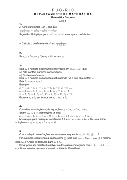
UNIVERSIDADE FEDERAL DE PERNAMBUCO
CENTRO DE CIÊNCIAS BIOLÓGICAS
MESTRADO EM BIOQUÍMICA E FISIOLOGIA
EMMANUEL VIANA PONTUAL
EXTRATO DE FLORES DE Moringa oleifera: ATIVIDADE LARVICIDA E EFEITO
SOBRE TRIPSINA E ACETILCOLINESTERASE DE LARVAS DE Aedes aegypti
ORIENTADORA: PROFª. DRª. PATRÍCIA M. G. PAIVA
CO-ORIENTADORA: PROFª. DRª. LUANA C. B. B. COELHO
RECIFE
AGOSTO, 2010
EMMANUEL VIANA PONTUAL
EXTRATO DE FLORES DE Moringa oleifera: ATIVIDADE LARVICIDA E EFEITO
SOBRE TRIPSINA E ACETILCOLINESTERASE DE LARVAS DE Aedes aegypti
Dissertação apresentada para o cumprimento
parcial das exigências para obtenção do título
de Mestre em Bioquímica e Fisiologia pela
Universidade Federal de Pernambuco.
Orientadora: Profa. Dra. Patrícia Maria Guedes Paiva
Co-orientadora: Profa. Dra. Luana Cassandra Breitenbach Barroso Coelho
RECIFE
AGOSTO, 2010
Pontual, Emmanuel Viana
Extrato de flores de Moringa oleifera: atividade larvicida e efeito sobre
tripsina e acetilcolinesterase de larvas de Aedes aegypti / Emmanuel
Viana pontual. – Recife: O Autor, 2010.
70 folhas : il., fig., tab.
Orientadora: Patrícia Maria Guedes Paiva.
Co-orientadora: Luana Cassandra Breitenbach Barroso
Coelho.
Dissertação (mestrado) – Universidade Federal de
Pernambuco. CCB. Bioquímica e Fisiologia, 2010.
Inclui bibliografia.
1. Dengue 2. Aedes aegypti 3. Inibidores de tripsina I. Título.
616.91852
CDD (22.ed.)
UFPE/CCB-2010-195
Dedico este trabalho à companheira de
afeto e de luta Inabelle, amada esposa, a
minha razão.
AGRADECIMENTOS
Ao maravilhoso Deus por jamais ter me desamparado e por me fazer compreender que
todas as coisas que possuo são efêmeras, exceto os meus sentimentos e o aprendizado ao qual
me permito; que nada carrego além de minhas idéias e que não há nada sob meus pés, a não
ser o chão que suporta todos nós igualmente.
À minha Orientadora Dra. Patrícia Paiva pela sua dedicação, pelo investimento na
minha formação, pelo seu respeito e carinho de sempre e acima de tudo pela sua amizade
querida; por ter, através do seu exemplo, me ensinado que a dedicação pelo que fazemos é
que faz o nosso trabalho importante e que quando nos entregamos, amamos, realizamos!
À querida Dra. Luana Coelho por ter me conquistado para as Flores de Moringa, pelas
suas orientações que me proporcionam crescimento e maturidade, por me ensinar a olhar
sempre mais longe, pelo seu exemplo de pessoa cristã e por estar sempre presente.
À Dra. Daniela Navarro pela colaboração indispensável na avaliação da atividade
larvicida contra Aedes aegypti, pela sua disponibilidade, pelo interesse em nosso projeto e
pela maneira doce com que sempre nos recebe.
Ao Dr. Haroudo Xavier pelo compromisso com a ciência e com a educação, pela
colaboração imprescindível na determinação da composição fitoquímica do extrato de flores.
Ao Dr. Ranilson Bezerra por ter me recebido em seu laboratório e pela contribuição
nos ensaios de atividade enzimática de acetilcolinesterase, que muito enriqueceu os nossos
resultados.
A Inabelle, mulher e amiga, companheira de todos os momentos; obrigado pelo seu
cuidado, pelo seu amor, por todo o crescimento que a nossa relação tem me proporcionado;
por segurar a minha mão quando fraquejo me dando condições para uma retomada, e todas as
vezes que existe uma conquista fazendo valer a pena. Nada teria o mesmo brilho se não
existisse a sua doce e forte presença.
Aos amados José, meu pai e Graça, minha mãe. Ele, o primeiro modelo de homem,
ela, o exemplo de doação e principalmente de luta que contribuiu para o mergulho na busca
dos meus objetivos. Aos dois, obrigado pelo amor e pelos valores que hoje me constituem.
À minha tia Sônia, pelo seu apoio imensurável, pelo amor materno, por me ajudar a
acreditar, por ser sempre tão presente e importante.
Às minhas irmãs Amanda e Emanuelle e minha prima Fátima, embora muitas vezes
não estando juntos fisicamente, pois, como escreveu Antoine de Saint-Exupéry “O essencial é
invisível aos olhos, só se vê bem com o coração”.
Ao amigo Thiago Napoleão pela importância que dedicou a esse trabalho, pela sua
ajuda indispensável para torná-lo realizado, pelo tanto que me ensinou, mas, mais ainda pela
sua amizade incondicional e pela mão amiga nas horas em que mais precisei.
À Camila, Geanne e Luana do Laboratório de Ecologia Química pelo apoio e
dedicação durante os bioensaios com A. aegypti.
Ao MSc. Caio Dias pelo tanto que me ensinou e pela sua proximidade durante os
experimentos no Laboratório de Enzimologia.
À amiga Nataly pela ideia inicial de investigar a atividade larvicida, pela amizade,
apoio e presença nos momentos mais importantes.
À amiga Lidiane pelas vezes que me socorreu no corre-corre dos experimentos, pela
sua presença nos momentos de descontração, por seu carinho e por escutar meus desabafos
com a paciência que só uma verdadeira amizade explica.
Às queridas Luciana e Tatiana pela amizade e pelos momentos compartilhados. Amo
vocês.
A Adriana Argolo, Andréa Santos, Mariana Fernandes, Michele Dalvina, Regina
Araújo, Roberto Sá e Rodrigo Ferreira, pessoas queridas que muito contribuíram com a minha
formação.
A todos que fazem o Laboratório de Glicoproteínas da UFPE, em especial aos
queridos Bernardo, Carlos Sales, Cynarha, Dalila, Felipe, Fernando, Francis, Giselly, Ídila,
Kaleen, Kézia, Marília, Mercia, Thamara e Thâmarah.
A Belany e Maiara pelo desafio que foi lidar com a realidade e a necessidade de cada
uma, por me colocar frente aos meus limites e à necessidade de transpor cada um deles
contribuindo para que me tornasse mais profissional.
A todos que fazem parte do Laboratório de Enzimologia da UFPE, especialmente a
Carol, Douglas, Fábio, Fernanda, Guto, Helane, Janilson, Kelma, Renata e Verla.
À Dra. Vera Menezes pelo compromisso com o Programa de Pós Graduação em
Bioquímica e Fisiologia.
À Dra. Tereza Correia pelo apoio e consideração a mim destinados.
Aos queridos Maria Reis e João Virgínio pelo apoio técnico e convivência amável.
Aos inesquecíveis Alice, Analice, Isla, Jailson, Lídia, Hernando e Rafa Guerra pela
parte de vocês guardada dentro de mim.
A todos que direta ou indiretamente contribuíram para a realização deste trabalho.
“Cada um de nós compõe a sua história e cada ser
em si carrega o dom de ser capaz, de ser feliz”
(Almir Sater e Renato Teixeira)
RESUMO
Dengue é uma arbovirose transmitida pelo Aedes aegypti e o controle do mosquito é
fundamental para reduzir a propagação da doença. As larvas de A. aegypti têm desenvolvido
resistência a inseticidas organofosforados. O uso de compostos naturais que promovam
mortalidade pode evitar a emergência de larvas resistentes, devido à rotatividade dos
inseticidas. Este trabalho relata a atividade larvicida (CL50 de 0.925%, p/v) do extrato de
flores de Moringa oleifera sobre o quarto instar larval (L4) de A. aegypti. Inibidor de tripsina
de natureza protéica (MoFTI, 169,9 kDa, Ki: 0,38 nM), triterpeno (β-amirina), esteróide (βsitosterol) e flavonóides (kaempferol e quercetina) foram detectados no extrato; lectina não
foi detectada. Tripsina do extrato do intestino de L4 foi inibida por MoFTI (Ki: 0,6 nM);
entretanto, a atividade de acetilcolinesterase (AChE) do extrato de L4 inteiras não foi alterada.
Ensaio em condições in vivo mostrou que a atividade de tripsina do intestino de L4 tratadas
com o extrato de flores de M. oleifera diminuiu ao longo do tempo (0 a 1440 min) e foi
fortemente inibida (98,6 %) após 310 min de incubação; a atividade de AChE do extrato de
L4 inteiras não foi afetada neste período. O estudo aponta o extrato de flores de M. oleifera
como uma ferramenta biodegradável para o controle de larvas de A. aegypti e sugere que o
mecanismo larvicida envolve a inibição da tripsina do intestino das L4 por MoFTI.
Palavras-chave: Moringa oleifera; Aedes aegypti; atividade larvicida; inibidor de tripsina;
tripsina; acetilcolinesterase.
ABSTRACT
Dengue Fever is an arboviruses transmitted by Aedes aegypti and mosquito control is
fundamental to reduce disease spreading. A. aegypti larvae have developed resistance to
organophosporous insecticides and the use of natural compounds that promote mortality may
avoid emergence of resistant larvae due to rotation of insecticides. This work reports the
larvicidal activity (LC50 of 0.925% w/v) of Moringa oleifera flower extract on fourth larval
instars (L4) of A. aegypti. Proteinaceous trypsin inhibitor from M. oleifera flower (MoFTI,
169.9 kDa, Ki: 0.38 nM), triterpene (β-amyrin), sterol (β-sitosterol) and flavonoids
(kaempferol and quercetin) were detected in the extract; lectin was absent. Trypsin from L4
gut extract was inhibited by MoFTI (Ki: 0.6 nM), however acetylcholinesterase (AChE)
activity from total L4 extract was not altered. In vivo assay showed that gut trypsin activity
from L4 treated with M. oleifera flower extract decreased along the time (0 to 1440 min) and
was strongly inhibited (98.6 %) after 310 min incubation; AChE activity from total L4 extract
was not affected in this period. The study points out M. oleifera flower extract as a
biodegradable tool for A. aegypti larvae control and suggests that larvicidal mechanism
involves inhibition of L4 gut trypsin by MoFTI.
Keywords: Moringa oleifera; Aedes aegypti; larvicidal activity; trypsin inhibitor; gut trypsin;
acethylcholisnesterase.
LISTA DE FIGURAS
Figura 1. Países com risco de transmissão de dengue. ............................................................ 16
Figura 2. Ciclo biológico do mosquito A. aegypti. .................................................................. 18
Figura 3. Atividade Hemaglutinante de lectina. ...................................................................... 24
Figura 4. Aspectos de M. oleifera ........................................................................................... 28
ARTIGO
Figure 1. Mortality of A. aegypti L4 in incubation with M. oleifera flower extract………....53
Figure 2. Characterization of M. oleifera flower extract…………………………………….54
Figure 3. Effect of M. oleifera flower extract on gut trypsin and AChE activities from A.
aegypti L4………………………………………………………………………...57
Figure 4. Effect of M. oleifera flower extract on trypsin and AChE activities from live A.
aegypti L4………………………………………………………………………...58
LISTA DE TABELAS
Table 1. Bovine trypsin and L4 gut trypsin-like activities in presence of M. oleifera flower
extract and quercetin................................................................................................................. 55
Table 2. Electric eel AChE and L4 AChE activities in presence of M. oleifera flower extract
and organophosphorous insecticides. ....................................................................................... 55
LISTA DE ABREVIATURAS
AChE – Acetilcolinesterase
AH – Atividade hemaglutinante
BApNA - N-benzoil-DL-arginil-ρ-nitroanilida
Bti – Bacillus turingiensis var. israelensis
ClaveLL – Lectina do líquen Cladonia verticillaris (do inglês Cladonia verticillaris Lichen
Lectin)
cMoL – Lectina coagulante de Moringa oleifera (do inglês coagulant M. oleifera lectin)
DDT – Dicloro difenil tricloro etano
DTNB – Ácido 5,5'-dithiobis-(2-nitrobenzóico)
L1, L2, L3 e L4 – Primeiro, segundo, terceiro e quarto ínstares larvais de Aedes aegypti
MoFTI – Inibidor de tripsina de flores de Moringa oleifera (do inglês Moringa oleifera flower
trypsin inhibitor)
MuBL – Lectina de entrecasca de Myracrodruon urundeuva (do inglês Myracrodruon
urundeuva bark lectin)
MuHL – Lectina de cerne de Myracrodruon urundeuva (do inglês Myracrodruon urundeuva
heartwood lectin)
MuLL – Lectina de folha de Myracrodruon urundeuva (do inglês Myracrodruon urundeuva
leaf lectin)
OMS – Organização Mundial de Saúde
PMSF – Fluoreto de Fenilmetilsulfonil
SDS-PAGE – Eletroforese em gel de poliacrilamida contendo dodecil sulfato de sódio (do
inglês sodium dodecyl sulphate polyacrylamide gel electrophoresis)
TLC – Cromatografia em camada delgada (do inglês thin layer chromatography)
WSMoL – Lectina solúvel em água de M. oleifera (do inglês water-soluble M. oleifera lectin)
SUMÁRIO
1. INTRODUÇÃO ................................................................................................................................14
2. FUNDAMENTAÇÃO TEÓRICA ..................................................................................................16
2.1 Dengue ................................................................................................................ 16
2.2 O mosquito Aedes aegypti .................................................................................. 17
2.3 Controle do A. aegypti ........................................................................................ 19
2.4 Resistência a inseticidas ..................................................................................... 20
2.5 Inseticidas naturais.............................................................................................. 21
2.5.1 Metabólitos secundários .............................................................................. 22
2.5.2 Lectinas ........................................................................................................ 23
2.5.3 Inibidores de proteases ................................................................................ 25
2.6 Moringa oleifera ................................................................................................. 27
3. OBJETIVOS .....................................................................................................................................30
3.1 Objetivo geral ..................................................................................................... 30
3.2 Objetivos específicos .......................................................................................... 30
4. REFERÊNCIAS ...............................................................................................................................31
5. ARTIGO............................................................................................................................................39
6. CONCLUSÃO ..................................................................................................................................70
14
1. INTRODUÇÃO
Cerca de 40% da população mundial encontra-se hoje em risco de dengue, doença
infecciosa comum em regiões tropicais e subtropicais; o agente etiológico, o arbovírus
DENV, existe em quatro sorotipos distintos (HUHTAMO et al., 2008; OMS, 2009). A dengue
é transmitida principalmente através da picada de fêmeas do mosquito Aedes aegypti (SILVA
et al., 2008); o controle de populações do inseto é de grande importância desde que não existe
ainda vacina contra a doença (OMS, 2009).
O uso de inseticidas tem induzido a seleção de linhagens de mosquitos resistentes, o
que tem impulsionado a busca por inseticidas naturais isentos de toxicidade ao meio ambiente
(BRAGA e VALLE, 2007; SILVA et al., 2008). Atividade inseticida contra A. aegypti tem
sido descrita para compostos de plantas como os metabólitos secundários e as proteínas
(BROUSSALIS et al., 2010; OCHIENG et al., 2010; OLIVEIRA et al., 2010; PRASAD et
al., 2010).
O ciclo de vida do mosquito A. aegypti envolve as fases de ovo, larva (quatro instares:
L1, L2, L3 e L4), pupa e adulto; tripsina e enzimas com atividade semelhante estão presentes
em todas as fases do ciclo do mosquito, embora com diferentes níveis de expressão de acordo
com o estágio de desenvolvimento (BOROVSKY e MEOLA, 2004). A tripsina, devido à sua
presença indiscriminada e envolvimento nos processos de digestão em insetos, é sugerida
como interessante alvo de agentes inseticidas (HILDER et al., 1987; BROADWAY, 1995).
Os inibidores de tripsina são capazes de induzir a morte de insetos por inibir a
atividade catalítica da enzima, prejudicando a digestão de alimentos de natureza protéica e,
conseqüentemente, reduzindo a biodisponibilidade e absorção dos nutrientes (HILDER et al.,
1987; BROADWAY, 1995; CARLINI e GROSSI-DE-SÁ, 2002; BHATTACHARYYA et
al., 2007a, 2007b). Atividade inseticida de inibidores de tripsina tem sido descrita; larvas
15
alimentadas com ração contendo inibidores de proteases podem apresentar perda de peso,
atraso no desenvolvimento e morte por inanição (CARLINI e GROSSI-DE-SÁ, 2002;
BHATTACHARYYA et al., 2007a, 2007b).
Moringa oleifera, planta nativa da Índia, tem despertado interesse devido ao seu
potencial na indústria e medicina (MAKKAR e BECKER, 1996; MCCONNACHIE et al.,
1999; FOIDL et al., 2001; KARADI et al., 2006). As sementes de moringa apresentam
moléculas com atividade coagulante e são capazes de promover clarificação em águas turvas.
As flores apresentam atividade antioxidante, devido à presença de α e γ-tocoferol, e
antimicrobiana, devido à presença do alcalóide pterigospermina (MAKKAR e BECKER,
1996; GUEVARA et al., 1999; SÁNCHEZ-MACHADO et al., 2006; ONG, 2008).
Coelho et al. (2009) relataram que extrato aquoso de sementes de M. oleifera atrasou o
desenvolvimento de larvas de A. aegypti, bem como apresentou atividade larvicida contra L4.
A busca por substâncias naturais e biodegradáveis com propriedades inseticidas justifica a
investigação do efeito larvicida do extrato de flores de M. oleifera sobre A. aegypti.
16
2. FUNDAMENTAÇÃO TEÓRICA
2.1 Dengue
A dengue é uma doença infecciosa que ocorre em regiões tropicais e subtropicais
(Figura 1); nas décadas recentes, a dengue tem se tornado um importante problema de saúde
pública internacional desde que aproximadamente 2,5 bilhões de pessoas, ou seja, dois
quintos da população mundial, vivem em áreas de risco de transmissão da doença. Cerca de
50 milhões de casos são registrados mundialmente a cada ano, o que caracteriza a dengue
como uma pandemia (OMS, 2009).
Figura 1. Países com risco de transmissão de dengue.
Fonte: OMS (2006)
O agente etiológico da dengue é um arbovírus pertencente ao gênero Flavivirus
(família Flaviviridae) que ocorre em quatro sorotipos: DENV-1 a DENV-4 (HUHTAMO et
17
al., 2008). A dengue é transmitida pela picada de mosquitos do gênero Aedes, tendo sido
descrita a ocorrência do vírus nas espécies A. aegypti, A. albopictus e A. polynesiensis,
pertencentes ao subgênero Stegomyia (ROBHAIN e ROSEN, 1997). Devido à não existência
de uma vacina contra a dengue, o controle de populações do mosquito vetor é fundamental
para prevenir a transmissão da doença (OMS, 2009).
Segundo a Organização Mundial de Saúde (OMS), a dengue pode afetar crianças,
adolescentes e adultos, raramente levando à morte (OMS, 2009). Os sintomas aparecem de 3 a
15 dias após a picada pelo inseto infectado e envolvem dor de cabeça, febre alta, forte dor no
corpo e, em alguns casos, vômito. É frequente a ocorrência de manchas avermelhadas na pele
(semelhantes ao sarampo ou rubéola) que aparecem 3 a 4 dias após o início da febre,
acompanhadas de prurido; pessoas que desenvolvem a forma severa da doença (dengue
hemorrágica) podem apresentar uma excessiva permeabilidade capilar responsável pelo
sangramento de gengivas (gengivorragia), nariz (epistaxe), sangramento gastrointestinal,
hematúria
(sangue
na
urina)
e
aumento
(http://www.cives.ufrj.br/informacao/dengue/den-iv.html).
do
As
fluxo
hemorragias
menstrual
variam
de
intensidade e podem desencadear choque por diminuição da pressão sanguínea seguido de
morte (HUBERT e HALSTEAD, 2009).
2.2 O mosquito Aedes aegypti
A. aegypti (Diptera: Culicidae), cujo nome significa “o indesejável do Egito”, é
originário da África, de onde migrou para as Américas e Ásia. O A. aegypti é o principal vetor
da dengue e febre amarela e foi provavelmente introduzido no Brasil na metade do século
XIX, através de navios negreiros (SILVA et al., 2008).
18
Medindo menos de 1 cm, o A. aegypti possui coloração preta com listras brancas no
corpo e nas pernas. Seu ciclo de vida compreende os estágios de ovo, larva, pupa e adulto
(Figura 2). A fase larval de A. aegypti compreende quatro instares denominados L1, L2, L3 e
L4 (SILVA et al., 2008).
Figura 2. Ciclo biológico do mosquito A. aegypti.
(Fonte: Secretaria de Saúde e Defesa Civil do Estado do Rio de Janeiro, 2007).
Estudos desenvolvidos com populações de A. aegypti oriundas de quatro regiões
bioclimáticas da Paraíba (municípios de Remígio, Boqueirão, Brejo dos Santos e Itaporanga)
mostraram a duração média de cada fase do ciclo (BEZERRA e CASTRO JR., 2008). Com
pequena variação, os quatro dias iniciais correspondem ao período de desenvolvimento
embrionário (ovo) após os quais ocorre a eclosão. O primeiro instar larval dura de 1,4 a 2 dias
passando para L2. A muda que origina o terceiro instar ocorre entre 1 e 2 dias após a
emergência de L2. A fase de L3 dura 1 dia ao fim do qual aparece o último instar (L4) que
19
pode durar de 2 a 3 dias até se desenvolver em pupa. Esta ultima pode permanecer cerca de 2
dias após os quais o vetor da dengue atinge a fase reprodutiva, o mosquito adulto; os autores
verificaram que os adultos podem viver de 42 a 44 dias (machos) ou até 46 dias (fêmeas).
A espécie A. aegypti alimenta-se de seiva vegetal, contudo, após o acasalamento, as
fêmeas desenvolvem o hábito hematofágico desde que existem proteínas no sangue que são
extremamente necessárias para o desenvolvimento dos ovos. A picada do mosquito ocorre
durante as primeiras horas do dia e as últimas da tarde e não causa dor e nem coceira (SILVA
et al., 2008).
As enzimas digestivas mais importantes presentes no intestino de A. aegypti em todas
as fases de seu ciclo biológico são a tripsina e a quimotripsina, ambas muito expressas nas
fases de larva e pupa (YANG et al., 2003). Segundo Venancio et al. (2009), existem pelo
menos 66 genes em A. aegypti que codificam tripsinas: 12 em larvas, 15 em adultos e 39 em
ambos; as tripsinas expressas apenas em larvas ou em adultos diferem entre si e, segundo os
autores, isso resulta provavelmente da emergência do hábito hematofágico após a maturidade
sexual.
2.3 Controle do A. aegypti
Inseticidas químicos têm sido bastante utilizados em programas de controle de
doenças transmitidas por vetores; no entanto sabe-se que possuem alta toxicidade ao ambiente
(BRAGA e VALLE, 2007). Os organoclorados e piretróides mantêm abertos os canais de
sódio das membranas de neurônios e os organofosforados e carbamatos atuam como
inibidores da acetilcolinesterase (AChE), enzima que catalisa a hidrólise da acetilcolina nas
sinapses colinérgicas após a propagação do impulso nervoso. A fosforilação da AChE pelo
organofosforado leva à inibição irreversível da enzima e promove a morte do inseto por
20
paralisia devido a contrações musculares intermináveis. O combate às larvas de A. aegypti
tem sido feito principalmente pela utilização do organofosforado temefós; a genotoxicidade e
mutagenicidade do temefós, em concentrações similares àquelas utilizadas no combate ao A.
aegypti, foram descritas (AIUB et al., 2002).
O controle biológico dos insetos tem sido realizado pela utilização de invertebrados
aquáticos ou larvas de peixes que se alimentam de insetos nas fases larvais e de pupa ou ainda
utilizando fungos ou bactérias patogênicas como o Bacillus thuringiensis (Bti). No Brasil, o
Programa Nacional de Controle da Dengue já utiliza o Bti (Fundação Nacional de Saúde,
2002). Dois mecanismos envolvidos no controle pelo Bti são sugeridos: A) a digestão de
endotoxinas produzidas durante a esporulação da bactéria libera no intestino do inseto
derivados com ação larvicida e B) interação entre glicoconjugados da membrana peritrófica
do intestino dos insetos e endotoxinas que possuem domínios com atividade de lectina
(proteína ligadora de carboidratos) prejudica os processos de digestão e absorção, levando as
larvas à morte (GILL et al., 1992; BURTON et al., 1999). Araújo et al. (2007) avaliaram a
atividade larvicida do Bti contra A. aegypti; tabletes contendo 15% (p/p) de esporos e cristais
de endotoxinas causaram 100 % de mortalidade de L1 e L4.
2.4 Resistência a inseticidas
Populações de insetos consistem em uma mistura de indivíduos com susceptibilidade
variável de acordo com suas características genéticas; uma mudança na resposta de uma
população ao tratamento com um inseticida ou o aumento da concentração letal necessária
para matar 50% de insetos (CL50) podem ser devidos à redução na proporção de insetos
susceptíveis (DAVISON, 1992).
21
De acordo com a OMS, resistência é a habilidade de uma população de insetos em
tolerar uma dose de inseticida que, em condições normais, causaria sua morte. O dicloro
difenil tricloro etano (DDT), um dos primeiros xenobióticos utilizados no controle de
populações de insetos, mostrou uma grande eficiência que decaiu rapidamente devido ao
surgimento de linhagens resistentes. Os mecanismos que conferem resistência aos insetos
incluem diminuição da taxa de penetração do xenobiótico pela cutícula, detoxificação
metabólica aumentada e diminuição da sensibilidade do sítio-alvo (BRAGA e VALLE, 2007).
A ocorrência de populações de A. aegypti resistentes ao temefós, único larvicida
empregado no controle do mosquito até o ano 2000, tem sido comprovada; o desenvolvimento
de resistência pelas larvas tem sido atribuído a alterações da enzima AChE, bem como a uma
alta atividade das enzimas glutationa S-transferases e α- e β-esterases, responsáveis pelo
metabolismo dos xenobióticos e conseqüente detoxificação do organismo (LIMA et al., 2003;
BRAGA e VALLE, 2007; MELO-SANTOS et al., 2010).
2.5 Inseticidas naturais
A busca por inseticidas naturais e biodegradáveis tem como objetivos minimizar os
danos ambientais, evitar o surgimento de larvas resistentes devido à rotatividade dos
compostos utilizados no combate ao mosquito e identificar substâncias ativas contra linhagens
resistentes (BRAGA e VALLE, 2007; SILVA et al., 2008). As plantas produzem fitotoxinas
em resposta a ataques de fitopatógenos e herbívoros como uma estratégia de defesa; uma
estratégia economicamente vantajosa e ecologicamente viável é a utilização de inseticidas
extraídos de plantas, que podem ser produtos do seu metabolismo primário ou secundário
(BROUSSALIS et al., 2010; OCHIENG et al., 2010; OLIVEIRA et al., 2010; PRASAD et
al., 2010; SHI et al., 2010).
22
2.5.1 Metabólitos secundários
Os metabólitos secundários presentes em vegetais superiores não estão envolvidos de
modo direto no crescimento e desenvolvimento da planta, mas atuam na defesa contra
fitopatógenos (herbívoros e microrganismos), como atraentes de polinizadores e dispersores
de sementes (TAIZ e ZEIGER, 2004). Os metabólitos secundários podem ser nitrogenados
(alcalóides, aminoácidos não protéicos, aminas, alcamidas, glicosídeos cianogênicos e
glicosinolatos) e não-nitrogenados (monoterpenos, diterpenos, triterpenos, tetraterpenos,
sesquiterpenos, saponinas, flavonóides, esteróides, cumarinas) e são precursores de diversos
derivados produzidos de acordo com as necessidades do tecido ou órgão da planta e o estágio
de desenvolvimento (WINK, 2003). Quando produzidos na forma de precursores inativos, os
metabólitos secundários se tornam ativos em caso de ferimento, infecções ou quando
ingeridos por herbívoros.
Esses compostos promovem respostas celulares por diferentes mecanismos. Os
alcalóides podem atuar como agonistas ou antagonistas de neurotransmissores e
neuroreceptores ou acarretar distúrbios na replicação e transcrição. Taninos e outros fenóis
formam pontes de hidrogênio e ligações iônicas com proteínas induzindo modificações
conformacionais que podem levar à perda de função das proteínas. Os compostos lipofílicos,
tais como terpenos e saponinas, interagem com membranas formando poros e induzindo
distúrbios na permeabilidade celular (WINK, 2003).
Metabólitos secundários com atividade inseticida têm sido descritos. Compostos
fenólicos derivados do ácido elágico presentes em galhos e casca de Laguncularia racemosa
quando incorporados a uma dieta artificial oferecida a larvas de Spodoptera littoralis, após
seis dias de exposição, inibiram em até 90% o crescimento das larvas (SCHI et al., 2010).
Extratos orgânicos de Hybanthus parviflorus, contendo β-sitosterol, foram ativos contra larvas
23
de Ceratitis capitata diminuindo a formação de pupa e emergência de adultos ou induzindo
mortalidade das larvas (BROUSSALIS et al., 2010).
Atividade larvicida contra A. aegypti tem sido descrita para extratos de plantas
contendo óleos essenciais (PITASAWAT et al., 2007; AUTRAN et al., 2009) e outros
metabólitos secundários como limonóides (WANDSCHEER et al., 2004), quinonas (IOSET
et al., 2000) e saponinas (CHAPAGAIN et al., 2008). O esteróide β-sitosterol extraído de
Abutilon indicum apresentou atividade larvicida com CL50 de 11,49 ppm e os flavonóides
quercetina (35,7 µg/mL) e kaempferol (30,65 µg/mL) isolados de folhas de Gardenia
ternifolia promoveram mortalidade de L2 (RAHUMAN et al., 2008; OCHIENG et al., 2010).
2.5.2 Lectinas
Lectinas, proteínas ou glicoproteínas de origem não imune, interagem com
carboidratos através de no mínimo dois sítios de ligação e, por isso, aglutinam células e
precipitam polissacarídeos, glicoproteínas ou glicolipídeos, sem ocasionar modificações em
suas estruturas (GOLDSTEIN et al., 1980). A palavra lectina, proveniente do latim (Lectus,
selecionar/escolher), reflete a especificidade com que ocorre a ligação reversível entre uma
lectina e o seu carboidrato específico (KENNEDY et al., 1995; MATSUI et al., 2001). A
presença de lectinas em determinada amostra é verificada através do ensaio de atividade
hemaglutinante (AH) realizado em suspensão de eritrócitos e confirmado pela inibição da AH
quando em presença de carboidratos (Figura 3).
24
Pontual, E.V. (2008)
Figura 3. Atividade Hemaglutinante de lectina. A) Esquema da malha formada pela ligação
da lectina aos carboidratos da superfície dos eritrócitos. B) Inibição da AH por carboidrato e
C) Aspecto do ensaio de AH em microplaca. lectina –
; eritrócito –
e carboidrato –
.Controle: NaCl 0,15 M.
Atividade inseticida de lectinas contra espécies de pragas de importância econômica
tem sido descrita; ClaveLL, a lectina extraída do líquen Cladonia verticillaris e as lectinas
ligadoras de quitina extraídas do cerne (MuHL), da casca (MuBL) e da folha (MuLL) de
Myracrodruon urundeuva (Aroeira do Sertão) possuem atividade termiticida contra soldados
e operários de Nasutitermes corniger (SÁ et al., 2008; SILVA et al., 2009; NAPOLEÃO et
al., 2010). Sugere-se que o efeito deletério ocorre devido à interação entre domínios ligadores
de carboidratos das lectinas e unidades de N-acetil-glicosamina da matriz peritrófica dos
insetos (SÁ et al., 2009). As lectinas que são resistentes a proteases do trato digestivo de
homens e outros animais podem se ligar a carboidratos da superfície de células da mucosa
intestinal e interferir nos processos de digestão e absorção dos alimentos reduzindo a
eficiência do aproveitamento dos nutrientes (VASCONCELOS e OLIVEIRA, 2004;
25
KANSAL et al., 2006). Napoleão et al. (2010) descreveram a resistência de MuHL, MuBL e
MuLL à digestão pela tripsina do intestino de N. corniger; adicionalmente os autores
sugeriram que a atividade termiticida pode ter sido decorrente da atividade bacteriostática e
bactericida sobre simbiontes do intestino das térmitas.
O controle de populações de A. aegypti utilizando preparações contendo lectinas
também pode representar uma alternativa econômica e viável; MuHL e MuBL possuem
atividade larvicida contra L4 de A. aegypti com CL50 de 0,04 e 0,125 mg/mL,
respectivamente. Atividade larvicida (CL50 de 0,197 mg/mL), bem como atraso no
desenvolvimento larval de A. aegypti, foram descritos para WSMoL, a lectina solúvel em
água extraída de sementes de M. oleifera; as larvas apresentaram ausência da camada epitelial
que delimita o lúmen do intestino bem como aumento do lúmen e hipertrofia de segmentos
(COELHO et al., 2009).
2.5.3 Inibidores de proteases
A interação entre inibidores de proteases e enzimas proteolíticas pode resultar em
modificações conformacionais na molécula da protease decorrentes da formação de
complexos estáveis inativos ou com baixa atividade (LIAO et al., 2007).
Compostos fenólicos do tipo flavonóides podem inibir tripsina pela formação de duas
ou mais pontes de hidrogênio e interações eletrostáticas com a região S1 da molécula da
enzima; quercetina, miricetina, morina e kaempferol inibem tripsina sendo as concentrações
que inibem 50% da atividade máxima (CI50) iguais a 0,010, 0,015, 0,027 e 0,06 mM,
respectivamente (MALIAR et al., 2004).
Inibidores de proteases de natureza protéica são classificados em inibidores de serino,
treonino, cisteíno, aspártico e metaloproteases, de acordo com o resíduo nucleofílico presente
26
no sítio ativo da enzima sobre a qual ocorre inibição (FEAR et al., 2007). Os inibidores de
serinoproteases interagem de maneira estável com o sítio ativo das enzimas impedindo sua
ligação com o substrato e, conseqüentemente, sua atividade hidrolítica (BODE e HUBER,
2000).
Devido à especificidade e versatilidades dos inibidores de proteases, as ciências
médica e farmacêutica têm explorado seu potencial aplicativo como agentes antifúngicos,
antiprotozoários, antivirais e terapêuticos no tratamento de doenças, entre as quais estão o
câncer e a diabetes mellitus (KOBLINSKI et al., 2000; FEAR et al., 2007).
Os inibidores de proteases são comumente expressos em tecidos vegetais e sua
biossíntese pode ser regulada em resposta ao ataque de pragas e herbívoros
(BHATTACHARYYA et al., 2007a). A atividade inseticida de inibidores de tripsina ocorre
devido à diminuição na biodisponibilidade de aminoácidos e pobre absorção de nutrientes que
levam o inseto à morte por inanição (CARLINI e GROSSI-DE-SÁ, 2002; MACEDO et al.,
2002; BHATTACHARYYA et al., 2007a, 2007b; OLIVEIRA et al., 2007; RAMOS et al.,
2009).
O controle de pragas de interesse econômico através do bloqueio da digestão pela
utilização de inibidores de tripsina (serinoprotease) tem despertado considerável interesse
devido à ocorrência generalizada dessa enzima no intestino de insetos (HILDER et al., 1987;
BROADWAY, 1995). Larvas de Anagasta kuehniella, praga que se alimenta de produtos em
estoque, alimentadas com inibidor de tripsina de sementes de Adenanthera pavonina (1% p/p)
apresentaram atividade de tripsina diminuída, alteração no ciclo biológico (aumento do
período larval e de pupa) e redução de emergência e sobrevivência de adultos (MACEDO et
al., 2010). Os inibidores de tripsina de sementes de Cajanus cajan e Vigna mungo também
inibiram a tripsina do intestino de Achaea janata, Helicoverpa armigera e Spodoptera litura e
foram resistentes à digestão por proteinases intestinais das larvas; alimentação artificial
27
contendo os inibidores acarretou em diminuição do peso e da taxa de sobrevivência das larvas
seguindo uma curva dose-resposta (PRASAD et al., 2010).
Inibidores de tripsina de Glycine max e Archidendron ellipticum causaram inibição do
crescimento e diminuição do peso corporal de larvas de S. litura (BHATTACHARYYA et al.,
2007a). Kansal et al. (2008) verificaram que o inibidor de tripsina isolado de sementes de
Vigna radiata causou morte e atraso no crescimento de larvas de Helicoverpa armigera.
Tratamento do besouro Anthonomus grandis (Bicudo-do-algodoeiro) com o inibidor de
tripsina isolado de Glycine max resultou em larvas com peso e tamanho reduzidos, indução
de mortalidade em adultos e deformidades em todos os estágios de desenvolvimento; em
adultos, as deformidades observadas incluíram ausência de tórax e asas (FRANCO et al.,
2004).
2.6 Moringa oleifera
M. oleifera (Figura 4), lírio branco ou quiabo de quina, pertence à família das
Moringaceae. É nativa da Índia e amplamente cultivada nos trópicos desde que sobrevive por
longo período em solos pobres e com baixo teor de umidade (MCCONNACHIE et al., 1999).
A moringa desperta grande interesse devido às suas propriedades medicinais e utilização
como planta forrageira, bem como por ser uma fonte promissora de óleos e biogás
(MAKKAR e BECKER, 1996; FOIDL et al., 2001; KARADI et al., 2006).
28
Figura 4. Aspectos de M. oleifera: (A) árvore, (B) sementes e (C) inflorescência.
Fotos: Pontual, E.V., 2008 (A e C) e www.mfrural.com.br (B)
Sementes de M. oleifera possuem propriedades coagulantes devido à presença de
proteínas e de um polieletrólito orgânico de 3 kDa hábeis em clarificar águas turvas
(OKUDA, et al., 2001; GHEBREMICHAEL et al., 2005; SANTOS et al., 2009); sua
utilização no tratamento de água para consumo humano em países em desenvolvimento tem
sido descrita. Dentre as proteínas envolvidas no mecanismo de coagulação promovido pelas
sementes de M. oleifera foi descrita a lectina cMoL (SANTOS et al., 2009).
As flores de M. oleifera cruas ou após cozimento brando são utilizadas como alimento
constituindo uma rica fonte de íons cálcio e potássio e dos antioxidantes naturais α- e γ-
29
tocoferol (RAMACHANDRAN et al., 1980; MAKKAR e BECKER, 1996; GUEVARA et
al., 1999; FOIDL et al., 2001; SÁNCHEZ-MACHADO et al., 2006). As flores de moringa
são utilizadas com fins medicinais como colagogo, diurético, hipoglicemiante e tônico
(KHARE, 2007; PARROTTA, 2009). Pterigospermina, um alcalóide com atividade
antifúngica e antibacteriana, é encontrado nas flores (LIZZY et al., 1968; ONG, 2008).
30
3. OBJETIVOS
3.1 Objetivo geral
Investigar extrato aquoso de flores de M. oleifera quanto à presença de compostos
inseticidas (metabólitos secundários, lectinas, inibidor de tripsina e inibidor de AChE) e
atividade larvicida contra A. aegypti. Avaliar as atividades de tripsina e AChE de L4 tratadas
com o extrato de flores em condições in vivo.
3.2 Objetivos específicos
•
Determinar a taxa de mortalidade de L1 e L4 após incubação com o extrato de flores
de M. oleifera.
•
Investigar o extrato de flores de M. oleifera quanto à presença de metabólitos
secundários.
•
Determinar o perfil eletroforético, em gel de poliacrilamida, de proteínas do extrato de
flores de M. oleifera sob condições desnaturantes (sulfato sódico de dodecila) e
redutoras (β-mercaptoetanol).
•
Avaliar a presença de lectinas no extrato de flores de M. oleifera, através da
determinação de atividade hemaglutinante.
•
Investigar o extrato de flores de M. oleifera quanto à presença de inibidores de tripsina
e de AChE utilizando enzimas comerciais e substratos sintéticos.
•
Determinar a atividade de tripsina e AChE em extratos de L4.
•
Determinar o efeito de quercetina comercial sobre tripsina de L4.
•
Avaliar o efeito do extrato de flores de M. oleifera sobre tripsina e AChE de L4.
•
Determinar as atividades de tripsina e AChE em L4 tratadas com o extrato de flores.
31
4. REFERÊNCIAS
AIUB, C.A.F.; COELHO, E.C.A.; SODRÉ, E.; PINTO, L.F.R.; FELZENSZWALB, I.
Genotoxic evaluation of the organophosphorous pesticide temephos. Genetics and
Molecular Research, v. 1, p. 159–166, 2002.
ARAÚJO, A.P.; MELO-SANTOS, M.A.V.; CARLOS, S.O.; RIOS, E.M.M.M.; REGIS, L.
Evaluation of an experimental product based on Bacillus thuringiensis sorovar. israelensis
against Aedes aegypti larvae (Diptera:Culicidae). Biological Control, v. 41, p. 339-347, 2007.
AUTRAN, E.S.; NEVES, I.A.; DA SILVA, C.S.B.; SANTOS, G.K.N.; DA CÂMARA,
C.A.G.; NAVARRO, D.M.A.F. Chemical composition, oviposition deterrent and larvicidal
activities against Aedes aegypti of essential oils from Piper marginatum Jacq. (Piperaceae),
Bioresource Technology, v. 100, p. 2284-2288 , 2009.
BEZERRA, E.B.; CASTRO JR., F.P. Biologia comparada de populações de Aedes
(Stegomyia) aegypti (L.) (Díptera; Culicidae) da Paraíba. Neotropical Entomology, v. 37, p.
081-085, 2008.
BHATTACHARYYA, A.; LEIGHTON, S.M.; BABU, C.R. Bioinsecticidal activity of
Archidendron ellipticum trypsin inhibitor on growth and serine digestive enzymes during
larval development of Spodoptera litura. Comparative Biochemistry and Physiology part
C, v. 145, p. 669-677, 2007a.
BHATTACHARYYA, A.; RAI, S.; BABU, C.R. A trypsin and chymotrypsin inhibitor from
Caesalpinia bonduc seeds: isolation, partial characterization and insecticidal properties. Plant
Physiology and Biochemistry, v. 45, p. 169-177, 2007b.
BODE, W.; HUBER, R. Structural basis of the endoproteinase-protein inhibitor interaction.
Biochimica et Biophysica Acta, v. 1477, p. 241-252, 2000.
BOROVSKY, D.; MEOLA, S. M. Biochemical and cytoimmunological evidence for the
control of Aedes aegypti larval trypsin with Aea-TMOF. Archives of Insect Biochemistry
and Physiology, v. 55, p. 124-139, 2004.
BRAGA, I.A.; VALLE, D. Aedes aegypti: surveillance, resistance monitoring, and control
alternatives in Brazil. Epidemiologia e Serviços de Saúde, v. 16, p. 295-302, 2007.
BROADWAY, R.M. Are insects resistant to plant proteinase inhibitors? Journal of Insect
Physiology, v. 41, pp. 107–116, 1995.
32
BROUSSALIS, A.M.; CLEMENTE, S.; FERRARO, G.E.; Hybanthus parviflorus
(violaceae): insecticidal activity of a South American plant. Crop Protection, v. 29, p. 953956, 2010.
BURTON, S.; ELLAR, D.J.; LI, J.; DERBYSHIRE, D.J. N-Acetylgalactosamine on the
putative insect receptor aminopeptidase N is recognised by a site on the domain III lectin-like
fold of a Bacillus thuringiensis insecticidal toxin. Journal of Molecular Biology, v. 287, p.
1011-1022, 1999.
CARLINI, C.R.; GROSSI-DE-SÁ, M.F. Plant toxic proteins with insecticidal properties. A
review on their potential as bioinsecticides. Toxicon, v. 40, p. 1515–1539, 2002.
CHAPAGAIN, B.P.; SAHARAN, V.; WIESMAN, Z. Larvicidal activity of saponins from
Balanites aegyptiaca callus against Aedes aegypti mosquito. Bioresource Technology, v. 99,
p. 1165-1168, 2008.
COELHO, J.S.; SANTOS, N.D.L.; NAPOLEÃO, T.H.; GOMES, F.S.; FERREIRA, R.S.;
ZINGALI, R.B.; COELHO, L.C.B.B.; LEITE, S.P.; NAVARRO, D.M.A.F.; PAIVA, P.M.G.
Effect of Moringa oleifera lectin on development and mortality of Aedes aegypti larvae.
Chemosphere, v. 77, p. 934-938, 2009.
DAVISON, E.W. Development of insect resistance to biopesticides. Pesquisa Agropecuária
Brasileira, v. 27, p. 47-57, 1992.
FEAR, G.; KOMARNYTSKY, S.; RASKIN, I. Protease inhibitors and their peptidomimetic
derivatives as potential drugs. Pharmacology & Therapeutics, v. 113, p. 354-368, 2007.
FOIDL, N.; MAKKAR, H.P.S.; BECKER, K. The potential of Moringa oleifera for
agricultural and industrial uses, in: Fuglie, L.J. (Ed.), The Miracle Tree/ The Multiple
Attributes of Moringa. CTA, New York, p. 45–76, 2001.
FRANCO, O.L.; DIAS, S.C.; MAGALHÃES C.P.; MONTEIRO, A.C.S.; BLOCH JR., C.;
MELO, F.R.; OLIVEIRA-NETO, O.B.; MONNERAT, R.G.; GROSSI-DE-SÁ, M.F. Effects
os soybean kunitz trypsin inhibitoron the cotton boll weevil (Anthonomus grandis).
Phrytochemistry, v. 65, p. 81-89, 2004.
FUNDAÇÃO NACIONAL DE SAÚDE (FUNASA). Roteiro para capacitação de agentes
do PACS/PSF nas ações de controle da dengue. Brasilia (DF); 2002.
33
GHEBREMICHAEL, K.A.; GUNARATNA, K.R.; HENRIKSSON, H.; BRUMER, H.;
DALHAMMAR, G. A simple purification and activity assay of the coagulant protein from
Moringa oleifera seed. Water Research, v. 39, p. 2338-2344, 2005.
GILL, S.S.; COWLES, E.A.; PIETRANTONIO, P.V. The mode of action of Bacillus
thuringiensis endotoxins. Annual Review of Entomology, v. 37, p. 615-634 , 1992.
GOLDSTEIN, I.J.; HUGHES, C.E.; MONSIGNY, M.; OSAWA, T.; SHARON, N. What
should be called a lectin? Nature, v. 285, p. 66, 1980.
GUEVARA, A.P.; VARGAS, C.; SAKURAI, H.; FUJIWARA, Y.; HASHIMOTO, K.;
MAOKA, T.; KOZUKA, M.; ITO, Y.; HARUKUNI, T.; NISHINO, H. An antitumor
promoter from Moringa oleifera Lam. Mutation Research,v. 440,p. 181-188, 1999.
HILDER, V.A.; GATEHOUSE A.M.R.; SHEERMAN S.E.; BAKER R.F.; BOULTER D. A
novel mechanism of insect resistance engineered into tobacco. Nature, v. 330, p. 160–163,
1987.
HUBERT, B.; HALSTEAD, S.B. Dengue 1 virus and dengue hemorrhagic fever, French
Polynesia. Emerging Infectious diseases, v. 15, p. 1265-1270, 2009.
HUHTAMO, E.; UZCÁTEGUI, N.Y.; SIIKAMAKI, H.; SAARINEN, A.; PIIPARINEN, H.;
VAHERI, A.; VAPALAHTI, O. Molecular epidemiology of Dengue virus strains from
finnish travelers. Emerging Infectious diseases, v. 14, p. 80-83, 2008.
IOSET, J.R.; MARSTON, A.; GUPTA, M.P.; HOSTETTMANN, K. Antifungal and
larvicidal compounds from the root bark of Cordia alliodora. Journal of Natural Products,
v. 63, p. 424–426, 2000.
KANSAL, R.; Gupta, R.N.; KOUNDAL, K.R.; KUHAR, K.; GUPTA, V.K. Purification,
characterization and evaluation of insecticidal potential of trypsin inhibitor from mungbean
(Vigna radiate L. Wilczek) seeds. Acta Physiologiae Plantarum, v. 30, p. 761-768, 2008.
KANSAL, S.; SHARMA, A.; GUPTA, M. N. An integrated process for obtaining oil,
protease inhibitors and lectin from soybean flour. Food Research International, v. 39, p.
499-502, 2006.
KARADI, R.V.; GADGE, N.B.; ALAGAWADI, K.R.; SAVADI, R.V. Effect of Moringa
oleifera Lam. root-wood on ethylene glycol induced urolithiasis in rats. Journal of
Ethnopharmacology, v. 105, p. 306-311, 2006.
34
KENNEDY, J. F.; PAIVA, P. M. G.; CORREIA, M. T. S.; CAVALCANTE, M. S. M.;
COELHO, L. C. B. B. Lectins, versatile proteins of recognition: a rewiew. Carbohydrate
Polymers, v. 26, p. 219-230, 1995.
KHARE, C.P.
Indian Medicinal Plants - An Illustrated Dictionary. Springer,
Berlin/Heidelberg, 2007.
KOBLINSKI, J. E.; AHRAM, M.; SLOANE, B. F. Unraveling the role of proteases in cancer.
Clinica Chimica Acta, v. 291, p. 113-135, 2000.
LIAO, H.; REN, W.; KANG, Z.; ZHAO, X. J.; DU, L. F. A trypsin inhibitor from Cassia
obtusifolia seeds: isolation, characterization and activity against Pieris rapae. Biotechnology
Letters, v. 29, p. 653-658, 2007.
LIZZY, K.S.; NARASHIMA RAO, P.L.; PUTTASWAMY, T.L. Chemotherapy of bacterial
infections. Part 4: potential anticholera agents. Indian Journal of Experimental Biology v.
6, p. 168–169, 1968.
MACEDO, M.L.R.; MELLO, G.C.; FREIRE, M.G.M.; NOVELLO, J.C.; MARANGONI, S.;
MATOS, D.G.G. Effect of a trypsin inhibitor from Dimorphandra mollis seeds on the
development of Callosobruchus maculatus. Plant Physiology and Biochemistry, v. 40, p.
891-898, 2002.
MACEDO, M.L.R.; DURIGAN, R.A.; SILVA, D.S.; MARANGONI, S.; FREIRE, M.G.M.;
PARRA, J.R.P. Adenanthera pavonina trypsin inhibitor retard growth of Anagasta kuehniella
(Lepidoptera; Pyralidae). Archives of Insect Biochemistry and Physiology, v. 73, p. 213231, 2010.
MAKKAR, H. P. S.; BECKER, K. Nutritional value and antinutritional components of whole
and ethanol extracted Moringa oleifera. Animal Feed Science and Technology, v. 63, p.
211-228, 1996.
MALIAR, T.; JEDINÁK, A.; KADRABOVÁ, J.; STURDÍK. E. Structural aspects of
flavonoids as trypsin inhibitors. European Journal of Medicinal Chemistry, v. 39, p. 241248, 2004.
MATSUI, T.; HAMAKO, J.; OZEKI, Y.; TITANI, K. Comparative study of blood grouprecognizing lectins toward ABO blood group antigens on neoglycoproteins, glycoproteins and
complex-type oligosaccharides. Biochimica et Biophysica Acta, v. 1525, p. 50-57, 2001.
35
MCCONHACHIE, G. L.; FOLKARD, G. K.; MTAWALI, M. A.; SUTHERLAND, J. P.
Field trials of apropriate hydraulic flocculation processes. Water Research, v. 33, p. 14251434, 1999.
MELO-SANTOS, M.A.V.; VARJAL-MELO, J.J.M.; ARAÚJO, A.P.; GOMES T.C.S.;
PAIVA, M.H.S.; REGIS, L.N.; FURTADO, A.F.; MAGALHÃES, T.; MACORIS, M.L.G.;
ANDRIGHETTI, M.T.M.; AYRES, C.F.J. Resistance to the organophosphate temephos:
mechanisms, evolution and reversion in an Aedes aegypti laboratory strain from Brazil. Acta
Tropica, v. 113, p. 180 –189, 2010.
NAPOLEÃO, T.H.; GOMES, F.S.; LIMA, T.A.; SANTOS, N.D.L.; SÁ, R.A.;
ALBUQUERQUE, A.C.; COELHO, L.C.B.B.; PAIVA, P.M.G. Termiticidal activity of
lectins from Myracrodruon urundeuva against Nasutitermes corniger and its mechanisms.
International Biodeterioration and Biodegradation, doi:10.1016/j.ibiod.2010.05.015,
2010.
OCHIENG, C.O.; MIDIWO, J.O.; OWUOR, P.O. Anti-plasmodial and larvicidal effects of
surface exudates of Gardenia ternifolia aerial parts. Research Journal of Pharmacology, v.
4, p. 45-50, 2010.
OKUDA, T.; BAES, A.U.; NISHIJIMA, W.; OKADA, M. Isolation and characterization of
coagulant extracted from Moringa oleifera seed by salt solution. Water Research, v. 35, n.
15, p. 405-410, 2001.
OLIVEIRA, A.S.; MIGLIOLO, L.; AQUINO, R.O.; RIBEIRO, J.K.C.; MACEDO, L.L.P.;
ANDRADE, L.B.S.; BEMQUERER, M.P.; SANTOS, E.A.; KIYOTA, S.; SALES, M.P.
Purification and characterization of a trypsin-papain inhibitor from Pithecelobium dumosum
seeds and its in vitro effects towards digestive enzymes from insect pests. Plant Physiology
and Biochemistry, v. 45, p. 858-856, 2007.
OLIVEIRA, D.H.; SOUSA D.O.B.; OLIVEIRA, J.T.A.; CARLINI, C.R.; OLIVEIRA, P.H.;
PEREIRA, M.L.; ROCHA, R.O.; MORAIS, J.K.S.; GOMES-FILHO, E.; VASCONCELOS,
I.M. Gm-TX, a new toxic protein from soybean (Glycine max) seeds with potential for
controlling insect pests. Process Biochemistry, v. 45, p. 634-640, 2010.
ONG, H. C. Drumstick, in: ONG, H. C. (Ed.), Vegetables for health and healing. Utusan
Publications & Distributors Sdn Bhd, Kuala Lumpur, p. 94-95, 2008
ORGANIZAÇÃO MUNDIAL DE SAÚDE. Dengue and dengue hemorrhagic fever. Fact
sheet, p. 117, 2009.
36
PARROTTA, J.A. Moringa oleifera LAM., 1785, IN: ROLOFF, A., WEISGERBER, H.,
LANG, U., STIMM, B. (Eds.), Enzyklopädie der Holzgewächse, Handbuch und Atlas der
Dendrologie. Enzyklopädie der Holzgewächse – 40. WILEY-VCH Verlag GmbH & Co.
KGaA, Weinheim, 2009.
PITASAWAT, B.; CHAMPAKAEW, D.; CHOOCHOTE, W.; JITPAKDI, A.;
CHAITHONG,
U.;
KANJANAPOTHI,
D.;
RATTANACHANPICHAI,
E.;
TIPPAWANGKOSOL, P.; RIYONG, D.; TUETUN, B.; CHAIYASIT, D. Aromatic plantderived essential oil: An alternative larvicide for mosquito control. Fitoterapia, v. 78, p. 205210, 2007.
PRASAD, E.R.; DUTTA-GUPTA, A.; PADMASREE, K.Insecticidal potential of BowmanBirk proteinase inhibitors from red gram (Cajanus cajan) andblack gran (Vigna mungo)
against lepdppteran insect pests. Pesticide Biochemistry and Physiology, v. 98, p. 80-88,
2010.
RAHUMAN, A.A.; GOPALAKRISHNAN, G.; VENKATESAN, P.; GEETHA, K. Isolation
and identification of mosquito larvicidal compound from Abutilon indicum (Linn.) Sweet.
Parasitology Research, v. 102, p. 981-988, 2008.
RAMACHANDRAN, C.; PETER, K.V.; GOPALAKRISHNAN, P.K. Drumstick (Moringa
oleifera): a multipurpose Indian vegetable. Economic Botany, v. 34, p. 276-283, 1980.
RAMOS, V.S.; FREIRE, M.G.M.; PARRA, J.R.P.; MACEDO, M.L.R. Regulatory effects of
an inhibitor from Plathymenia foliolosa seeds on the larval development of Anagasta
kuehniella (Lepidoptera). Comparative Biochemistry and Physiology part A, v. 152, p.
255-261, 2009.
RODHAIN F.; ROSEN, L. Mosquito vectors and dengue virus-vector relationships. In:
Gubler DJ; Kuno G. Dengue and dengue hemorragic fever. New York: CAB International,
1997
SÁ, R. A.; NAPOLEÃO, T. H.; SANTOS, N. D. L.; GOMES, F. S.; ALBUQUERQUE, A.
C.; XAVIER, H. S.; COELHO, L. C. B. B.; BIEBER, L. W.; PAIVA, P. M. G. Induction of
mortality on Nasutitermes corniger (Isoptera, Termitidae) by Myracrodruon urundeuva
heartwood lectin. International Biodeterioration & Biodegradation, v. 62, p. 460-464,
2008.
SÁ, R.A.; SANTOS, N.D.L.; SILVA, C.S.B.; NAPOLEÃO, T.H.; GOMES, F.S.; CAVADA,
B.S.; COELHO, L.C.B.B.; NAVARRO, D.M.A.F.; BIEBER, L.W.; PAIVA, P.M.G.
Larvicidal activity of lectins from Myracrodruon urundeuva on Aedes aegypti. Comparative
Biochemistry and Physiology C, v. 149, p. 300-306, 2009.
37
SÁNCHEZ-MACHADO, D. I.; LÓPEZ-CERVANTES, J.; VÁZQUEZ, N. J. R. Highperformance liquid chromatography method to measure α and γ-tocopherol in leaves, flowers
and fresh beans from Moringa oleifera. Journal of Chromatography A, v. 1105, p. 111-114.
2006.
SANTOS, A.F.S.; LUZ, L.A.; ARGOLO, A.C.C.; TEIXEIRA, J.A.; PAIVA, P.M.G.;
COELHO, L.C.B.B. Isolation of a seed coagulant Moringa oleifera lectin. Process
Biochemistry, v. 44, p. 504-508, 2009.
SHI, C.; XU, M.J.; BAYER, M.; DENG, Z.W.; KUBBUTAT, M.H.G.; WAEJEN, W.;
PROKSCH, P.; LIN, W.H. Phenolic compounds and their anti-oxidative properties and
protein kinase inhibition fron the Chinese mangrove plant Laguncularia racemosa.
Phytochemistry, v. 71, p. 435-442, 2010.
SILVA, J. S.; MARIANO Z.F.; SCOPEL, I. A dengue no Brasil e as políticas de combate ao
Aedes aegypti: da tentativa de erradicação às políticas de controle. Hygeia, v. 3, p. 163-175,
2008.
SILVA, M. D. C.; SÁ, R. A.; NAPOLEÃO, T. H.; GOMES, F. S.; SANTOS, N. D. L.;
ALBUQUERQUE, A. C.; XAVIER, H. S.; PAIVA, P. M. G.; CORREIA, M. T. S.;
COELHO, L. C. B. B. Purified Cladonia verticillaris lichen lectin: insecticidal activity on
Nasutitermes corniger (Isoptera: Termitidae). International Biodeterioration and
Biodegradation, v. 63, p. 334-340, 2009.
TAIZ, L.; ZEIGER, E. Metabólitos secundários e defesa vegetal. In: TAIZ, L.; ZEIGER, E.
Fisiologia Vegetal. Porto Alegre: Artmed, 2004.
VASCONCELOS, I. M.; OLIVEIRA, J. T. A. Antinutritional properties of plant lectins.
Toxicon, v. 44, p. 385-403, 2004.
VENANCIO, T.M.; CRISTOFOLETTI, P.T.; FERREIRA, C.; VERJOVSKI-ALMEIDA, S.;
TERRA, W.R. The Aedes aegypti larval transcriptome: a comparative perspective with
emphasis on trypsins and the domain structure of peritrophins. Insect Molecular Biology, v.
18, p. 33-44, 2009.
WANDSCHEER, C.B.; DUQUE, J.E.; DA SILVA, M.A.N.; FUKUYAMA, Y.; WOHLKE,
J.L.; ADELMANN, J.; FONTANA, J.D. Larvicidal action of ethanolic extracts from fruit
endocarps of Melia azedarach and Azadirachta indica against the dengue mosquito Aedes
aegypti. Toxicon, v. 44, p. 829-835, 2004.
38
WINK, M. Evolution of secondary metabolites from an ecological and molecular
phylogenetic perspective. Phytochemistry, v. 64, p. 3-19, 2003.
YANG, Y.J.; DAVIES, D.M. Trypsin and chymotrypsin during metamorphosis in Aedes
aegypti and properties of the chymotrypsin. Journal of Insect Physiology, v. 17, p. 117-131,
1971.
39
5. ARTIGO
EFFECT OF LARVICIDAL EXTRACT FROM Moringa oleifera FLOWERS ON GUT
TRYPSIN AND ACETHYLCHOLINESTERASE ACTIVITY FROM Aedes aegypti
LARVAE
A ser submetido ao periódico “Insect Biochemistry and Molecular Biology”
(Impacto: 3.117)
40
Effect of larvicidal extract from Moringa oleifera flowers on gut trypsin and
acethylcholinesterase activity from Aedes aegypti larvae
Emmanuel V. Pontuala, Thiago H. Napoleãoa, Caio R.D. Assisa, Haroudo S. Xavierb, Ranilson
S. Bezerraa, Daniela M.A.F. Navarroc, Luana C.B.B. Coelhoa, Patrícia M.G. Paivaa,*
a
Departamento de Bioquímica, Centro de Ciências Biológicas, Universidade Federal de
Pernambuco, Avenida Prof. Moraes Rego, S/N, Cidade Universitária, 50670-420, Recife-PE,
Brazil.
b
Departamento de Ciências Farmacêuticas, Centro de Ciências da Saúde, Universidade
Federal de Pernambuco, 50740-521, Recife-PE, Brazil.
c
Departamento de Química Fundamental, Centro de Ciências Exatas e da Natureza,
Universidade Federal de Pernambuco, 50670-901, Recife-PE, Brazil.
*Corresponding author: Tel: +558121268540; Fax: +558121268576
E-mail address: [email protected] (P.M.G. Paiva).
41
Abstract
Dengue Fever is an arboviruses transmitted by Aedes aegypti and mosquito control is
fundamental to reduce disease spreading. A. aegypti larvae have developed resistance to
organophosporous insecticides and the use of natural compounds that promote mortality may
avoid emergence of resistant larvae due to rotation of insecticides. This work reports the
larvicidal activity (LC50 of 0.925% w/v) of Moringa oleifera flower extract on fourth larval
instars (L4) of A. aegypti. Proteinaceous trypsin inhibitor from M. oleifera flower (MoFTI,
169.9 kDa, Ki: 0.38 nM), triterpene (β-amyrin), sterol (β-sitosterol) and flavonoids
(kaempferol and quercetin) were detected in the extract; lectin was absent. Trypsin from L4
gut extract was inhibited by MoFTI (Ki: 0.6 nM), however acetylcholinesterase (AChE)
activity from total L4 extract was not altered. In vivo assay showed that gut trypsin activity
from L4 treated with M. oleifera flower extract decreased along the time (0 to 1440 min) and
was strongly inhibited (98.6 %) after 310 min incubation; AChE activity from total L4 extract
was not affected in this period. The study points out M. oleifera flower extract as a
biodegradable tool for A. aegypti larvae control and suggests that larvicidal mechanism
involves inhibition of L4 gut trypsin by MoFTI.
Keywords: Moringa oleifera; Aedes aegypti; larvicidal activity; trypsin inhibitor; gut trypsin;
acethylcholisnesterase.
42
1. Introduction
Dengue Fever is a mosquito-borne infection that has become a major international
public health concern, mainly in tropical and sub-tropical regions. Today approximately 2.5
billion people (two-fifths of the world population) are in risk to be infected and 50 million
cases of dengue fever are recorded every year worldwide, characterizing a pandemia (World
Health Organization, 2009). The Dengue Fever is caused by an arbovirus and the main vector
is the predominantly urban mosquito Aedes aegypti (Culicidae). Mosquito control is
fundamental to reduce the disease spreading since there is no vaccine for Dengue Fever.
A. aegypti development occurs through stages of egg, larvae (four instars: L1, L2, L3
and L4), pupa and adult. A. aegypti larvae have been controlled in Public Health Programs
mainly using the organophosphorous temephos which target is the serine hydrolase
acetylcholinesterase (AChE); this enzyme, after propagation of nervous impulse, hydrolyses
the
acetylcholine
from
cholinergic
synapses.
Phosphorylation
of
AChE
by
organophosphorous leads to irreversible inhibition of enzyme and promotes insect death by
paralysis due to interminable muscular contractions. Resistance of A. aegypti larvae to
insecticides has been attributed to insensitive AChE as well as higher activity of glutathione
S-transferases and α- and β-esterases (Braga and Valle, 2007; Melo-Santos et al., 2010).
Genotoxicity and mutagenicity of temephos was detected by single cell gel electrophoresis
(comet assay), SOS/umu, and Ames/Salmonella assays in concentrations similar to those
routinely used to combat A. aegypti (Aiub et al., 2002).
A strategy used in Brazil as part of the National Program of Dengue Control
(Fundação Nacional de Saúde, 2002) is the biological control with Bacillus thuringiensis
serovar israelensis (Bti). The endotoxin Cry1AC, produced during Bti sporulation, is digested
by enzymes of larvae midgut releasing larvicidal toxins; tablet containing spore and crystals
(15%, w/w) of B. thuringiensis was able to cause 100 % mortality of larvae and was
43
suggested for use in programs to control dengue vector (Araújo et al., 2007). Cry1AC has a
N-acetylgalactosamine-specific lectin domain that binds glycoconjugates at insect midgut
(Gill et al., 1992; Burton et al., 1999).
The use of natural and biodegradable insecticides to minimize environmental damage,
can promote mortality of resistant larvae and avoid emergence of resistant larvae due to
rotation of compounds used in A. aegypti control. Lectins, hemagglutinating proteins with
carbohydrate-binding property, from Myracrodruon urundeuva bark and heartwood were able
to kill A. aegypti L4 and it was suggested that binding of lectins to peritrophic membrane of
larvae was involved in the larvicidal activity (Sá et al., 2009). Larvicidal activity against A.
aegypti was reported for plant extracts containing secondary metabolites such as essential oils,
limonoids, quinones and saponins (Ioset et al., 2000; Wandscheer et al., 2004; Pitasawat et al.,
2007; Chapagain et al., 2008; Autran et al., 2009).
Larval stages in A. aegypti development correspond to phagoperiod and the digestive
process is critical and highly active in larvae (Yang et al., 1971; Ho et al., 1992). Kunz (1978)
reported that larvae of A. aegypti possess at least 12 serine proteinases with molecular mass of
20 to 25 kDa, some with trypsin-like and others with chymotrypsin-like activity. Borovsky
and Meola (2004) reported that in A. aegypti the amount of trypsin synthesized in the larval
gut is higher than chymotrypsin and that trypsin biosynthesis increases during larval
development. Analysis of A. aegypti genome revealed that 51 genes that encode trypsin are
expressed in larval stage (Borovsky and Meola, 2004; Venancio et al., 2009).
Damage to digestion process can be a strategy to control insect population. Borovsk
and Meola (2004) showed that inhibition of trypsin biosynthesis in A. aegypti larvae reared
with a decapeptide named trypsin-modulating oostatic factor resulted in larval mortality. Plant
trypsin inhibitors are able to reduce larval survival rate by decreasing in the essential amino
acids bioavailability and poor nutrient absorption (Carlini and Grossi-de-Sá, 2002; Macedo et
44
al., 2002; Macedo et al., 2003; Bhattacharyya et al., 2007a, 2007b; Oliveira et al., 2007;
Ramos et al., 2009).
Moringa oleifera (Moringaceae family) is a tree widely cultivated throughout the
tropics and subtropics due to its medicinal properties, used as a forage plant and a source of
biogas and oil with industrial applications (Foidl et al., 2001). The seeds contain coagulant
proteins and organic polyelectrolyte of 3 kDa able to remove water turbidity and have been
used in developing countries to water treatment for human consumption (Okuda et al., 2001;
Ghebremichael et al., 2005; Santos et al., 2009). The water-soluble M. oleifera lectin
(WSMoL) isolated from seeds promoted A. aegypti L4 mortality (LC50 of 0.197 mg/ml);
morphological changes including hypertrophy of the segments and absence of epithelial layer
that delimits the larval gut were observed (Coelho et al., 2009). The flowers contain
antioxidants (α and γ-tocopherol) and pterigospermin, alkaloid with fungicidal and
bactericidal activities (Lizzy et al., 1968; Foidl et al., 2001; Sánchez-Machado et al., 2006;
Ong, 2008). The flowers are eaten raw or after lightly blanched and have medicinal use as
cholagogue diuretic, hypoglycemic and tonic (Khare, 2007; Parrotta, 2009).
This work determined the effect of M. oleifera flower extract on survival of A. aegypti
L1 and L4, the first and latest larval instars; the flower extract was evaluated for secondary
metabolites, lectin as well as trypsin and AChE inhibitors. The effect of M. oleifera flower
extract on gut trypsin and AChE from L4 was investigated; trypsin and AChE activities from
L4 treated with M. oleifera flower extract at in vivo conditions were also determined.
2. Materials and Methods
2.1 Plant material
45
M. oleifera Lam. (Division Magnoliophyta, Class Magnoliopsida, Subclass
Dilleniidae, Order Capparidales, Family Moringaceae) has the vernacular names “moringa” in
Portuguese, “árbol del ben” in Spanish and horseradish tree in English. Flowers were
collected in Recife City, State of Pernambuco, Northeastern Brazil. A voucher specimen is
deposited under number 73345 at the herbarium “Dárdano de Andrade Lima” (Empresa
Pernambucana de Pesquisa Agropecuária, IPA, Recife, Brazil).
2.2 M. oleifera flower extract
M. oleifera fresh flowers (50 g) were added to distilled water (100 ml) and after
homogenisation in a blender (10 min at 27°C), followed by filtration through gauze and
centrifugation (9,000 g, 15 min, 4 ºC), the extract (clear supernatant) was obtained. The
extract was dried by lyophilization, ressuspended in a concentration of 4.5 % (dry
weight/volume) in distilled water and evaluated for protein concentration according to Lowry
et al. (1951) using serum albumin (31.25-500 µg/ml) as standard.
2.3 A. aegypti larvae
A. aegypti eggs were hatched in distilled water at a temperature in the range 25-27 ºC.
Cat food (Whiskas®) was offered to larvae; L1 and L4 were separated and used in the
larvicidal assays. For identification of larval stage, color of the anterior region (head) and
length of larvae (L1: 0.11 cm ± 0.02; L2: 0.39 cm ± 0.02; L3: 0.47 cm ± 0.04; L4: 0.63 cm ±
0.10) were observed (Coelho et al., 2009).
2.4 Larvicidal activity from M. oleifera extract
Larvicidal activity was performed according to an adaptation of the World Health
Organization (1981) method described by Navarro et al. (2003); bioassay with L1 (25) used
46
tissue culture plate due to reduced dimensions of larvae while L4 (25) were placed into glass
beaker. Larvae were incubated with M. oleifera flower extract at 0.669-1.115 % (w/v),
corresponding to 1.336-2.227 mg/ml in protein concentration, and distilled water (negative
control); the final volume of each assay was 2 (L1) and 20 mL (L4). Mortality rate (%) was
determined after 24 h of incubation at 27 ±2 °C and 12–12 (light–dark) photoperiod using a
stereomicroscope (Leica MZ6). Three independent experiments were run in quadruplicate.
2.5 Investigation of M. oleifera extract for secondary metabolites
Phytochemical evaluation of M. oleifera flower extract (15 µl) was performed by thin
layer chromatography (TLC) on silica sheet (Merck, Germany). The presence of alkaloids
(mobile phase: 100:11:11:26 [v/v] EtOAc/HCOOH/AcOH/H2O; revealer: Dragendorff’s
reagent),
terpenoids
and
steroids
(mobile
phase:
100:0.5:0.5:0.5
[v/v]
EtOAc/HCOOH/AcOH/H2O; revealer: Liebermann-Burchard’s reagent), saponins (mobile
phase: 100:11:11:26 [v/v] EtOAc/HCOOH/AcOH/H2O; revealer: anisaldehyde),
iridoids
(mobile phase: 100:11:11:26 [v/v] EtOAc/HCOOH/AcOH/H2O; revealer: vanillin-sulphuric
acid), coumarins (mobile phase: 50:50:50 [v/v] Et2O/toluene/10% AcOH; detection: UV 365
nm), cinnamic derivatives, phenylpropanoglucosides, flavonoids and phenolic acids (mobile
phase: 100:11:11:26 [v/v] EtOAc/HCOOH/AcOH/H2O; revealer: Neu’s reagent), condensed
proanthocyanidins
and
leucoanthocyanidins
(mobile
phase:
100:11:11:26
[v/v]
EtOAc/HCOOH/AcOH/H2O; revealer: vanillin-chloridric acid), and hydrolysable tannins
(mobile phase: 40:50:10 [v/v] n-BuOH/Me2CO/phosphate buffer pH 5.0; revealer: 1% iron
alum) were investigated (Stiasny, 1912; Wallenfels, 1950; Neu, 1956; Roberts et al., 1956;
Markhan, 1982; Wagner and Bladt, 1996; Harborne, 1998).
47
2.6 Polyacrylamide gel electrophoresis (PAGE) of M. oleifera extract
M. oleifera flower extract (100 μg of protein) was evaluated by PAGE (7-18%, w/v,
gradient gel) under denaturing conditions containing sodium dodecyl sulphate (SDS-PAGE)
according to Laemmli (1970) in presence or absence of the reducing agent β-mercaptoethanol.
Polypeptides and molecular mass markers (myosin, 198.8 kDa; β-galactosidase, 115.7 kDa;
bovine serum albumin, 96.7 kDa; ovalbumin, 53.5 kDa; carbonic anhydrase, 37.1 kDa;
soybean trypsin inhibitor, 29.1 kDa; lysozyme, 19.5 kDa from Bio-Rad, USA) were stained
with 0.02% (v/v) Coomassie Brilliant Blue in 10% (v/v) acetic acid.
2.7 Hemagglutinating assay for lectin detection
Hemagglutinating activity was carried out in microtiter plates (Kartell S.P.A., Italy)
according to Paiva and Coelho (1992) using rabbit erythrocyte suspension (2.5%, v/v) treated
with glutaraldehyde (Bing et al., 1967). One hemagglutination unit was defined as the
reciprocal of the highest dilution of sample that promotes full agglutination of erythrocytes
(Chumkhunthod et al., 2006).
2.8 Investigation of trypsin inhibitor activity from M. oleifera flower extract
2.8.1. Inhibition of bovine trypsin by M. oleifera flower extract
Trypsin inhibitor activity was assayed using 0.1 mg/ml bovine trypsin (Sigma-Aldrich,
USA) in 0.1 M Tris-HCl pH 8.0 containing 0.02 M CaCl2. Bovine trypsin (5 μl) was
incubated (5 min, 37 ºC) with M. oleifera flower extract (50 μl, 135-244 µg of protein) in
Tris-HCl pH 8.0 (140 μl). Following, the synthetic substrate N-benzoyl-DL-arginyl-ρnitroanilide (BApNA) dissolved in dimethyl sulfoxide was added (5 μl) and the mixture was
incubated (30 min, 37 ºC). The substrate hydrolysis was followed by measurement of
48
absorbance at 405 nm (A405nm) and the inhibitory activity was determined by remaining
hydrolytic activity towards BApNA.
A Dixon plot analysis was employed to determine the constant of inhibition (Ki) for
bovine trypsin (Segel, 1975). Enzyme inhibition was carried out at two different BApNA
concentrations ([BApNA]; 4 mM and 8 mM). Samples were prepared to achieve inhibitor
concentrations (nM) of 0.794-1.44 nM. The initial slope v was determined for each inhibitor
concentration. Dixon plots were generated using the reciprocal velocity (1/v) versus inhibitor
concentration. Intersection of the two regression lines for each [BApNA] yielded the Ki.
2.8.2. Reverse zymography
M. oleifera flower extract was submitted to SDS-PAGE on 7-18% (w/v) gradient gel
containing 0.1% (w/v) casein and the electrophoresis was performed at 4 ºC. After running,
the gel was removed and placed in 2.5% Triton X-100 with continuous stirring for 45 min at
25 ºC to remove SDS. The gel was washed with distilled water three times and then incubated
with the development buffer (10 mM Tris–HCl pH 7.6, 200 mM NaCl, 10 mM CaCl2, 0.02%
Brij-35, and 450 µg/100 ml trypsin) at 37 ºC for 9 h to digest out the background substrate.
After proteolysis by incubation with trypsin, the gel was stained with 0.02% (v/v) Coomassie
Brilliant Blue in 10% (v/v) acetic acid. The undigested bands corresponded to the proteins
with inhibitory activity (Le and Katunuma, 2004).
2.9 Investigation of AChE inhibitor activity from M. oleifera flower extract
AChE inhibitor activity was assayed using 1 µg/ml electric eel (Electrophorus
electricus) type VI-S acetylcholinesterase (Sigma-Aldrich, USA) in Tris-HCl 0.5 M, pH 8.0.
Enzyme activity was determined using 0.062 M acetylthiocholine (Sigma-Aldrich, USA) as
substrate and 0.25 mM 5,5'-dithiobis-(2-nitrobenzoic) acid (DTNB) as colour-developing
49
agent. The enzyme (20 µl) was incubated with the substrate (20 µl) and DTNB (200 µl)
during 3 min at 25 ºC. The thiocholine generation in the presence of DTNB was determined
by measurement of the increase of absorbance at 405 nm resulting from the formation of
thiolate dianion of DTNB. The effect of M. oleifera flower extract (0.1-20,000 μg/ml of
protein) on AChE was determined by previous incubation (60 min, 25 ºC) of the extract (10
µl) with the enzyme (10 µl; 1.0 µg/ml). Assays were performed in quadruplicate. The
organophosphorous (0.1-1,000 µg/ml) dichlovors (Sigma-Aldrich, USA) and temephos
(Sigma-Aldrich, USA) were used as positive controls.
2.10 Trypsin activity from L4 gut extract
Groups of 50 live L4 were immobilized by placing them at 4 ºC for 10 min. The gut of
each larva was removed using a needle (8 mm length; 0.3 mm caliber) and immediately
homogenized in tissue grinder with 1 ml of 0.1 M Tris-HCl pH 8.0 containing 0.15 M NaCl.
The homogenate was centrifuged (9,000 g, 4 °C, 15 min) and the supernatant (L4 gut extract)
was evaluated for protein concentration and trypsin activity. L4 gut extract (20 µl, 28 µg of
protein) was incubated (30 min, 37 ºC) with 8 mM BApNA (5 μl) in Tris buffer (175 μl). The
enzyme activity was followed by measurement of absorbance at 405 nm (A405nm). One unit
of trypsin activity was defined as the amount of enzyme that hydrolyzes 1 µg of BApNA per
minute. Control of substrate hydrolysis was performed by incubation (30 min, 37 °C) of
bovine trypsin (0.5 μg) with 8 mM BApNA (5 μl).
Zymography for proteases was carried out according to the method described by
Garcia-Carreño et al. (1993). Sample of L4 gut extract (20 µg of protein) was submitted to
SDS-PAGE using a 12.5% (w/v) gel at 4 ºC. After electrophoresis, the gel was immersed in
2.5% Triton X-100 in 0.1 M Tris-HCl pH 8.0 to remove SDS and incubated (30 min, 4 °C)
with 3% casein (w/v) in 0.1 M Tris-HCl pH 8.0. The temperature was then raised to 37 ºC and
50
kept for 90 min to allow the digestion of casein by the active polypeptides. Finally, the gel
stained for protein with 0.02% (v/v) Coomassie Brilliant Blue in 10% (v/v) acetic acid was
washed with destaining solution (40% methanol, 10% acetic acid, and 50% distilled water).
Light bands against the dark background indicated proteolytic activity.
2.11 Acetylcholinesterase activity from total L4 extract
Groups of 50 L4 were immobilized by placing them at 4 ºC for 10 min and following
were homogenized in tissue grinder with 1 ml of 0.1 M Tris-HCl pH 8.0 containing 0.15 M
NaCl. The homogenate was centrifuged (9,000 g, 4 °C, 15 min) and the supernatant (L4
extract) was evaluated for protein concentration and AChE activity. Total L4 extract (10 µl,
37 µg of protein) was incubated with 0.062 M acetylthiocholine (20 µl) and 0.25 mM DTNB
(200 µl) during 3 min at 25 ºC and the increase of the absorbance at 405 nm was monitored.
One unit of AChE activity was defined as the amount of enzyme capable of converting 1
µmol of substrate per minute.
Zymography for AChE was performed according to Mohamed et al. (2007). Total L4
extract (100 µg of protein) was submitted to SDS-PAGE on 6-10% (w/v) gradient gel. After
electrophoresis, the gel was washed three times with 50 mM phosphate buffer pH 7.5. The gel
was incubated (16 h, 27 °C) in substrate buffer (50 mg of acetylthiocholine iodide dissolved
in 65 ml of 100 mM sodium phosphate buffer pH 7.5, 5 mL of 100 mM sodium citrate, 10 ml
of 30 mM copper sulfate, 10 mL H2O and 10 mL of 5 mM potassium ferricyanide). After
visualization of AChE bands, the gel was incubated in 10% (v/v) acetic acid. AChE activity
appeared as polypeptide bands of brown color.
2.12 Effect of quercetin on trypsin activity from L4 gut extract
51
L4 gut extract (20 μl, 28 µg of protein) was incubated (5 min, 37 ºC) with 0.01-0.1
mM quercetin (Merck, Germany) in 0.1 M Tris-HCl, pH 8.0. Following, 8 mM BApNA (5 μl)
was added and after incubation (30 min, 37 ºC) trypsin activity was recorded as described in
2.10. Assay was performed in triplicate.
2.13 Effect of M. oleifera flower extract on trypsin activity from L4 gut extract
L4 gut extract (20 μl, 28 µg of protein) was incubated (5 min, 37 ºC) with M. oleifera
flower extract (135-480 μg of protein corresponding to inhibitor concentration of 0.794-2.23
nM) in 0.1 M Tris-HCl, pH 8.0. Following, 8 mM BApNA (5 μl) was added and after
incubation (30 min, 37 ºC) trypsin activity was recorded as described in 2.10. Assay was
performed in triplicate. Inhibition curve was plotted using the GraFit 3.0 software and the Ki
for L4 gut trypsin was determined using the Dixon plot analysis as described in 2.8.1 item
using different BApNA concentrations (4 mM and 8 mM).
Zymography of the L4 gut extract (20 µg of protein) after incubation (30 min, 27 ºC)
with M. oleifera flower extract (210 µg of protein) was performed on SDS-PAGE 12.5%
(w/v) gel as described in item 2.10. L4 gut extract (20 µg) was also incubated (30 min, 27 °C)
with the serine protease inhibitor PMSF (1 mM; 5 µl) which was used as positive control in
zymography.
2.14 Effect of M. oleifera flower extract on AChE activity from total L4 extract
M. oleifera flower extract (0.1-20,000 µg/ml of protein) was incubated (60 min, 25 ºC)
with total L4 extract (26 μl, 37 µg of protein). Following, AChE activity was determined as
described in 2.11 and a curve was plotted using the GraFit 3.0 software. Assays were
performed in quadruplicate. The organophosphorous dichlovors and temephos at
concentrations of 0.1-1,000 µg/ml were used as positive controls. The Ki were calculated
52
using the formula Ki= IC50/(1+([acetylthiocoline]/Km)), where IC50 is the concentration of
inhibitor required to produce 50 % inhibition of the enzymatic reaction and Km is the
Michaelis-Menten constant (Cheng and Prusoff, 1973).
Zymography of total L4 extract (100 µg of protein) after incubation (60 min, 27 ºC)
with M. oleifera flower extract (500 µg of protein) was performed on SDS-PAGE 6-10%
(w/v) gel as described in item 2.11.
2.15 Trypsin and AChE activities from L4 treated with M. oleifera flower extract
To assess trypsin and AChE activities from live larvae treated with M. oleifera flower
extract, larvicidal bioassay was performed as described in 2.9. L4 were incubated with the
extract (0.925%, w/v) during different times (20, 40, 60, 120, 215, 310 and 1440 min). After
each time of incubation live larvae were used to prepare L4 gut and total L4 extracts that were
evaluated for trypsin and AChE activities as described in 2.10 and 2.11, respectively. Trypsin
and AChE activities from dead larvae found after 1440 min incubation and live larvae from
control (distilled water) were also determined. Three independent assays were performed in
triplicate.
2.16 Statistical analysis
Standard deviations (SD) were calculated using GraphPad Prism version 4.0 for
Windows (GraphPad Software, San Diego, California, USA) and data were expressed as a
mean of replicates ± SD. Significant differences between treatment groups were analysed by
Student´s t-test (significance at p<0.05) using Origin 6.0 program. The lethal concentrations
required to kill 16% (LC16), 50% (LC50) and 84% (LC84) of larvae in 24 h were calculated by
probit analysis with a reliability interval of 95% using the computer software StatPlus® 2006
(AnalystSoft, Canada).
53
3. Results
Bioassay using L1 revealed that survival rate (92%) determined after incubation of
larvae with M. oleifera extract at all tested concentrations was the same as that detected in the
negative control. M. oleifera flower extract was toxic on A. aegypti L4 (Figure 1) and the
LC16, LC50, and LC84 values calculated by probit analysis were 0.675, 0.925 and 1.159 %;
these values correspond in protein concentrations to 1.384, 1.851, and 2.319 mg/ml,
respectively. L4 was the selected stage to next assays due to the deleterious effect of M.
oleifera extract on this larval instar.
Figure 1. Mortality of A. aegypti L4 in incubation with M. oleifera flower extract. Lethal
protein concentration required to kill 50% (LC50) of larvae in 24 h was determined by probit
analysis with a reliability interval of 95%.
M. oleifera flower extract was evaluated for presence of secondary metabolites by
TLC. Fluorescent spots with yellow and green colors were observed when Neu’s reagent was
used indicating the presence of the flavonoids kaempferol and quercetin in the extract. The
presence of β-amyrin (triterpene) and β-sitosterol was detected after reaction of the extract
with acetic anhydride in the presence of concentrated sulfuric acid (Liebermann-Burchard’s
reagent).
54
M. oleifera flower extract containing 8.91 mg/ml of protein showed a single
polypeptide band of 169.9 kDa on SDS-PAGE (Figure 2A); the same electrophoretic profile
was detected in presence of the reducing agent β-mercaptoethanol. The extract did not present
hemagglutinating activity on rabbit erythrocytes revealing that the conditions used for protein
extraction did not solubilize lectin.
Figure 2. Characterization of M. oleifera flower extract. (A) SDS-PAGE of molecular mass
standards and extract peptides (100 µg of protein) in absence (1) and presence (2) of βmercaptoethanol stained with Coomassie Brilliant Blue. (B) Bovine trypsin inhibitor activity
and reverse zymography (inset). (C) Electric eel AChE activity at presence of M. oleifera
flower extract.
55
The extract was able to inhibit bovine trypsin (Table 1) with Ki of 0.38 nM (Figure
2B); the M. oleifera flower trypsin inhibitor was called MoFTI. The reverse zymography
shows that the polypeptide band of 169.9 kDa detected on SDS-PAGE was stained by
Coomassie Blue (Figure 2B, inset) revealing that it was not digested by bovine trypsin. This
result agrees with the presence of MoFTI in the extract. The activity of electric eel AChE was
slightly inhibited by extract (Figure 2C) whereas the enzyme was inhibited by positive
controls dichlovors and temephos (Table 2).
Table 1. Bovine trypsin and L4 gut trypsin-like activities in presence of M. oleifera flower
extract and quercetin.
Sample
Enzyme activity*
Bovine trypsin (control)
Bovine trypsin + M. oleifera flower extract
Bovine trypsin + quercetin
0.205 ± 0.019 a
0.091 ± 0.011 b
0.112 ± 0.021 c
L4 gut extract (control)
L4 gut extract + M. oleifera flower extract
L4 gut extract + quercetin
0.319 ± 0.006 d
0.220 ± 0.010 e
0.325 ± 0.068 d
Concentrations of bovine trypsin and quercetin were 0.1 mg/ml and 0.1 mM, respectively. Protein in M. oleifera
flower extract and L4 gut extract were 200 and 28 µg, respectively.*Absorbance at 405 nm. Enzyme activity
corresponds to the increase of absorbance due to BAPNA hydrolysis and reduction in absorbance reveals
enzyme inhibition. Different letters indicate significant differences between treatments.
Table 2. Electric eel AChE and L4 AChE activities in presence of M. oleifera flower extract
and organophosphorous insecticides.
Sample
Enzyme activity*
Electric eel AChE (control)
Electric eel AChE + M. oleifera flower extract
Electric eel AChE + dichlorvos
Electric eel AChE + temephos
0.0270 ± 0.0009 a
0.0235 ± 0.0015 a
0
0
Total L4 extract (control)
Total L4 extract + M. oleifera flower extract
Total L4 extract + dichlorvos
Total L4 extract + temephos
0.0094 ± 0.0005 b
0.0090 ± 0.0003 b
0.0048 ± 0.0001 c
0.0056 ± 0.0001 d
Concentrations of electric eel AChE, dichlovors and temephos were 1 µg/ml, 100 µg/ml and 1000 µg/ml,
respectively. Protein amount in M. oleifera flower extract and Total L4 extract were 200 and 37 µg,
respectively.*Absorbance at 405 nm. AChE activity corresponds to the increase of absorbance resulting from the
formation of the thiolate dianion of DTNB and reduction in absorbance reveals enzyme inhibition. Different
letters indicate significant differences between treatments.
56
L4 gut extract (28 µg of protein) showed trypsin activity (234 mU/ml). This activity
was not inhibited by quercetin but reduction of activity was detected after incubation of L4
gut extract with MoFTI (Table 1); Ki of 0.6 nM was determined (Figure 3A). Zymography of
L4 gut extract showed multiple polypeptide bands (Figure 3B1) and protease activities were
abolished when L4 gut extract was incubated with PMSF indicating the presence of serine
protease in larval preparation (Figure 3B2). Zymography of L4 gut extract incubated with M.
oleifera flower extract shows the absence of three polypeptide bands in comparison with
untreated L4 gut extract revealing that activities of three enzymes were inhibited by MoFTI
(Figure 3B3).
Total L4 extract (75 μg) contains AChE (1.59 mU/ml) activity since promoted
acetylthiocholine hydrolysis revealed by DTNB reaction; this activity was not inhibited by M.
oleifera flower extract (Figure 3C). Zymography also revealed that AChE activity from total
L4 extract (Figure 3D1) was not inhibited by M. oleifera flower extract (Figure 3D2).
Dichlovors and temephos inhibit L4 AChE with Ki of 8.64 x 10⁻¹⁰ M and 2.19 x 10⁻⁸ M,
respectively (Table 2; Figure 3E).
Treatment of L4 with M. oleifera extract at LC50 concentration resulted in inhibition of
trypsin activity from L4 gut extract. Exponential correlation (y = 564.69e-0.0143x; R2: 0.9798,
y= incubation time in min and x= L4 gut trypsin activity in mU/ml) was detected between
increasing period of incubation and loss of enzyme activity (Figure 4A). The enzyme activity
was almost completely inhibited (98.6 %) after 310 min incubation in comparison with
control (0 min). Gut extracts of live and dead larvae incubated with M. oleifera flower extract
by 1440 min were not able to promote BApNA hydrolysis. Incubation of A. aegypti L4 with
M. oleifera flower extract did not result in significant reduction of AChE from total L4 extract
(Figure 4A). Trypsin and AChE activities from untreated L4 remained actives during all time
of experiment (Figure 4B).
57
Fig. 3. Effect of M. oleifera flower extract on gut trypsin and AChE activities from A. aegypti
L4. (A) Inhibition of trypsin activity from L4 gut extract by M. oleifera flower extract. (B)
Zymography for proteases of L4 gut extract (1) and L4 gut extract incubated with PMSF (2)
and M. oleifera flower extract (3). (C) Effect of M. oleifera flower extract on AChE activity
from total L4 extract. (D) Zymography for AChE from total L4 extract (1) and total L4 extract
after incubation with M. oleifera flower extract (2). (E) AChE activity from total L4 extract at
presence of positive controls dichlorvos and temephos. The points are the mean of four
experiments ± SD. Arrows indicate the polypeptide bands absent in L4 gut extract incubated
with M. oleifera flower extract in comparison with untreated L4 gut extract.
58
Fig. 4. Effect of M. oleifera flower extract on trypsin and AChE activities from live A. aegypti
L4. Enzyme activities from (A) L4 treated with M. oleifera flower extract at LC50 (1.851
mg/ml) and (B) L4 from control (distilled water) measured after different incubation times.
The bars are represented as the mean of three experiments ± SD. Data were analyzed with a
Student´s t-test (Origin 6.0 program) to determine significant differences (p<0.05) among
treatments. The different letters indicate significant differences between treatments.
4. Discussion
The search for alternative insecticides to control of A. aegypti population is of great
importance in dengue endemic countries. In this work an aqueous extract of M. oleifera
flowers was investigated for larvicidal activity on A. aegypti L1 and L4. To search for
larvicidal agents in the flower extract, assays were conducted to determine the presence of
secondary metabolites, lectin as well as trypsin and AChE inhibitors. The activities of trypsin
59
and AChE from L4 treated with the extract were also evaluated in an attempt to explain
larvicidal mechanism.
Larvicidal activity of M. oleifera flower extract was only detected on L4. Unlike M.
oleifera flower extract, those from Albizzia amara and Ocimum basilicum leaves were toxic
on L1 and L4. M. oleifera flower extract was more efficient in promoting L4 mortality (LC50
of 0.925 %) than A. amara and O. basilucum extracts which LC50 were 7.515% and 5.124%,
respectively (Murugan et al., 2007). Ferreira et al. (2009) also demonstrated that a water M.
oleifera seed extract was larvicidal on A. aegypti L3 (LC50 of 1,260 µg/ml) and it was
suggested the involvement of protein in the larvicidal activity.
M. oleifera flower extract contains the secondary metabolites β-amyrin, β-sitosterol,
kaempferol and quercetin which were already described as larvicidal agents. A mixture of βamyrin and 12-oleanene 3β, 21β-diol was highly effective against Culex quinquefasciatus
larvae (Nikkon et al, 2010) and β-sitosterol from Abutilon indicum was a potential new
mosquito larvicidal compound with LC50 of 11.49, 3.58 and 26.67 ppm against A. aegypti, A.
stephensi and C. quinquefasciatus, respectively (Rahuman et al, 2008). Quercetin (35.7
µg/ml) and kaempferol (30.65 µg/ml) isolated from Gardenia ternifolia leaves showed
larvicidal effect against A. aegypti L2 (Ochieng et al., 2010). Flavonoids bind to trypsin S1
region through hydrogen bonds and electrostatic interactions; porcine trypsin was inhibited by
quercetin, myricetin, morin and kaempferol with inhibitory concentrations (IC) of 0.010 mM,
0.015 mM, 0.027 mM and 0.06 mM, respectively (Maliar et al., 2004).
Santos et al. (2009) detected lectin in saline extract of M. oleifera flowers but the M.
oleifera flower extract here evaluated was prepared with water and did not show
hemagglutinating activity; this result indicates that solubilization of flower lectin depends on
favorable electrostatic interactions between the charged residues of the lectin and salt ions
present in extraction solution similar to other plant lectins (Sá et al., 2009; Napoleão et al.,
60
2010). The presence of lectin in the M. oleifera flower extract was investigated since
larvicidal activity on A. aegypti has been reported for plant lectins (Coelho et al., 2009; Sá et
al., 2009). The absence of lectin indicates that larvicidal activity of M. oleifera flower extract
did not involve the action of this group of insecticidal proteins.
M. oleifera flower extract contains trypsin inhibitor activity. The presence of a single
polypeptide band in SDS-PAGE reveals that one polypeptide was water extracted and reverse
zymography showed that this polypeptide corresponds to a trypsin inhibitor (MoFTI). SDSPAGE also revealed that MoFTI did not contain disulfide bridges in its structure since the
same electrophoretic pattern was detected in presence or absence of reducing agent able to
break this interaction force. MoFTI may consist in a flower defensive strategy against
pathogens as well as may act in the control of protease activity, similarly to other protease
inhibitors (García-Carreño, 1996).
The larvicidal activity of M. oleifera extract on L4 stimulated the evaluation of L4
enzymes. Trypsin activity from L4 gut was inhibited by M. oleifera flower extract.
Zymography revealed that activities of three proteases were inhibited and this result is in
accordance to the presence of several trypsin-like enzymes in A. aegypti larvae as described
by Kunz (1978) and Venancio et al. (2009). The sensibility of L4 gut trypsin to commercial
quercetin was tested aiming to investigate if the larvicidal activity of M. oleifera flower
extract was due to trypsin inhibitory activity of this constituent. The enzyme activity was not
altered and this result rules out inhibition of A aegypti trypsin by the flavonoid as larvicidal
mechanism. Larvicidal activity of M. oleifera extract may be due to inhibition of gut trypsin
by MoFTI. Damage to digestion process by insecticides inhibitors of trypsin and trypsin-like
enzymes found in insect guts has been associated to insect mortality and can be a strategy for
control of insect population (Carlini and Grossi-de-Sá, 2002). It has been reported that effects
of trypsin inhibitor on larvae include reduction in body weight, decreasing in the survival rate
61
as well as delay and disruption of development (Macedo et al., 2002; Macedo et al., 2003;
Bhattacharyya et al., 2007a, 2007b; Oliveira et al., 2007; Ramos et al., 2009). The absence of
larvicidal activity of M. oleifera flower extract on L1 may be explained by low expression of
trypsin in this larval stage. The increasing of trypsin biosynthesis in A. aegypti midgut during
larval development was reported by Borovsky and Meola (2004) that determined trypsin
activities (ng/gut) of 2.4, 17.4, 58.6 and 60 in L1, L2, L3 and L4, respectively.
L4 extract contains AChE activity, enzyme target of inorganic insecticides (Braga and
Valle, 2007). Flavonoids are able to reversibly inhibit human butyryl- and acetylcholinesterases (Khan et al., 2009; Katalinić et al., 2010) and presence of quercetin and
kaempferol in M oleifera flower extract stimulated us to investigate if AChE activity from L4
would be sensitive to M. oleifera extract. L4 AChE was not inhibited by extract and this result
is in according with the absence of electric eel AChE inhibitor in the flower extract
demonstrated here. The detected L4 AChE inhibition by organophosphorous of recognized
action on A. aegypti larvae assegurates that assay conditions were proper for detection of
enzyme activity. The data shows that larvicidal activity of M. oleifera flower extract was not
due to AChE inhibition by flavonoids.
L4 were treated with M. oleifera flower extract aiming to determine if the effect on L4
gut trypsin and L4 AChE activities will be also detected by assay at in vivo conditions.
Increase in incubation period was accompanied by progressive reduction of L4 gut trypsin
activity and no alteration in AChE activity. Similarly to M. oleifera flower extract, a trypsin
inhibitor from Archidendron ellipticum seeds promoted a decrease of trypsin-like activity on
Spodoptera litura larvae reared on diet containing the inhibitor in comparison to larvae fed
with control diet, demonstrating the influence of trypsin inhibitor on larval gut physiology
(Bhattacharyya et al., 2007a). The results from in vivo assay corroborates with the hypothesis
62
that larvicidal mechanism of flower extract involves the inhibition of gut trypsin and no
interference on L4 AChE activity.
5. Conclusions
This study points out M. oleifera flower aqueous extract as a new biodegradable tool
for control of A. aegypti larvae. The extract contains MoFTI, a protein with trypsin inhibitor
activity; in vivo assay indicates that the larvicidal mechanism involves gut trypsin inhibition
by MoFTI.
References
Aiub, C.A.F., Coelho, E.C.A., Sodré, E., Pinto, L.F.R., Felzenszwalb, I., 2002. Genotoxic
evaluation of the organophosphorous pesticide temephos. Genet. Mol. Res. 1, 159–166.
Araújo, A.P., Melo-Santos, M.A.V., Carlos, S.O., Rios, E.M.M.M., Regis, L., 2007.
Evaluation of an experimental product based on Bacillus thuringiensis sorovar.
israelensis against Aedes aegypti larvae (Diptera:Culicidae). Biol. Control 41, 339-347.
Autran, E.S., Neves, I.A., da Silva, C.S.B., Santos, G.K.N., da Câmara, C.A.G., Navarro,
D.M.A.F., 2009. Chemical composition, oviposition deterrent and larvicidal activities
against Aedes aegypti of essential oils from Piper marginatum Jacq. (Piperaceae)
Bioresour. Technol. 100, 2284-2288.
Bhattacharyya, A., Leighton, S.M., Babu, C.R., 2007a. Bioinsecticidal activity of
Archidendron ellipticum trypsin inhibitor on growth and serine digestive enzymes
during larval development of Spodoptera litura. Comp. Biochem. Physiol. C 145, 669677.
63
Bhattacharyya, A., Rai, S., Babu, C.R., 2007b. A trypsin and chymotrypsin inhibitor from
Caesalpinia bonduc seeds: isolation, partial characterization and insecticidal properties.
Plant. Physiol. Biochem. 45, 169-177.
Bing, D.H., Weyand, J.G., Stavinsky, A.B., 1967. Hemagglutination with aldehyde-fixed
erythrocytes for assay of antigens and antibodies. Proc. Soc. Exp. Biol. Med. 124, 11661170.
Braga I.A., Valle D., 2007. Aedes aegypti: surveillance, resistance monitoring, and control
alternatives in Brazil. Epidemiol. Serv. Saúde 16, 295-302.
Burton, S., Ellar, D.J., Li, J., Derbyshire, D.J., 1999. N-Acetylgalactosamine on the putative
insect receptor aminopeptidase N is recognised by a site on the domain III lectin-like
fold of a Bacillus thuringiensis insecticidal toxin. J. Mol. Biol. 287, 1011-1022.
Carlini, C.R., Grossi-de-Sá, M.F., 2002. Plant toxic proteins with insecticidal properties. A
review on their potential as bioinsecticides. Toxicon 40, 1515–1539.
Chapagain, B.P., Saharan, V., Wiesman, Z., 2008. Larvicidal activity of saponins from
Balanites aegyptiaca callus against Aedes aegypti mosquito. Bioresour. Technol. 99,
1165-1168.
Cheng Y., Prusoff W.H., 1973. Relationship between the inhibition constant (Ki) and the
concentration of inhibitor which causes 50 per cent inhibition (I50) of an enzymatic
reaction. Biochem. Pharmacol. 22, 3099–108.
Chumkhunthod, P., Rodtong, S., Lambert, S.J., Fordham-Skelton, A.P., Rizkallah, P.J.,
Wilkinson, M.C., Reynolds, C.D., 2006. Purification and characterization of na Nacetyl-D-galactosamine-specific lectin from the edible mushroom Schizophyllum
commune. Biochim. Biophys. Acta – Gen. Subj. 1760, 326–332.
Coelho, J.S., Santos, N.D.L., Napoleão, T.H., Gomes, F.S., Ferreira, R.S., Zingali, R.B.,
Coelho, L.C.B.B., Leite, S.P., Navarro, D.M.A.F., Paiva, P.M.G., 2009. Effect of
64
Moringa oleifera lectin on development and mortality of Aedes aegypti larvae.
Chemosphere 77, 934-938.
Ferreira, P.M.P., Carvalho, A.F.U., Farias, D.F., Cariolano, N.G., Melo, V.M.M., Queiroz,
M.G.R., Martins, A.M.C., Machado-Neto, J.G., 2009. Larvicidal activity of Moringa
oleifera seeds against Aedes aegypti and its toxicity upon laboratory animals. Ann. Braz.
Acad. Sci. 81, 207-216.
Foidl, N., Makkar, H.P.S., Becker, K., 2001. The potential of Moringa oleifera for
agricultural and industrial uses, in: Fuglie, L.J. (Ed.), The Miracle Tree/ The Multiple
Attributes of Moringa. CTA, New York, pp. 45–76.
Fundação Nacional de Saúde, 2002. Programa Nacional de Controle da Dengue (PNCD).
FUNASA, Brasília.
García-Carreño, F.L., Dimes, L.E., Haard, N.F., 1993. Substrate gel electrophoresis for
composition and molecular weight of proteinases and proteinaceous proteinase
inhibitors. Anal. Biochem. 214, 65–69.
García-Carreño, F.L., 1996. Proteinase inhibitors. Trends Food Sci. Technol. 7, 197-204
Ghebremichael, K.A., Gunaratna, K.R., Henriksson, H., Brumer, H., Dalhammar, G., 2005. A
simple purification and activity assay of the coagulant protein from Moringa oleifera
seed. Water Res. 39, 2338-2344
Gill, S.S., Cowles, E.A., Pietrantonio, P.V., 1992. The mode of action of Bacillus
thuringiensis endotoxins. Ann. Rev. Entomol. 37, 615-634.
Harborne, J.B., 1998. Phytochemical Methods: a guide to modern techniques of plant
analysis. Chapman & Hall, London.
Ho, B.C., Khoo, H.G.N., Chew, L.-M., Wong, K.P., Ewert, A., 1992. Food ingestion and
digestive enzymes in larval Aedes aegypti and Ae. albopictus (Diptera: Culicidae), J.
Med. Entomol. 29, 960–964.
65
Ioset, J.R., Marston, A., Gupta, M.P., Hostettmann, K., 2000. Antifungal and larvicidal
compounds from the root bark of Cordia alliodora. J. Nat. Prod. 63, 424–426.
Katalinić, M., Rusak, G., Barović, J.D., Sinko, G., Jelić, D., Antolović, R., Kovarik, Z., 2010.
Structural aspects of flavonoids as inhibitors of human butyrylcholinesterase. Eur. J. of
Med. Chem. 45, 186 –192.
Khan., M.T.H., Orhan, I., Senol F.S., Kartal, M.¸ Sener, B., Dvorská, M., Smejkal, K.,
Slapetová, T., 2009. Cholinesterase inhibitory activities of some flavonoid derivatives
and chosen xanthone and their molecular docking studies. Chem.-Biol. Interac. 181, 383
–389.
Khare, C.P., 2007.
Indian Medicinal Plants - An Illustrated Dictionary. Springer,
Berlin/Heidelberg.
Laemmli, U.K., 1970. Cleavage of structural proteins during the assembly of the head of
bacteriophage T4. Nature 227, 680-685.
Le, Q.T., Katunuma, N., 2004. Detection of Protease inhibitors by a reverse zymography
method, performed in a tris (hydroxymethyl) aminomethane-tricine buffer system. Anal.
Biochem. 324, 237–240.
Lizzy, K.S., Narashima Rao, P.L., Puttaswamy, T.L., 1968. Chemotherapy of bacterial
infections. Part 4: potential anticholera agents. Indian J. Exper. Biol. 6, 168–169.
Lowry, O.H., Rosebrough, N.J., Farr, A.L., Randall, R.J., 1951. Protein measurement with the
Folin phenol reagent. J. Biol. Chem.193, 265–275.
Macedo, M.L.R., Mello, G.C., Freire, M.G.M., Novello, J.C., Marangoni, S., Matos, D.G.G.,
2002. Effect of a trypsin inhibitor from Dimorphandra mollis seeds on the development
of Callosobruchus maculatus. Plant Physiol. Biochem. 40, 891-898.
Macedo, M.L.R., Freire, M.G.M., Cabrini, E.C., Toyama, M.H., Novello, J.C., Marangoni, S.,
2003. A trypsin inhibitor from Peltophorum dubium seeds active against pest proteases
66
and its effect on the survival of Anagasta kuehniella (Lepdoptera: Pyralidae). Biochim.
Biophys. Acta 1621, 170-182.
Maliar, T., Jedinák, A., Kadrabová, J., Sturdík. E., 2004. Structural aspects of flavonoids as
trypsin inhibitors. Eur. J. Med. Chem. 39, 241-248.
Markhan, K.R., 1982. Techniques of flavonoid identification. Academic Press, London.
Melo-Santos, M.A.V., Varjal-Melo, J.J.M., Araújo, A.P., Gomes T.C.S., Paiva, M.H.S.,
Regis, L.N., Furtado, A.F., Magalhães, T., Macoris, M.L.G., Andrighetti, M.T.M.,
Ayres, C.F.J., 2010. Resistance to the organophosphate temephos: mechanisms,
evolution and reversion in an Aedes aegypti laboratory strain from Brazil. Acta Trop.
113, 180 –189.
Mohamed, M.A., Abdel-Gawad, S.A., Ghazy, A.E.M., 2007. Purification and characterization
of
an
acetylcholinesterase
from
the
infective
juveniles
of
Heterorhabditis
bacteriophora. Comp. Biochem. Physiol. C, 146, 314–324.
Murugan, K., Murugan, P., Noortheen., A., 2007. Larvicidal and repellent potential of
Albizzia amara Boivin and Ocimum basilicum Linn against dengue vector, Aedes
aegypti (Insecta:Diptera:Culicidae). Bioresour. Technol. 98, 198-201.
Napoleão, T.H., Gomes, F.S., Lima, T.A., Santos, N.D.L., Sá, R.A., Albuquerque, A.C.,
Coelho, L.C.B.B., Paiva, P.M.G., 2010. Termiticidal activity of lectins from
Myracrodruon urundeuva against Nasutitermes corniger and its mechanisms. Int.
Biodeterior. Biodegrad., doi:10.1016/j.ibiod.2010.05.015.
Navarro, D.M.A.F., Oliveira, P.E.S., Potting, R.J.P., Brito, A.C., Fital, S.J.F., Sant’Ana,
A.E.G., 2003. The potential attractant or repellent effects of different water types on
oviposition in Aedes aegypti L. (Dipt., Culicidae). J. Appl. Entomol. 127, 46-50.
Neu, R.A., 1956. A new reagent for differentiating and determining flavones on paper
chromatograms. Naturwissenschaften 43, 82.
67
Ochieng, C.O., Midiwo, J.O., Owuor, P.O., 2010. Anti-plasmodial and larvicidal effects of
surface exudates of Gardenia ternifolia aerial parts. Res. J. Pharmacol. 4, 45-50.
Okuda, T., Baes, A.U., Nishijima, W., Okada, M., 2001. Isolation and characterization of
coagulant extracted from Moringa oleifera seed by salt solution. Water Res. 35, 405410.
Oliveira, A.S., Migliolo, L., Aquino, R.O., Ribeiro, J.K.C., Macedo, L.L.P., Andrade, L.B.S.,
Bemquerer, M.P., Santos, E.A., Kiyota, S., Sales, M.P., 2007. Purification and
characterization of a trypsin-papain inhibitor from Pithecelobium dumosum seeds and
its in vitro effects towards digestive enzymes from insect pests. Plant Physiol. Biochem.
45, 858-856.
Ong, H. C., 2008. Drumstick, in: Ong, H. C. (Ed.), Vegetables for health and healing. Utusan
Publications & Distributors Sdn Bhd, Kuala Lumpur, pp. 94-95.
Paiva, P.M.G., Coelho, L.C.B.B., 1992. Purification and partial characterization of two lectin
isoforms from Cratylia mollis Mart. (camaratu bean). App. Biochem. Biotechnol. 36,
113-118.
Parrotta, J.A., 2009. Moringa oleifera LAM., 1785, in: Roloff, A., Weisgerber, H., Lang, U.,
Stimm, B. (Eds.), Enzyklopädie der Holzgewächse, Handbuch und Atlas der
Dendrologie. Enzyklopädie der Holzgewächse – 40. WILEY-VCH Verlag GmbH & Co.
KGaA, Weinheim.
Pitasawat, B., Champakaew, D., Choochote, W; Jitpakdi, A., Chaithong, U., Kanjanapothi,
D., Rattanachanpichai, E., Tippawangkosol, P., Riyong, D., Tuetun, B., Chaiyasit, D.,
2007. Aromatic plant-derived essential oil: An alternative larvicide for mosquito
control. Fitoterapia 78, 205-210.
68
Rahuman, A.A., Gopalakrishnan, G., Venkatesan, P.,
Geetha, K., 2008. Isolation and
identification of mosquito larvicidal compound from Abutilon indicum (Linn.) Sweet.
Parasitol. Res. 102, 981-988.
Ramachandran, C., Peter, K.V., Gopalakrishnan, P.K., 1980. Drumstick (Moringa oleifera): a
multipurpose Indian vegetable. Econ Bot. 34, 276-283.
Ramos, V.S., Freire, M.G.M., Parra, J.R.P., Macedo, M.L.R., 2009. Regulatory effects of an
inhibitor from Plathymenia foliolosa seeds on the larval development of Anagasta
kuehniella (Lepidoptera). Comp. Biochem. Physiol. A, 152, 255-261.
Roberts, E.H., Cartwright, R.A., Wood, D.J., 1956. The flavonols of tea. Journal of the
Science of Food and Agriculture 7, 637-646.
Sá, R.A., Santos, N.D.L., Silva, C.S.B., Napoleão, T.H., Gomes, F.S., Cavada, B.S., Coelho,
L.C.B.B., Navarro, D.M.A.F., Bieber, L.W., Paiva, P.M.G., 2009. Larvicidal activity of
lectins from Myracrodruon urundeuva on Aedes aegypti. Comp. Biochem. Physiol. C,
149, 300-306.
Sánchez-Machado, D.I., López-Cervantes, J., Vázquez, N.J.R., 2006. High-performance
liquid chromatography method to measure α and γ-tocopherol in leaves, flowers and
fresh beans from Moringa oleifera. J. Chromatogr. A 1105, 111-114.
Santos, A.F.S., Luz, L.A., Argolo, A.C.C., Teixeira, J.A., Paiva, P.M.G., Coelho, L.C.B.B.,
2009. Isolation of a seed coagulant Moringa oleifera lectin. Process Biochem. 44, 504508.
Segel, I.H., 1975. Enzyme kinetics: Behavior and analysis of rapid equilibrium and steady
state enzyme systems. Wiley-Interscience Publication, New York.
Stiasny, E., 1912. The qualitative detection and differentiation of vegetable tannins.
Collegium, Haltingen.
Wagner, H., Bladt, S., 1996. Plant drug analysis. Springer, New York.
69
Wallenfels K., 1950. Detection of reducing sugars in paper chromatogram and quantitative
evaluation. Naturwissenschaften 37, 491-492.
Wandscheer, C.B., Duque, J.E., da Silva, M.A.N., Fukuyama, Y., Wohlke, J.L., Adelmann, J.,
Fontana, J.D., 2004. Larvicidal action of ethanolic extracts from fruit endocarps of
Melia azedarach and Azadirachta indica against the dengue mosquito Aedes aegypti.
Toxicon 44, 829-835.
World Health Organization, 1981. Instructions for determining the susceptibility or resistance
of mosquito larvae to insecticides. WHO/VBC/81.807, pp. 1–6.
World Health Organization, 2009. Dengue and dengue hemorrhagic fever. Fact sheet 117.
Yang, Y.J., Davies, D.M., 1971. Trypsin and chymotrypsin during metamorphosis in Aedes
aegypti and properties of the chymotrypsin. J. Insect Physiol. 17, 117-131.
70
6. CONCLUSÃO
Os resultados deste trabalho apontam o extrato aquoso de flores de M. oleifera como
uma nova ferramenta biodegradável para uso no controle de populações de larvas de A
aegypti. O extrato contem MoFTI, uma proteína com atividade inibidora de tripsina; ensaio
em condições in vivo indica que o mecanismo larvicida do extrato envolve a inibição da
tripsina do intestino das larvas por MoFTI.
Baixar