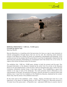
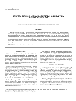
REVIEW ARTICLES
AAEM
Ann Agric Environ Med 1998, 5, 1–5
AMERICAN CUTANEOUS LEISHMANIASIS IN THE STATE OF SÃO PAULO, BRAZIL
- EPIDEMIOLOGY IN TRANSFORMATION
Joao Pupo Nogueira Neto, Gisele Basso, Ana Paula Cipoli, Laila El Kadre
Department of Dermatology of the Pontifícia Universidade Católica de Campinas, Campinas, Brazil
Pupo Nogueira Neto J, Basso G, Cipoli A, El Kadre L: American cutaneous leishmaniasis
in the State of São Paulo, Brazil - epidemiology in transformation. Ann Agric Environ
Med 1998, 5, 1-5.
Abstract: American cutaneous leishmaniasis, a disease of great worldwide importance,
especially on the American continent, has had its epidemological modifications. These
are revised and discussed in view of the current situation of recent increase in its
incidence in regions with low morbidity since 1950, where no apparent cause that
would justify an epidemic outbreak could be found. We analysed the causes of these
alterations and the profile now seen in the State of São Paulo, Brazil.
Address for correspondence: Dr Joao Pupo Nogueira Neto, Rua Maria Monteiro 1572,
POBox 13025-152, Cambui, Campinas - SP, Brazil.
Key words: tropical skin diseases, leishmamasis, Leishmania spp., Brazil,
epidemiology.
INTRODUCTION
American cutaneous leishmaniasis is an anthropozoonosis
caused by a protozoan of the genus Leishmania, whose
vector is an insect Phlebotomus [19].
It afflicts man and different species of wild and
domestic animals in hot, underdeveloped regions of the
Old and New World [29].
It is characterized by the parasitisation of the cells of
the mononuclear phagocytic system of the vertebrate host
and by cutaneous lesions arising at the point of the
parasite´s innoculation, possibly developing into mucosa
lesions after dissemination [29].
The importance of leishmaniasis relies on its high
incidence. The sickness can show various severity in
particular cases and can lead to severe permanent
mutilation in thousands of people every year around the
world, with repercussions in the public health of many
nations [19].
At present in Brazil the disease presents two characteristic
epidemiological patterns: epidemic outbreaks associated
with deforestation for the construction of highways and
access to villages in pioneer regions. This pattern is
characterized by zoonosis of wild animals, which attack
Received: 27 November 1997
anyone who comes into contact with zoonotic foci, which
is the pattern initially described. The second pattern is
leishmaniasis in old colonial regions, not associated with
deforestation, where dogs, horses and rodents seem to
play an important role as reservoirs of the parasite [34].
This paper attempts to show the current situation of this
illness in Brazil, specifically in the State of São Paulo.
HISTORIC
The first researcher to observe the parasites belonging
to the genus Leishmania in cases of visceral leishmaniasis
was Wright, in 1885 in India [51]. After that, in 1898,
Borovsky, the Russian scientist, described in detail these
parasites in cases of cutaneous leishmaniasis, but he did
not name them [24]. In 1903 they were observed by
Leishman, who attributed to them the etiology of the
Indian disease kala-azar [8].
It is accepted that cutaneous leishmaniasis is an
autochtonous disease of the American continent [8]. In
Peru, the pre-columbian indians sculptered these destructive
lesions, especially on the nostrils and upper lips on
ceramics with human shapes called huacos, that when
analysed with our current knowledge leave no doubt as to
2
Pupo Nogueira Neto J, Basso G, Cipoli A, El Kadre L
Figure 3. Phlebotomus (Lutzomia). Species involved in the transmission
in our region (by courtesy of the Secretaria de Saúde do Estado de São
Paulo).
Figure 1. Environment classically described as endemic for leishmaniasis
(by courtesy of the Secretaria de Saúde do Estado de São Paulo).
its leishmaniatic nature [39]. Already in 1908, Escomel
indicated the great similarity existing between the
physiognomy presented by the huaco and people afflicted
with cutaneous leishmaniasis [14].
In Brazil in 1895, Moreira observed the existence of this
illness, clinically identifying it with Biskra’s nodule [35].
In Italy in 1895, Breda described the disease in Italians
who had returned from São Paulo to their homeland [8].
In 1908, in the Santa Casa de São Paulo a great number of
sick people with leishmaniasis appeared [36]. The disease
received various denominations (Bauru ulcer, sharp wound,
Northeast wound) without the etiological cause being
known [36]. Then, on 30 March 1909, Adolfo Lindenberg
announced the discovery of the leishmaniasis parasite [27,
28]. In 1911, Pedroso and Dias da Silva, using the Neal,
Nory and Nicolle medium, obtained Leishmania cultures
from material from Bauru ulcers [37].
The idea that cutaneous leishmaniasis was transmitted
by man-biting insects of the genus Phlebotomus was
suggested for the first time in 1905 by Sergent et al. [43].
In 1922 in Brazil, Aragão succeeded in reproducing ulceration
in a dog by injecting squashed infected insects [5, 34].
Leishmania brasiliensis was experimentally inoculated
in man by Montenegro in 1923 and later by Herrer and
Batistini in 1951 [36]. Based upon these studies, Montenegro
introduced in 1926 the intradermal test, currently still in
use for the diagnosis of leishmaniasis [34, 36].
As classicaly described by Rey in 1973, leishmaniasis
is a wild animal zoonosis, especially affecting rodents,
transmission of which depends on Phlebotomus spp.
living in primitive tropical forests (Fig. 1) [36].
Lately, however, leishmaniasis in Brazil has presented
certain epidemological aspects that are distinct from the
classical concept, with the appearance of endemic foci
apparently not connected to the forest. This pattern is
observed by the Secretary of Health in our region (Fig. 2).
NATURAL HISTORY OF THE DISEASE
Figure 2. City of Campinas, State of São Paulo, Brazil, with autochtonous
cases of leishmaniasis described in recent years.
The transmission occurs through bites of various insect
species of Phlebotomus sensu lato belonging to different
genera (Psychodopygus, Lutzomia), depending on the
geographical localization (Fig. 3) [34]. These small insects,
measuring from 2 to 3 mm, take cover during the day in
humid and dark hiding places [29]. They start their activities
at nightfall, and only the female is hematophagous [42].
Forest species bite also during the day when disturbed by
Cutaneous leishmaniasis in Brazil
3
Figure 4. Leishmaniatic ulcer on the nose with edges in frame and
granulated background.
man, and peridomestic species such as Lutzomia intermedia
can penetrate houses [29].
Human leishmaniasis is evoked by promastigote forms
of the parasite transmitted by the bite of Phlebotomus to
uncovered parts of the skin [36]. After the incubation
phase, which can last from 1 to 12 months, the parasites
multiply in the form of amastigotes. The culmination of
this process is the appearance of the leishmaniatic lesions
(Figures 4–5) [34].
EPIDEMIOLOGY
The importance of leishmaniasis in the world increases.
In 1989, WHO [50] estimated that 350 million people
were exposed to the risk of acquiring the disease, and 12
million people were infected in 1992. This places
leishmaniasis as one of the six most important infectoparasitary diseases in the world.
Its importance in America is great, the area of
appearance of the illness extending from the south of the
USA down to the north of Argentina [42]. In Brazil, cases
have been noted in practically all the States. In recent
years, the Ministry of Health has registered an average of
Figure 5. Leishmaniatic lesion on the thigh.
Figure 6. Geographical distribution of leishmaniasis in Brazil. Areas
with higher frequency of cases and proportion of occurrence are indicated
by region.
25,000 new cases of American cutaneous and visceral
leishmaniasis each year, thus the disease in this area
presents the highest prevalence in the world (Fig. 6) [23].
After a general reduction in the occurrence of the
different forms of leishmaniasis in the 1950’s, the number
of registrations has been progressively growing in the last
20 years. Epidemic outbreaks can be observed in the
south-east region in which the State of São Paulo is
located [34].
The epidemiologic analysis of the incidence of
American cutaneous leishmaniasis in various decades
reveals the predominantly endemic nature of the disease
[19]. Special circumstances that have driven a large
human contingency to come into contact with virgin
forests have led to the above quoted outbreaks [19]. The
natural history of this parasitosis in Brazil has registered
in numerous events of this kind, which first occurred
some time ago, when the Atlantic Forest (Mata Atlântica)
in the south-east region was extensively destroyed (Fig. 7).
Nowadays, this scenario is being repeated in a more
expressive form with the destruction of the Amazon
Forest, but occurs on a smaller scale over the rest of the
country, due to the destruction of the residual forest
(Mata) [6, 12, 18, 49].
The environmental alterations that arise from the
devastation of forests has shown that the species
Leishmania amazonensis and L. brasiliensis are apt to
survive in an alternative environment [17, 26]. The first
species presents a growing distribution in the country
[10]. However, as it has a vector with zoophilic
characteristics, it hardly signifies an real risk to humans
[19]. On the other hand, L. brasiliensis has great ecological
importance, made evident by its vast distribution in Brazil
and the Americas, in areas of ancient colonization where
there was great deforestation, thus beginning to bear
distinct epidemiological characteristics in the course of
4
Pupo Nogueira Neto J, Basso G, Cipoli A, El Kadre L
Figure 7. The advance of deforestation in the State of São Paulo. Modified from Victor, 1975. A. Forest covering in the State of São Paulo at the
beginning of the century; B. the same area in the 50’s - fall in the incidence of leishmaniasis; C. Current situation.
time according to the distribution of the different species of
Phlebotomus involved in the transmission [25]. In the 1930’s
and 1940’s, during the process of colonization of the
south and south-east regions, the transmission was associated
with vectors Lutzomia whitmani, Lu. pessoai and Lu.
mingonei, of wildlife behaviour [34].
Nowadays, the responsible vector is Lu. intermedia,
which is found to be associated with the endemic coastal
areas, and with the valleys of great rivers of the interior of
the country [20, 22] where Phlebotomus is found in
shelters of domestic animals and surrounding habitations
[1, 9, 12, 15, 21, 26, 30, 45, 46, 47]. This occurs because
there persists a rigid environmental law which does not
allow the deforestation of the banks of rivers in order to
avoid obstruction.
The adaptation of Lu. intermedia to altered ecosystems
is mainly observed when close to forested areas, its
presence in forests being quite rare [2, 22].
Up until the present moment, no wild animal has been
pointed out as a reservoir of L. brasiliensis [29]. However,
the encounter of various domestic species that are carriers
of the parasite such as dogs, horses and mules is frequent
in various Brazilian States [3, 4, 10, 13, 15, 16, 30, 31, 32,
33, 38, 40, 41, 49, 52].
In areas in which Lu. intermedia prevails, the endemic
has lost its characteristic forest transmission, is not related
to occupational activities, and occurs in individuals of
both sexes [23] and all ages [19].
In areas where L. brasiliensis is present, children and
women, as well as domestic dogs, are frequently afflicted,
reflecting the dominant character of domestic transmissions
[34]. The afflicted population is generally of a low social
and economic level [7, 33].
Dogs and horses are the only animals that reproduce an
infection that resembles the human disease, and respond
to treatment with antimonials [30, 38].
FINAL COMMENTS
American cutaneous leishmaniasis, once considered to
be practically eradicated from places of ancient colonization,
has again taken on an endemic nature due to the adaptation
of the infectious agents and vectors to environments greatly
altered by man. Leishmaniasis must still be remembered as
one of the most important infecto-contagious pathologies in
the worldwide public health. It is worthwhile remembering
that its control is neither difficult nor expensive, therefore
the outbreaks of leishmaniasis and their consequences
which still occur in our region - supposedly the richest
and most developed in Brazil - cannot be justified.
REFERENCES
1. Aguiar GM, Vilela ML, Lima RB: Ecology of the sandflies of Rio
de Janeiro - food preferences (Diptera, Psychodidae, Phlebotominae).
Mem Inst Oswaldo Cruz 1987, 82, 583-584.
2. Aguiar GM, Santos JLP, Galatti EAP: Ecologia dos flebótomos em
um recente foco ativo de leishmaniose tegumentar no norte do Estado do
Paraná (Diptera, Psychodidae, Phlebotominae). Mem Inst Oswaldo Cruz
1989, 84 (Supl IV), 7-8.
3. Aguiar GM, Rangel EF, et al.: Leishmania (Viannia) braziliensis
associated with domestic animals in Venezuela and Brazil. Mem Inst
Oswaldo Cruz 1989, 84, 19-28.
4. Alencar JE: Um caso de leishmaniose tegumentar em Equus
assinus. Annais do 16o. Congr Bras Hig. Niterói-Brasil (manuscript).
5. Aragão HB: Transmissão da leishmaniose no brasil pelo Phlebotomus
intermedius. Brazil Med 1922, 36, 129-130.
6. Aragão MB, Lima LC: Sobre a dispersão da L. intermedia.
(Diptera, Psychodidae) Cad Saúde Publ 1987, 4, 473-479.
7. Araújo Filho MB, Coura JR: Leishmaniose tegumentar americana
na Ilha Grande, Rio de Janeiro. 1. Investigação epidemiológica, clínica e
laboratorial. Rev Soc Bras Med Trop 1981, 14, 135-143.
8. Azulay RB: Leishmaniose Tegumentar. Rio de Janeiro 1952.
9. Barbosa-Santos, EGA, Marzochi, MCA et al.: Inquérito epidemiológico e
HQWRPROyJLFR HP iUHD HQG PLFD GH OHLVKPDQLRVH FXWkQHD QR 5LR GH
Janeiro (Pacuí-Jacarepaguá). Rev Bras Med Trop 1988, 21 (Suppl), 81-82.
10. Barreto AC, Cuba CC, et al.: Características epidemiológicas da
leishmaniose tegumentar americana, em região end
PLFD do Estado da
Bahia, Brasil. 11. Leishmaniose canina. Rev Soc Med Trop 1984, 17, 59-65.
11. Borgonovi M, Chiarini JV, Amaral AZ, Souza Coelho AG, de
Oliveira D: De Cobertura vegetal do Estado de São Paulo. 1: Levantamento
por fotointerpretação das áreas cobertas com cerrado, cerradão, e campo,
em 1962. Bragantia Campinas 1965, 14, 159-179.
12. Coutinho SG, Marzochi MCA, et al. Leishmaniose tegumentar
americana. J Bras Med 1981, 41, 104-118.
13. Dias M, Mayrink W, et al.: Endemologia da leishmaniose
tegumentar americana. 1. Estudo de reservatórios do Estado de Minas
Gerais. Rev Inst Med Trop 1977, 19, 403-410.
14. Escomel E: La leishmaniose américaine et les leishmanioses en
amériques. Bull Soc Path Exot 1929, 22, 35-46.
15. Falqueto A, Coura JR, et al.: Participação do cão no ciclo de
transmissão de leishmaniose tegumentar no município de Viana, Espírito
Santo. Mem Inst Oswaldo Cruz 1986, 81, 155-163.
16. Falqueto A, Varejão JBM, Sessa PA: Cutaneous leishmanioses in
a horse from endemic area in the State of Espírito Santo, Brazil. Mem
Inst Oswaldo Cruz 1987, 82, 443.
5
Cutaneous leishmaniasis in Brazil
17. Forratini OP: Entomologia Médica. Edgard Blucher, USP, São
Paulo 1973.
18. Forratini OP, Pattoli DBG, et al.: Infecção natural de flebotomíneos
em foco enzoótico de leishmaniose tegumentar no Estado de São Paulo.
Rev Saúde Publ 1972, 6, 431-433.
19. Gomes AC: Perfil epidemiológico da leishmaniose tegumentar no
Brasil. An Bras Dermatol 1992, 67(Suppl 2), 55-60.
20. Gomes AC, Barata JMS, et al.: Aspectos ecológicos da leishmaniose
tegumentar americana. 6. Fauna flebotomínica antropofílica em ambiente
florestal primário da Serra do Mar, região do Vale do Ribeira, Estado de
São Paulo- Brasil. Rev Saúde Publ 1989, 23, 89-97.
21. Gomes AC, Galatti EAB: Aspectos ecológicos da leishmaniose
tegumentar. 7. Capacidade vetorial flebotomínica em ambiente florestal
primário da Serra do Mar, região do Vale do Ribeira, Estado de São
Paulo- Brasil. Rev Saúde Publ 1989, 23, 89-97.
22. Gomes AC, Santos JLF, Galatti EAB: Ecological aspects of
american cutaneous leishmaniasis-observations on the endophilic behaviour
of the sandfly and the vectorial role of Psychodopigus intermedius in the
Ribeira Valley region of the São Paulo State, Brazil. Rev Saúde Publ
1986, 20, 280-287.
23. Grimaldi G, Tesh RB, Pratt DM: A rewiew of the geographic
distribution and epidemiology of leishmaniasis in the new world. Am J
Trop Med Hyg 1989, 41, 687-725.
24. Hoare CA. Early discoveries redarding the parasite of oriental
sore. Trans Roy Soc Trop Med Hyg 1938, 32, 67-92.
25. Jones TC, Johnson Jr AC, et al. Epidemiology of american
cutaneous leishmaniasis due to Leishmania braziliensis braziliensis. J
Inf Dis 1987, 153, 73-83
26. Laison R: Demographic changes and their influence on the
epidemiology of the American leishmaniasis. In: Demography and
Vector - borne Diseases. CRC Boca Raton, Florida 1989.
27. Lindenberg A: A úlcera de bauru e seu micróbio. Comunicação
preventiva. Rev Med S Paulo 1909, 12, 166-120.
28. Lindenberg A: L´ulcére de bauru au le bouton d’orient au Brésil.
Bull Soc Path Exot 1909, 2, 252-254.
29. Marzochi MCA: As leishmanioses no Brasil- as leishmanioses
tegumentares. J Bras Med 1992, 63, 82-89.
30. Mazorchi MCA, Barbosa-Santos EGO, et al.: Forma mucocutânea
e disseminada da infecção natural por Leishmania (v.) braziliensis em
uma égua (Equus caballus). 11. aspectos clínicos, imunopatológicos e
terapêuticos. Rev Soc Bras Med Trop 1991, 24 (Suppl II), 99.
31. Marzochi MCA, Souza WJS, et al.: Evaluation of diagnostic
criteria in human, canine and equine (Equus caballus) leishmaniasis due
to Leishmania braziliensis braziliensis in the south west region of São
Paulo State, Brazil. Mem Inst Oswaldo Cruz 1990, 85, 133-134.
32. Mayrink W, Willians P, et al.: Epidemiology of dermal
leishmaniasis in the Rio Doce Valley, State of Minas Gerais, Brazil. Ann
Trop Med Paras 1979, 73, 124-137.
33. Menezes JÁ, Reis VLL, Vasconcelos JÁ: Pequeno surto de
leishmaniose tegumentar americana em Macuco (Cordeiro-RJ). Rev Soc
Bras Med Trop 1974, 8, 133-151.
34. Ministério da SaúDe: Guia de Controle da Leishmaniose
Tegumentar Americana. Brasília 1991.
35. Moreira J: Existe na Bahia o botão de biskra. Gaz Med Bahia
1895, 254.
36. Pessoa SB, Martins AV: Parasitologia Médica, 1988.
37. Pedroso AE, Silva PD: Botão do oriente (leishmaniose ulcerosa).
cultura da Leishmania tropica (Leishmania wrightii). Arch Soc Med &
Cir São Paulo 1911, 1, 305, 314.
38. Pirmez C, Coutinho SG, et al.: Canine American cutaneous
leihmaniasis: a clinical and imunological study in dogs naturally
infected with leishmania of Rio de Janeiro, Brazil. Am J Trop Med Hyg
1988, 38, 52-58.
39. Rabello E: Contribuições ao estudo da leishmaniose tegumentar
no Brasil 1. Origens, históricos, sinonímia. Ann Bras Dermat Sif 1925,
1, 3-29.
40. Sallenave SL, Valim C, et al.: Utilização de sondas específicas de
dna cinetoplástica na detecção de leishmania em hospedeiros
vertebrados e invertebrados. Rev Inst Med Trop São Paulo 1991,
33(Suppl), 2.
41. Santos GLP, Lima RMS, et al.: Estudo parasitológico de fauna
PXULQD HP iUHD GH RFRUU QFLD GH OHLVKPDQLRVH WHJXPHQWDU DPHULFDQD HP
município da região serrana do Estado do Rio de Janeiro. In: Anais do
24o. Congresso da Sociedade Brasileira de Medicina Tropical, Natal,
RN 1990, 238-239.
42. Secretaria de Estado da Saude: Manual de Vigilância Epidemiológica
Sobre Leishmaniose Tegumentar Americana - 1RUPDV H ,QVWUXoHV 6ão
Paulo 1995.
43. Sergent ED, Sergent ET, Parrot L, Donatien A, Beguel M:
Transmission du clou debiskra par le phlébotome (Phlebotomus
papatasil scop). C R Acad Sci 1921, 173, 1030-1032.
44. Souza MB, Marzochi MCA, Conceição NF: Estudo preliminar da
fauna flebotomínica da Ilha do Araújo, Município de Paraty, Rio de
Janeiro. Rev Soc Bras Med Trop 1991, 24(Suppl), 106.
45. Souza MB, Marzochi MCA, et al.: Estudo da fauna flebotomínica
no município de Santana do Deserto - Sossego, MG. Rev Soc Bras Med
Trop 1991, 24(Suppl II), 105.
46. Tolezano JE, Araujo MFL, et al.: Leishmaniose tegumentar
americana no município de São Roque, Estado de São Paulo, Brasil. Rev
Inst Med Trop São Paulo 1991, 81 (Suppl 33), 55.
47. Tolezano JE, Marcoris SA, Dinis JMP: Modificação na
epidemiologia da leishmaniose tegumentar americana no Vale do
Ribeira, Estado de São Paulo, Brasil. Rev Saude Publ 1989, 23, 89-97.
48. Venexat JÁ, Barreto AC, et al.: Infecção natural de Equus
assinus por Leishmania braziliensis braziliensis na Bahia, Brasil. Mem
Inst Oswaldo Cruz 1986, 81, 237- 238.
49. Victor MAM: A Devastação Florestal. Sociedade Brasileira de
Sivicultura, São Paulo 1975.
50. World Health Organization: Expert Committe on the Control of
the Leishmaniasis. Technical Report Series, 793, Geneva, 1989.
51. Wright JM. Protozoa in a case of tropical ulcer. J Cut Dis 1904,
22, 1-9.
52. Yoshida ELA, Correa FMA, et al.: Human, canine and equine
(Equus caballus) leishmaniasis due to in the south west region of São
Paulo State, Brazil. Mem Inst Oswaldo Cruz 1990, 85, 133-134.
Baixar