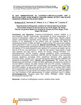
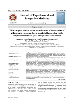
78
Journal of Exercise Physiologyonline
February 2014
Volume 17 Number 1
Official Research Journal of
Editor-in-Chief
Tommy
the American
Boone, PhD,
Society
MBA
of
Exercise
Physiologists
Review
Board
Todd Astorino, PhD
ISSN 1097-9751
Julien Baker,
PhD
Steve Brock, PhD
Lance Dalleck, PhD
Eric Goulet, PhD
Robert Gotshall, PhD
Alexander Hutchison, PhD
M. Knight-Maloney, PhD
Len Kravitz, PhD
James Laskin, PhD
Yit Aun Lim, PhD
Lonnie Lowery, PhD
Derek Marks, PhD
Cristine Mermier, PhD
Robert Robergs, PhD
Chantal Vella, PhD
Dale Wagner, PhD
Frank Wyatt, PhD
Ben Zhou, PhD
JEPonline
Residual Oil Fly Ash (ROFA) Inhalation Promotes Lung
and Heart Oxidative Stress without Hemodynamic
Effects in Exercising Rats
Thiago Gomes Heck1,3, Ramiro Barcos Nunes1, Marcelo Rafael
Petry1, Alexandre Maslinkiewicz1, Paulo Hilário Nascimento Saldiva2,
Pedro Dal Lago1,, Claudia Ramos Rhoden1
1
Programa de Pós-Graduação em Ciências da Saúde, Universidade
Federal de Ciências da Saúde de Porto Alegre (UFCSPA), Porto
Alegre, RS, Brasil, 2 Laboratório de Poluição Experimental, Faculdade
de Medicina– Universidade de São Paulo (USP), São Paulo, SP,
Brasil, 3Grupo de Pesquisa em Fisiologia (GPeF), Departamento de
Ciências da Vida, Programa de Pós-Graduação em Atenção Integral
à Saúde (PPGAIS), Universidade Regional do Noroeste do Estado
do Rio Grande do Sul (UNIJUÍ), Ijuí, RS, Brasil
ABSTRACT
Official Research Journal
of the American Society of
Exercise Physiologists
ISSN 1097-9751
Heck TG, Nunes RB, Petry MR, Maslinkiewicz A, Saldiva PHN, Dal
Lago P, Rhoden CR. ROFA Instillation Promotes Lung and Heart
Oxidative Stress without Hemodynamic Effects in Exercising Rats.
JEPonline 2014;17(1):78-89. The purpose of this study was to test the
hypothesis that exercise effort could increase oxidative damage
induced by residual oil fly ash (ROFA) inhalation that resulted in
hemodynamic alterations during exercise. Wistar rats that were
submitted to ROFA before 20 min of swimming exercise showed an
increase in lipid peroxidation (MDA and Chemiluminesnce) in the
lungs. The ROFA treatment also increased lipid peroxidation in the
heart. However, the treatment had no influence on heart rate or blood
pressure responses during exercise. These findings indicate that
particulate matter inhalation during exercise may exacerbate oxidative
stress in the heart and lungs without producing significant alterations
in the hemodynamic variables.
Key Words: Exercise, Oxidative Stress, Pollution, Particulate Matter
79
INTRODUCTION
Numerous epidemiological studies indicate that the exposure to particulate matter (PM) increases
cardiovascular complication. In fact, there are several indications that the exposure is associated with
the first cardiovascular event (22), the number of hospital readmissions (41), the increase in blood
pressure (48), and the increased risk of death for cerebrovascular and coronary heart diseases (3).
The mechanisms by which PM inhalation promotes cardiovascular injuries might be related to direct
or indirect actions. Ultrafine inhaled particles accumulated in the lungs are capable of translocating
from the airways into the bloodstream (23) causing systemic inflammation via oxidative stress that
mediates endothelial dysfunction and atherosclerosis (44) and increases blood coagulability by
activating platelets and coagulation factors such as fibrinogen. Furthermore, the interaction between
pollutants and lung cells may engage the heart by activating pulmonary neural reflexes that initiate
alterations in the autonomic nervous system and/or increase the levels of pro-inflammatory cytokines.
These inflammatory mediators result in an increase of lipid peroxidation and antioxidants enzyme
activity in the heart of rats (17,29) as well as dysfunction of the autonomic nervous system activity
(15).
Particulate matter has the ability to induce oxidative stress, which is largely mediated by the transition
metal adsorbed at its surface. These metals generate hydroxyl radicals that are able to induce cellular
damage(15). Similar effects after instillation of residual oil fly ash (ROFA –ultrafine particles rich in
transition metals) have been reported Rhoden et al. (29). These effects (i.e., the increase in
inflammation and oxidative stress markers) can also be found in humans (4). There are published
indications that pulmonary damage elicited by particle inhalation is augmented during exercise
through increased lung deposition, which may act in synergism with increased oxygen transportation
across the alveolar-capillary surface (5,32,39). Although the impact of PM on physical performance
has not been well documented, systemic oxidative stress and inflammation induced by the polluting
particles might have a negative impact on cardiovascular function and can be related to the decrease
in physical performance (11). As an example, chronic exposure to outdoor traffic pollution has been
suggested to be associated with the decrease in physical effort along with an increase in risk of
abnormal cardiovascular functionality during exercise (39).
Despite the extensive literature on oxidative stress induced by exercise and PM, relatively few studies
have investigated the cardiorespiratory effects of particles inhalation during exercise on oxidative
parameters. Hence, the purpose of this study was to test the hypothesis that exercise effort could
increase oxidative damage induced by residual oil fly ash (ROFA) instillation while not producing
significant alterations in the hemodynamic responses during exercise.
METHODS
Animals
Fifteen male Wistar rats aged 90 days from the Animal Breeding Unit of Universidade Federal de
Ciências da Saúde de Porto Alegre (UFCSPA) were subjects in this study. The animals were kept in
plastic cages (47 cm x 34 cm x 18 cm) under controlled humidity (75 to 85%) with a temperature of 22
± 2°C, a dark-light cycle of 12 hrs (lights on from 7 am to 7 pm). They were fed with a conventional
laboratory diet (Supra-lab, Alisul Alimentos S.A, Brazil) and water ad libitum.
Animal Care
The animals were handled humanely in the performance of this project to minimize the use of animals
and to prevent animal distress. All protocols were approved by the Universidade Federal de Ciências
da Saúde de Porto Alegre (UFCSPA) Ethical Committee for Research (CPA 017/053).
80
Adaptation to the Water
All rats were adapted to the water before the beginning of the study. The adaptation protocol
consisted of 3 consecutive days of keeping the animals 10 min in shallow water at 30°C to reduce the
rat behavior stress related to water contact. Physical training adaptations were not allowed with this
intervention.
Experimental Design
Heart Oxidative Stress and Hemodynamic Alterations Induced by Intratracheal ROFA
Inhalation: One day after the water adaptation period, the carotid of 15 additional rats was
cannulated (as described later). Twenty-four hrs after the cannulation, resting hemodynamic data
were recorded and blood samples were obtained for lactate concentration measurement. Later, the
rats were quickly and lightly anesthetized with ether for intratracheal instillation and divided into 2
groups: (a) Exercise + Saline 100 μL saline (EX-S, n = 8); and (b) Exercise + ROFA 500 μG/100μL
ROFA suspension (EX-RF, n = 7). After 10 min of the instillation, the rats from both groups were able
to perform 20 min of a swimming exercise in a chamber filled 45 cm with water at 30°C. The
hemodynamic parameters were recorded simultaneously during exercise. At the end of the exercise
session, blood samples were obtained for lactate concentration determinations and, then immediately
after, the rats were decapitated for heart extraction and posteriorly stored at -80°C for oxidative stress
analysis.
Characterization of the ROFA
The ROFA consisted of particles retained in the electrostatic precipitator installed in one of the
chimneys of a large steel plant localized in São Paulo city, Brazil. The element composition of ROFA
suspension particles was determined by neutron activation analysis that involved short irradiations of
5 min for the determination of chlorine (Cl), potassium (K), manganese (Mn), sodium (Na), and
strontium (Sr) using a pneumatic transfer system facility under a thermal neutron of 1.4x10¹² ncm-2s-1.
Longer irradiations of 16 hrs under a thermal neutron flux of about 1012 ncm-2s-1 were performed for
determinations of arsenic (As), barium (Ba), calcium (Ca), cerium (Ce), chromium (Cr), cobalt (Co),
potassium (K), lanthanum (La), manganese (Mn), sodium (Na), rubidium (Rb), antimony (Sb),
scandium (Sc), thorium (Th), vanadium (V), and zinc (Zn). After adequate decay times, the irradiated
samples and standards were measured using a hyperpure detector Model GX2020 coupled to Model
1510 (Perkin Elmer Ortec, Oak Ridge TN, USA) integrated signal processor, both from Canberra
Corporate Headquarters (Meriden CT, USA). Counting times from 200 to 50,000 sec were used,
depending on the half-lives or activities of radioisotopes considered. The radioisotopes measured
were identified according to their half-lives and γ-ray energies. The concentrations of the elements
were calculated using a comparative method. Measurements of the diameters of the particles (largest
diameter) were made using an integrating eyepiece with polarized light microscopy. Each line in the
eyepiece represented a 10 µm length at a magnification of 1000 x. About 230 events for each particle
were measured. ROFA presented 44.6+0.1% of iron; bromine, 1.482+19 µg g-1; cesium, 16.3+0.3 µg
g-1 ; cobalt, 9.90+0.25 µg g-1; chromium, 107.7+1.4 µg g-1; lanthanum, 10.3+0.1 µg g-1; manganese,
3,884+24 µg g-1; rubidium, 719.7+1.0 µg g-1; antimony, 2.27+0.9 µg g-1; arsenic, 154.4+0.8 µg g-1;
vanadium, 35+4 µg g-1; and zinc, 491.9+3.1 µg g-1. Almost all particles had a diameter of less than 10
µm; the means and standard deviations were 1.7+2.56 µm for carbon; 1.2+2.24 µm for transition
metals particles. About 78% of carbon particles and 98% of transition metals particles had a diameter
less than 2.5 µm. This preparation was used in previous studies (46,47).
Hemodynamic Data Acquisition and Analysis
One catheter (PE-10) filled with 0.06 mL saline was implanted in anesthetized rats (85 mg·kg-1
ketamine and 15 mg·kg-1 xylazine) into the carotid artery for direct arterial pressure measurements.
81
After surgery the rats were separated in individual cages and received food and water ad libitum. At
the day of experiment the arterial cannula was connected to a strain-gauge transducer (P23Db,
Gould-Statham), and blood pressure signals were recorded over 20 min at rest by a microcomputer
equipped with an analog-to-digital converter board (CODAS, 2-kHz sampling frequency; Dataq
Instruments, Inc). The recorded data were analyzed on a beat-to-beat basis to quantify changes in
systolic arterial pressure (SAP), diastolic arterial pressure (DAP), mean arterial pressure (MAP), and
heart rate (HR) (24). The hemodynamic data were also evaluated during the 20 min of exercise. We
used the following surgery procedure: Dopalen® (Ketamine 1.16 g·10 mL-1, Agribrands Ltda, Brazil)
and Anasedan® (Xylazine 2.3 g·100 mL-1, Agribrands Ltda, Brazil).
Measurement of Exercise Intensity by Blood Lactate Concentration
Caudal venous lactate concentrations were determined before and after exercise by Lactate Analyzer
(Accutrend®Plus System, Roche). The results were expressed as mmol·L-1.
Determination of Heart and Lung Oxidative Stress
Tissue Preparation: The heart and lungs from each rat was excised, washed in saline solution and
quickly frozen in liquid nitrogen. For determination of oxidative stress parameters, the heart and lung
samples were homogenized (1:5 and 1:7 w/v) in 120 mmol KCl, 30 mmol sodium phosphate buffer
(pH = 7.4) added with protease inhibitor 0.5 mmol PMSF (Phenylmethanesulfonyl Fluoride) at 0 to
4°C. The suspensions were centrifuged at 600 g for 10 min at 0 to 4°C to remove nuclei and cell
debris. The pellets discarded and the supernatants were used as homogenates (24,27).
Thiobarbituric Acid Reactive Substances Method (TBARS), Chemiluminescence, and Catalase
Enzyme Activity: Homogenates were precipitated with 10% TCA, centrifuged, and incubated with
thiobarbituric acid (T5500-Sigma) for 60 min at 1000C. TBARS were extracted using butanol (1:1
V/V). After centrifugation, the absorbance of the butanol layer was measured at 535 nm (6). The
amount of TBARS formed was expressed in nanomoles of malondialdehyde per milligram of protein
(nmol MDA/mg prot). Malondialdehyde standard was prepared from 1,1,3,3-tetramethoxypropane
(Fluka, USA) (24,46,47).
Chemiluminescence (CL) was measured in a liquid scintillation counter in the out-of-coincidence
mode (LKB Rack Beta Liquid Scintillation Spectrometer 1215, LKB - Produkter AB, Sweden). Heart
homogenates were placed in low-potassium vials at a protein concentration of 0.5 to 1.0 mg of
protein·mL-1 in a reaction medium consisting of 120 mmol·L-1 KCl, 30 mmol·L-1 sodium phosphate
buffer (pH = 7.4). Measurements were started by the addition of 3 mmol·L-1 tert-butyl hydroperoxide.
The data were expressed as counts per sec per milligram of protein. Catalase (CAT) activity was
determined by following the decrease at 240 nm-absorbance in a reaction medium containing 50
mmol·L-1 sodium phosphate buffer (pH = 7.2), and 10 mmol·L-1 hydrogen peroxide (H2O2 ) (1). It was
expressed as picomole of H2O2 reduced per sec per milligram of protein (pmol of H2O2/sec/mg prot).
Protein was measured by the method of Bradford (2), using bovine serum albumin (1 mg·mL-1 to 0.1
mg·mL-1 curve) as standard. The results were expressed in mg of protein/mL of homogenized tissue.
Statistical Analyses
The Student’s t-Test was used for the statistical analysis in blood lactate concentration and all
oxidative stress parameters (TBARS, CL, and CAT). The hemodynamic data (HR, MAP, DAP, and
SAP) were also analyzed by the Student’s t-Test and Paired t-Test to compare rest and exercise
hemodynamic data in the same group. The results were expressed in mean ± standard deviation. The
level of statistical significance was set at 5%. All statistical analyses were conducted using the SPSS
(V.18) Software for Windows.
82
RESULTS
ROFA Inhalation did not Influence the Blood Lactate Concentration during Exercise in Rats
Swimming exercise for 20 min promoted an increase in blood lactate levels (mmol·L-1) in both Saline
(EX-S = 1.82 ± 0.41 at rest vs. 4.01 ± 0.83 after exercise) and ROFA groups (EX-RF = 1.78 ± 0.63 at
rest vs. 4.08 ± 0.76 after exercise) (P<0.05). The ROFA instillation did not promote modifications in
the blood lactate concentration.
ROFA Inhalation Increased Oxidative Stress in Heart and Lungs of Rats Submitted to Exercise
As indicated in Figure 1, rats treated with the ROFA presented an increase in MDA levels (Figure 1A)
(P = 0.0047), and a significant decrease in CAT activity in lung tissue (Figure 1C) (P = 0.0007). The
Chemiluminescense of lung homogenates was not influenced by the ROFA treatment (refer to Figure
1B) (P = 0.3505). In the heart, rats treated with the ROFA presented an increase in MDA (Figure 2A)
(P = 0.03) and Chemiluminescense levels (Figure 2B) (P = 0.015). However, CAT activity (Figure 2C)
was not significantly different between the groups (P = 0.087).
Figure 1. Lung Oxidative Stress of Rats Submitted to Exercise after ROFA Instillation. Lipid peroxidation
levels measured by: (A) MDA concentration; (B) chemimiluminescence; and (c) CAT activity of lung
homogenates from rats submitted to 20 min of swimming exercise after Saline (EX-S, n = 8 ) or ROFA (EX-RF,
n = 7) intratracheal instillation. Results are expressed as mean ± standard deviation. *Significant differences
(P<0.05) between groups.
Figure 2. Heart Oxidative Stress of Rats Submitted to Exercise after ROFA Instillation. Lipid peroxidation
levels measured by: (A) MDA concentration: (B) chemimiluminescence; and (c) CAT activity of heart
homogenates from rats submitted to 20 min of swimming exercise after Saline (EX-S, n = 8 ) or ROFA (EX-RF,
n = 7) intratracheal instillation. Results are expressed as mean ± standard deviation. *Significant differences
(P<0.05) between groups.
83
ROFA Inhalation Had No Influence on Hemodynamic Response during Exercise in Rats
All the hemodynamic data are demonstrated in Table 1. The HR and DAP values and the SAP and
MAP values were similar between the groups during the rest condition. During exercise, there were
no significant differences in the hemodynamic data between the treatment groups. When the rest
condition was compared to the exercise condition, the HR values increased during the exercise
condition, while diastolic, systolic and mean arterial pressure decreased independently of treatment
(Saline or ROFA). The magnitude of the increase in HR and the decrease of the arterial pressure
were similar for the Saline and ROFA groups during the exercise.
Table 1. Hemodynamic Response during Exercise after ROFA Instillation.
Saline (n = 8)
Rofa (n = 7)
DAP (mmHg)
Rest
Exercise
delta
SAP (mmHg)
Rest
Exercise
delta
MAP (mmHg)
Rest
Exercise
delta
HR (beats·min-1)
Rest
Exercise
delta
101.1 ± 6.2
87.9 ± 8.3*
-13.2 ± 8.2
103.5 ± 7.8
87.3 ± 11.3*
-16.2 ± 7.5
136.3 ± 10.4
127.3 ± 9.7*
-9.0 ± 9.2
136.7 ± 9.8
128 ± 11*
-8.7 ± 3.9
120.7 ± 6.4
109.3 ± 9.7*
-11.4 ± 8.7
121.7 ± 6.8
109 ± 11.4*
-12.7 ± 5.8
370 ± 38
432 ± 15*
61 ± 43
348 ± 14
416 ± 36*
68 ± 24
DAP = diastolic arterial pressure, SAP = systolic arterial pressure, MAP = mean arterial pressure, HR = heart
rate at rest and during swimming exercise after Saline (EX-S, n = 8) or ROFA (EX-RF, n = 7) intratracheal
instillation. Results are expressed as mean ± standard deviation. *Significant differences (P<0.05) between
exercise and rest values in the same treatment group using the Paired Student’s t-Test.
DISCUSSION
It is well-recognized that the majority of the experimental work regarding adverse effects of PM is
conducted with rats in resting condition. In the present study we submitted rats to ROFA instillation
during exercise. Specifically, the acute response of particles on the rats’ hemodynamic response
during a swimming exercise was studied to evaluate the toxic effects of PM. This study found that
particles promoted heart and lung oxidative stress without modifying the hemodynamic response to
swimming.
In our study it was necessary to measure the intensity of exercise practiced by the animal groups. As
suggested by the literature, the blood lactate concentration was used to mark the intensity of exercise
(14,18,20,40). Kramer et al. (18) and Gobbato et al. (14) determined that values of blood lactate
concentration in rats around 2 mmol·L-1 are consistent with rest conditions while values of 5.5
mmol·L-1 indicate high intensity exercise. This study found an increase in lactate concentration after
84
exercise at values close to 4 mmol·L-1, indicating that the rats reached moderate intensity lactate
levels independently of treatment groups (14,40).
The administration of particles in the lungs of rodents is a reasonable approach to assess the
potential for and the profile of lung injury. In fact, it is common that a high dose of particles is used to
investigate the mechanisms of pathophysiological lesions (7) and, in this regards, the dose of ROFA
(500 μg) administered in the present study was well above the ambient concentration. In addition, it is
likely that the composition of the ROFA used may not have reflected the urban particles that present
a high contribution of mobile sources (17,28,46,47). Thus, we used ROFA to determine if the rats’
exercise hemodynamic responses would be impaired by heart lipid peroxidation damage induced by
particulate pollution.
Human studies have demonstrated that exposure to PM at rest produces changes in the subjects’
HR, HR variability, blood pressure, and electrical conduction system of the heart in patients with
preexisting cardiovascular disease. Since cardiorespiratory demand is increased during exercise, as
observed by the increase in oxygen consumption (VO2), there is a significant increase in pulmonary
ventilation and diffusion capacity. These physiological adjustments may be linked to an augmented
risk of inhaling particles that result in damage to the cardiorespiratory system during exercise (32,33).
This is an important point, given that the literature indicates that the total amount of ultrafine particles
deposited in respiratory tract of humans during moderate exercise is approximately 5 times higher
than at rest (9).
Given that the studies regarding heart oxidative stress and PM exposure were conducted with rats in
the resting condition, the present study is novel in that it was conducted with rats during exercise.
Thus, it was essential to determine the exercise intensity performed by the rats. Lactate concentration
(in all experiments) and cardiovascular parameters during experiment 2 (Table 1) showed that the
rats were submitted to a similar exercise intensity throughout the study. The findings support an
increase in oxidative stress in the rats’ heart that was induced by PM inhalation. Specifically, it was
observed that ROFA instillation promoted an increase of lipid peroxidation in heart tissue of rats
submitted to acute exercise. The fact that PM induced lipid peroxidation at rest is not novel. Previous
studies (10,27,28,30) have demonstrated that pulmonary and cardiac adverse effects of PM might be
mediated by mechanism dependent on oxidants and autononomic unbalance (13,29). But, the
increased oxidative risk without hemodynamic alterations during exercise after particles exposure is a
new and an important finding.
In the present study, the heart CAT activity of the rats was similar in the treated groups. This finding
disagrees with authors who demonstrated a protective effect of CAT in cardiac myocytes damage
induced by diesel exhaust (25). As an example, in an earlier study (10), we showed that 90 days of
ROFA treatment promoted an increase in heart CAT. Also, Gurgueira and colleagues (17) reported a
21% increase in CAT activity in heart tissue of rats exposed to concentrated ambient particles (CAPs)
for 5 hrs. In contrast, the explanation for the lack of an increase in CAT activity in the present study
might be explained to some extent by the different models (i.e., period and pollution protocols). Also,
the fact that acute exercise per se can increase mitochondrial CAT activity by 358% with a longer
exercise duration and a higher exercise intensity than used in the present study as compared with
rest condition (34). Therefore, it is reasonable to believe that the exercise may have masked the PM
effect on CAT activity.
The idea that oxidants generated by PM alter cardiac function is well documented (13,15,17). In fact,
it has been demonstrated that sympathetic and parasympathetic blockers prevent the CAPs-mediated
cardiac adverse effects, thus showing the critical role for autonomic nervous system signaling in
85
preventing damage (29). Similarly, with respect to the published literature (31,42) and ROFA
instillation, the present study indicates that the HR, SBP, DBP, and MAP responses are a function of
the exercise condition that masks the PM effect.
There are some controversial data in the literature regarding hemodynamic parameters in animal
models of exercise. For example, Kramer et al. (18) demonstrated an increase of HR in rats
submitted to a swim test. Yeung et al. (45) found an increase in HR and SAP without alteration in
DAP using a treadmill for 7 min. When evaluating cardiovascular responses during human exercise in
water, Park et al. (26) and Cider et al. (6) found an increase in HR and blood pressure when
compared to resting condition. During water immersion at 30° of temperature, Park et al. (26) showed
a 50% increase in cardiac output, although HR decreased 15%. The water immersion modified other
hemodynamic parameters such as an increase of 8% in SAP, 15% in DAP and decrease of 32% in
total peripheral resistance. Other studies found a decrease in blood pressure (8) or no significant
alterations in these parameters (6,12). Interestingly, a recent review showed that the HR response of
diving and proposed that the HR response of exercise in mammalians are variable because the
suprabulbar control over this reflex behavior. Moreover, underwater submersion results in
bradycardia mediated by parasympathetic neurons and the redistribution of blood flow away from
muscle followed by reperfusion during emersion (21).
In the present study, we observed that rats submitted to short-term moderate exercise activity
increased HR in both groups independently with or without the treatment with ROFA. However,
interestingly, all of the measurements regarding blood pressure were reduced with exercise. Also,
there were no differences between exposure groups. Similar changes in HR and PAM analysis in rats
during the swim exercise suggest that these effects occurred in response to gradual parasympathetic
influence that could be explained by the frequent submerges of the animal (19).
Although there are few studies in the literature associating air pollution adverse effects with exercise
(11,16,39,43), Sharman et al. (32) published a recommendation to avoid exercise practice in parks or
in recreational areas close to busy roadways or industrial sites, which is consistent with exercise
guidelines for people with high blood pressure (33). These authors suggest that it may be desirable to
limit the duration of the exercise session when air pollution is critical. Specifically, the PM may
decrease cardiorespiratory fitness (36) by influencing lung function after exposure (35). However, it is
also important to point out that the acute pollution exposure during a single exercise session is
possibly questionable especially in light of the protective effect that results from chronic exercise
(37,38).
CONCLUSIONS
The findings in this study indicate that the exposure of Wistar rats to ROFA before 20 min of
swimming exercise resulted in an increase in lipid peroxidation (MDA and Chemiluminesnce) in the
lungs. The ROFA treatment also increased lipid peroxidation in the heart. However, the treatment had
no influence on rats’ heart rate or blood pressure responses during exercise. Thus, it is reasonable to
conclude that the particulate matter inhalation during exercise may exacerbate oxidative stress in the
heart and lungs without producing significant alterations in the hemodynamic variables.
86
ACKNOWLEDGMENTS
This work was supported by Universidade Federal de Ciências da Saúde de Porto Alegre (UFCSPA).
T.G. Heck and M. Petry were supported by a fellowship from Coordenação de Aperfeiçoamento de
Pessoal de Nível Superior (CAPES). Dr C.R. Rhoden, Dr. P. Dal Lago and Dr. P.H.N. Saldiva are
supported by Conselho Nacional de Desenvolvimento Científico e Tecnológico (CNPq). The authors
declare they have no conflict of interest.
Address for correspondence: Heck TG, PhD, Department of Life Sciences (DCVida). Universidade
Regional do Noroeste do Estado do Rio Grande do Sul, Rua do comércio, Bairro Universitário, Ijuí,
RS. CEP: 98700-000. Brazil, Email: [email protected]
REFERENCES
1. Aebi H. Catalase in vitro. Methods Enzymol. 1984;105:121-126.
2. Bradford MM. A rapid and sensitive method for the quantitation of microgram quantities of
protein utilizing the principle of protein-dye binding. Anal Biochem. 1976;72:248-254.
3. Brook RD, Rajagopalan S, Pope CA, 3rd, Brook JR, Bhatnagar A, Diez-Roux AV, et al.
Particulate matter air pollution and cardiovascular disease: An update to the scientific
statement from the American Heart Association. Circulation. 2010;121(21):2331-2378.
4. Brucker N, Moro AM, Charao MF, Durgante J, Freitas F, Baierle M, et al. Biomarkers of
occupational exposure to air pollution, inflammation and oxidative damage in taxi drivers. Sci
Total Environ. 2013;463-464C:884-893.
5. Carlisle AJ, Sharp NC. Exercise and outdoor ambient air pollution. Br J Sports Med. 2001;35
(4):214-222.
6. Cider A, Svealv BG, Tang MS, Schaufelberger M, Andersson B. Immersion in warm water
induces improvement in cardiac function in patients with chronic heart failure. Eur J Heart Fail.
2006;8(3):308-313.
7. Costa DL, Lehmann JR, Winsett D, Richards J, Ledbetter AD, Dreher KL. Comparative
pulmonary toxicological assessment of oil combustion particles following inhalation or
instillation exposure. Toxicol Sci. 2006;91(1):237-246.
8. Craig AB, Jr., Dvorak M. Thermal regulation of man exercising during water immersion. J Appl
Physiol. 1968;25(1):28-35.
9. Daigle CC, Chalupa DC, Gibb FR, Morrow PE, Oberdorster G, Utell MJ, et al. Ultrafine particle
deposition in humans during rest and exercise. Inhal Toxicol. 2003;15(6):539-552.
10. Damiani RM, Piva MO, Petry MR, Saldiva PH, Tavares Duarte de Oliveira A, Rhoden CR. Is
cardiac tissue more susceptible than lung to oxidative effects induced by chronic nasotropic
instillation of residual oil fly ash (ROFA)? Toxicol Mech Method. 2012;22 (7):533-539.
87
11. Florida-James G, Donaldson K, Stone V. Athens 2004: The pollution climate and athletic
performance. J Sports Sci. 2004;22(10):967-980.
12. Gabrielsen A, Johansen LB, Norsk P. Central cardiovascular pressures during graded water
immersion in humans. J Appl Physiol. 1993;75(2):581-585.
13. Ghelfi E, Rhoden CR, Wellenius GA, Lawrence J, Gonzalez-Flecha B. Cardiac oxidative stress
and electrophysiological changes in rats exposed to concentrated ambient particles are
mediated by TRP-dependent pulmonary reflexes. Toxicol Sci. 2008;102(2):328-336.
14. Gobatto CA, de Mello MA, Sibuya CY, de Azevedo JR, dos Santos LA, Kokubun E. Maximal
lactate steady state in rats submitted to swimming exercise. Comp Biochem Physiol A Mol
Integr Physiol. 2001;130(1):21-27.
15. Gonzalez-Flecha B. Oxidant mechanisms in response to ambient air particles. Mol Aspects
Med. 2004;25(1-2):169-182.
16. Grievink L, Jansen SM, van't Veer P, Brunekreef B. Acute effects of ozone on pulmonary
function of cyclists receiving antioxidant supplements. Occup Environ Med. 1998;55(1):13-17.
17. Gurgueira SA, Lawrence J, Coull B, Murthy GG, Gonzalez-Flecha B. Rapid increases in the
steady-state concentration of reactive oxygen species in the lungs and heart after particulate
air pollution inhalation. Environ Health Perspect. 2002;110(8):749-755.
18. Kramer K, Dijkstra H, Bast A. Control of physical exercise of rats in a swimming basin. Physiol
Behav. 1993;53(2):271-276.
19. Kregel KC, Allen DL, Booth FW, Fleshner M, Henricksen E, Musch TI, et al. Resource Book
for the Animal Exercise Protocols. American physiological Society 2006.
20. Manchado Fde B, Gobatto CA, Voltarelli FA, Rostom de Mello MA. Non-exhaustive test for
aerobic capacity determination in swimming rats. Appl Physiol Nutr Metab. 2006;31(6):731736.
21. Michael Panneton W. The Mammalian diving response: an enigmatic reflex to preserve life?
Physiology. 2013;28(5):284-297.
22. Miller KA, Siscovick DS, Sheppard L, Shepherd K, Sullivan JH, Anderson GL, et al. Long-term
exposure to air pollution and incidence of cardiovascular events in women. N Engl J Med.
2007;356(5):447-458.
23. Nemmar A, Al-Maskari S, Ali BH, Al-Amri IS. Cardiovascular and lung inflammatory effects
induced by systemically administered diesel exhaust particles in rats. Am J Physiol Lung Cell
Mol Physiol. 2007;292(3):L664-L670.
24. Nunes RB, Tonetto M, Machado N, Chazan M, Heck TG, Veiga AB, et al. Physical exercise
improves plasmatic levels of IL-10, left ventricular end-diastolic pressure, and muscle lipid
peroxidation in chronic heart failure rats. J Appl Physiol. 2008;104(6):1641-1647.
88
25. Okayama Y, Kuwahara M, Suzuki AK, Tsubone H. Role of reactive oxygen species on diesel
exhaust particle-induced cytotoxicity in rat cardiac myocytes. J Toxicol Environ Health A.
2006;69(18):1699-1710.
26. Park KS, Choi JK, Park YS. Cardiovascular regulation during water immersion. Appl Human
Sci. 1999;18(6):233-241.
27. Pereira CE, Heck TG, Saldiva PH, Rhoden CR. Ambient particulate air pollution from vehicles
promotes lipid peroxidation and inflammatory responses in rat lung. Braz J Med Biol Res.
2007;40(10):1353-1359.
28. Rhoden CR, Lawrence J, Godleski JJ, Gonzalez-Flecha B. N-acetylcysteine prevents lung
inflammation after short-term inhalation exposure to concentrated ambient particles. Toxicol
Sci. 2004;79(2):296-303.
29. Rhoden CR, Wellenius GA, Ghelfi E, Lawrence J, Gonzalez-Flecha B. PM-induced cardiac
oxidative stress and dysfunction are mediated by autonomic stimulation. Biochim Biophys
Acta. 2005;1725(3):305-313.
30. Rhoden CR, Ghelfi E, Gonzalez-Flecha B. Pulmonary inflammation by ambient air particles is
mediated by superoxide anion. Inhal Toxicol. 2008;20(1):11-15.
31. Rivero DH, Soares SR, Lorenzi-Filho G, Saiki M, Godleski JJ, Antonangelo L, et al. Acute
cardiopulmonary alterations induced by fine particulate matter of Sao Paulo, Brazil. Toxicol
Sci. 2005;85(2):898-905.
32. Sharman JE, Cockcroft JR, Coombes JS. Cardiovascular implications of exposure to traffic air
pollution during exercise. QJM: Monthly Journal of the Association of Physicians. 2004;97
(10):637-643.
33. Sharman JE, Stowasser M. Australian association for exercise and sports science position
statement on exercise and hypertension. Aust J Sci Med Sport. 2009;12(2):252-257.
34. Somani SM, Frank S, Rybak LP. Responses of antioxidant system to acute and trained
exercise in rat heart subcellular fractions. Pharmacol Biochem Behav. 1995;51(4):627-634.
35. Steinvil A, Fireman E, Kordova-Biezuner L, Cohen M, Shapira I, Berliner S, et al.
Environmental air pollution has decremental effects on pulmonary function test parameters up
to one week after exposure. Am J Med Sci. 2009;338(4): 273-279.
36. Steinvil A, Shmueli H, Ben-Assa E, Leshem-Rubinow E, Shapira I, Berliner S, et al.
Environmental exposure to combustion-derived air pollution is associated with reduced
functional capacity in apparently healthy individuals. Clin Res Cardiol. 2013;102(8):583-591.
37. Veloso CF, Dalla Costa C, Rovani BT, Araldi ICC, Freitas RB, Cogo LA, et al. Effects of
exercise on cardiac oxidative stress in rats after exposure to cigarette smoke. JEPonline.
2013;16(5):21-27.
89
38. Vieira RP, Toledo AC, Silva LB, Almeida FM, Damaceno-Rodrigues NR, Caldini EG, et al.
Anti-inflammatory effects of aerobic exercise in mice exposed to air pollution. Med Sci Sports
Exerc. 2012;44(7):1227-1234.
39. Volpino P, Tomei F, La Valle C, Tomao E, Rosati MV, Ciarrocca M, et al. Respiratory and
cardiovascular function at rest and during exercise testing in a healthy working population:
Effects of outdoor traffic air pollution. Occup Med (Lond). 2004;54(7):475-482.
40. Voltarelli FA, Gobatto CA, de Mello MA. Determination of anaerobic threshold in rats using the
lactate minimum test. Braz J Med Biol Res. 2002;35(11):1389-1394.
41. von Klot S, Peters A, Aalto P, Bellander T, Berglind N, D'Ippoliti D, et al. Ambient air pollution
is associated with increased risk of hospital cardiac readmissions of myocardial infarction
survivors in five European cities. Circulation. 2005;112(20):3073-3079.
42. Wellenius GA, Saldiva PH, Batalha JR, Krishna Murthy GG, Coull BA, Verrier RL, et al.
Electrocardiographic changes during exposure to residual oil fly ash (ROFA) particles in a rat
model of myocardial infarction. Toxicol Sci. 2002;66(2):327-335.
43. Wong CM, Ou CQ, Thach TQ, Chau YK, Chan KP, Ho SY, et al. Does regular exercise protect
against air pollution-associated mortality? Prev Med. 2007;44(5):386-392.
44. Yamawaki H, Iwai N. Mechanisms underlying nano-sized air-pollution-mediated progression of
atherosclerosis: carbon black causes cytotoxic injury/inflammation and inhibits cell growth in
vascular endothelial cells. Circ J. 2006;70(1):129-140.
45. Yeung PK, Feng JD, Fice D. Exercise hemodynamic and neurohormone responses as
sensitive biomarkers for diltiazem in rats. J Pharm Pharm Sci. 2006;9(2):245-251.
46. Zanchi AC, Venturini CD, Saiki M, Nascimento Saldiva PH, Tannhauser Barros HM, Rhoden
CR. Chronic nasal instillation of residual-oil fly ash (ROFA) induces brain lipid peroxidation and
behavioral changes in rats. Inhal Toxicol. 2008;20(9):795-800.
47. Zanchi AC, Saiki M, Saldiva PH, Barros HM, Rhoden CR. Hippocampus lipid peroxidation
induced by residual oil fly ash intranasal instillation versus habituation to the open field.
Inhalation toxicology. 2010;22(1):84-88.
48. Zanobetti A, Canner MJ, Stone PH, Schwartz J, Sher D, Eagan-Bengston E, et al. Ambient
pollution and blood pressure in cardiac rehabilitation patients. Circulation. 2004;110(15):2184
-2189.
Disclaimer
The opinions expressed in JEPonline are those of the authors and are not attributable to JEPonline,
the editorial staff or the ASEP organization.
Baixar