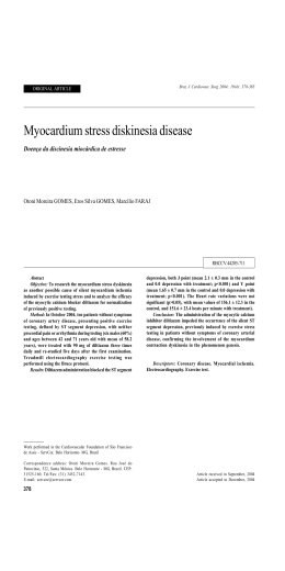
www.arquivosonline.com.br Sociedade Brasileira de Cardiologia • ISSN-0066-782X • Volume 101, Nº 6, December 2013 Figure 1 - 65-year-old male patient that came to the emergency room with atypical chest pain, nonspecific ECG and normal myocardial necrosis markers (enzymes). Page 567 Editorial Is Conventional Cardiac Pacing Harmful in Patients with Normal Implementation of Multicenter Records in the Therapeutic Ventricular Function? Cardiovascular Assessment in Brazil Brazilian Portuguese Validated Version of the Cardiac Anxiety Original Articles Questionnaire Predictors of Hospitalization in Patients with Syncope Assisted in Review Article Specialized Cardiology Hospital Coronary Computed Tomography Angiography in the Assessment of Prevalence of Ischemia on Myocardial Perfusion Scintigraphy of Pre- Acute Chest Pain in the Emergency Room and Postmenopausal Women Letter to the Editor Chromosomal Abnormalities in Patients with Congenital Heart Disease Clinical Significance of Histological Features of Thrombi in Patients Coronary Trunk Dissection with Stent Displacement: Points to Remember! The Year 2011 in Interventional Cardiology with Myocardial Infarction The Bleeding Risk Score as a Mortality Predictor in Patients with Acute Coronary Syndrome P Wave Indices to Predict Atrial Fibrillation Recurrences Post Pulmonary Vein Isolation Eletronic Pages Clinicoradiological Session Case 6/2013 – 56 years old Woman with Ebstein Anomaly in Heart Failure Mortality Impact of Thoracic Aortic Disease in São Paulo State from Case Report 1998 to 2007 Pericardial Synovial Sarcoma: Case Report and Literature Review Experimental Cardiac Arrest Treatment with Adrenaline, Vasopressin, Point of View or Placebo Cardiovascular Rehabilitation, Ballroom Dancing and Sexual Dysfunction REVISTA DA SOCIEDADE BRASILEIRA DE CARDIOLOGIA - Publicada desde 1948 Contents Editorial Implementation of Multicenter Records in the Therapeutic Cardiovascular Assessment in Brazil Luiz Felipe P. Moreira .....................................................................................................................................................................page 478 Original Articles Clinical Arrhythmia Predictors of Hospitalization in Patients with Syncope Assisted in Specialized Cardiology Hospital Leonardo Marques Fischer, João Pedro Passos Dutra, Augusto Mantovani, Gustavo Glotz de Lima, Tiago Luiz Luz Leiria .....................................................................................................................................................................page 480 Nuclear Cardiology Prevalence of Ischemia on Myocardial Perfusion Scintigraphy of Pre-and Postmenopausal Women Daniel Augusto Message dos Santos, Wendy Yasdin Sierraalta Navarro, Leonardo Machado Alexandre, Priscila Feitosa Cestari, Paola Emanuela Poggio Smanio .....................................................................................................................................................................page 487 Pediatric Cardiology Chromosomal Abnormalities in Patients with Congenital Heart Disease Patrícia Trevisan, Tatiana Diehl Zen, Rafael Fabiano Machado Rosa, Juliane Nascimento da Silva, Dayane Bohn Koshiyama, Giorgio Adriano Paskulin, Paulo Ricardo Gazzola Zen .....................................................................................................................................................................page 495 Acute Coronary Artery Disease Clinical Significance of Histological Features of Thrombi in Patients with Myocardial Infarction Juliana Canedo Sebben, Eduardo Cambruzzi, Luisa Martins Avena, Cristina do Amaral Gazeta, Carlos Antonio Mascia Gottschall, Alexandre Schaan de Quadros .....................................................................................................................................................................page 502 The Bleeding Risk Score as a Mortality Predictor in Patients with Acute Coronary Syndrome José Carlos Nicolau, Humberto Graner Moreira, Luciano Moreira Baracioli, Carlos Vicente Serrano Jr., Felipe Galego Lima, Marcelo Franken, Roberto Rocha Giraldez, Fernando Ganem, Roberto Kalil Filho, José Antônio Franchini Ramires, Roxana Mehran .....................................................................................................................................................................page 511 Arquivos Brasileiros de Cardiologia - Volume 101, Nº 6, December 2013 Electrocardiografía P Wave Indices to Predict Atrial Fibrillation Recurrences Post Pulmonary Vein Isolation Ahmed Salah, Shenghua Zhou, Qiming Liu, Hui Yan .....................................................................................................................................................................page 519 Epidemiology Mortality Impact of Thoracic Aortic Disease in São Paulo State from 1998 to 2007 Ricardo Ribeiro Dias, Omar Asdrubal Vilca Mejia, Fábio Fernandes, Félix José Alvarez Ramires, Charles Mady, Noedir Antonio Groppo Stolf, Fabio Biscegli Jatene .....................................................................................................................................................................page 528 Experimental Experimental Cardiac Arrest Treatment with Adrenaline, Vasopressin, or Placebo Manoel Ângelo Gomes Palácio, Edison Ferreira de Paiva, Luciano Cesar Pontes de Azevedo, Ari Timerman .....................................................................................................................................................................page 536 Pacemaker Is Conventional Cardiac Pacing Harmful in Patients with Normal Ventricular Function? Luiz Antonio Batista de Sá, Salvador Rassi, Márcia Andery Ludovico Batista .....................................................................................................................................................................page 545 Cardiovascular Rehabilitation Brazilian Portuguese Validated Version of the Cardiac Anxiety Questionnaire Aline Sardinha, Antonio Egidio Nardi, Claudio Gil Soares de Araújo, Maria Cristina Ferreira, Georg H. Eifert .....................................................................................................................................................................page 554 Review Article Coronary Computed Tomography Angiography in the Assessment of Acute Chest Pain in the Emergency Room Carlos Eduardo Elias dos Prazeres, Roberto Caldeira Cury, Adriano Camargo de Castro Carneiro, Carlos Eduardo Rochitte .....................................................................................................................................................................page 562 Letter to the Editor Coronary Trunk Dissection with Stent Displacement: Points to Remember! Marco Tulio Zanettini, Jacira Pisani Zanettini, João Otavio Zanettini .....................................................................................................................................................................page 570 The Year 2011 in Interventional Cardiology Pablo Avanzas, Magda Heras, Juan Sanchis .....................................................................................................................................................................page 571 Arquivos Brasileiros de Cardiologia - Volume 101, Nº 6, December 2013 Arquivos Brasileiros de Cardiologia - Eletronic Pages Clinicoradiological Session Case 6/2013 – 56 years old Woman with Ebstein Anomaly in Heart Failure Edmar Atik ................................................................................................................................................................ page e101 Case Report Pericardial Synovial Sarcoma: Case Report and Literature Review Sabrina Godoy Bezerra, Andrea Araújo Brandão, Denilson Campos Albuquerque, Rochelle Coppo Militão, Marcelo Souza Hadlich, Clerio Francisco Azevedo ................................................................................................................................................................ page e103 Point of View Cardiovascular Rehabilitation, Ballroom Dancing and Sexual Dysfunction Tales de Carvalho, Ana Inês Gonzáles, Sabrina Weiss Sties, Gabriela Maria Dutra de Carvalho ................................................................................................................................................................ page e107 * Indicate manuscripts only in the electronic version. To view them, visit: http://www.arquivosonline.com.br/2013/english/10106/edicaoatual.asp Arquivos Brasileiros de Cardiologia - Volume 101, Nº 6, December 2013 www.arquivosonline.com.br A JOURNAL OF SOCIEDADE BRASILEIRA DE CARDIOLOGIA - Published since 1948 Scientific Director Interventionist Cardiology Luiz Alberto Piva e Mattos Pedro A. Lemos Chief Editor Pediatric/Congenital Cardiology Luiz Felipe P. Moreira Associated Editors Clinical Cardiology José Augusto Barreto-Filho Surgical Cardiology Paulo Roberto B. Evora Antonio Augusto Lopes Arrhythmias/Pacemaker Mauricio Scanavacca Non-Invasive Diagnostic Methods Carlos E. Rochitte Basic or Experimental Research Leonardo A. M. Zornoff Epidemiology/Statistics Lucia Campos Pellanda Arterial Hypertension Paulo Cesar B. V. Jardim Ergometrics, Exercise and Cardiac Rehabilitation Ricardo Stein First Editor (1948-1953) † Jairo Ramos Editorial Board Brasil Adib D. Jatene (SP) Alexandre A. C. Abizaid (SP) Alfredo José Mansur (SP) Álvaro Avezum (SP) Amanda G. M. R. Sousa (SP) André Labrunie (PR) Andrei Sposito (DF) Angelo A. V. de Paola (SP) Antonio Augusto Barbosa Lopes (SP) Antonio Carlos C. Carvalho (SP) Antônio Carlos Palandri Chagas (SP) Antonio Carlos Pereira Barretto (SP) Antonio Cláudio L. Nóbrega (RJ) Antonio de Padua Mansur (SP) Ari Timerman (SP) Armênio Costa Guimarães (BA) Ayrton Klier Péres (DF) Ayrton Pires Brandão (RJ) Barbara M. Ianni (SP) Beatriz Matsubara (SP) Braulio Luna Filho (SP) Brivaldo Markman Filho (PE) Bruce B. Duncan (RS) Bruno Caramelli (SP) Carisi A. Polanczyk (RS) Carlos Alberto Pastore (SP) Carlos Eduardo Negrão (SP) Carlos Eduardo Rochitte (SP) Carlos Eduardo Suaide Silva (SP) Carlos Vicente Serrano Júnior (SP) Celso Amodeo (SP) Charles Mady (SP) Claudio Gil Soares de Araujo (RJ) Cleonice Carvalho C. Mota (MG) Dalton Valentim Vassallo (ES) Décio Mion Jr (SP) Denilson Campos de Albuquerque (RJ) Dikran Armaganijan (SP) Djair Brindeiro Filho (PE) Domingo M. Braile (SP) Edmar Atik (SP) Edson Stefanini (SP) Elias Knobel (SP) Eliudem Galvão Lima (ES) Emilio Hideyuki Moriguchi (RS) Enio Buffolo (SP) Eulógio E. Martinez Fº (SP) Evandro Tinoco Mesquita (RJ) Expedito E. Ribeiro da Silva (SP) Fábio Sândoli de Brito Jr. (SP) Fábio Vilas-Boas (BA) Fernando A. P. Morcerf (RJ) Fernando Bacal (SP) Flávio D. Fuchs (RS) Francisco Antonio Helfenstein Fonseca (SP) Francisco Laurindo (SP) Francisco Manes Albanesi Fº (RJ) Gilmar Reis (MG) Gilson Soares Feitosa (BA) Ínes Lessa (BA) Iran Castro (RS) Ivan G. Maia (RJ) Ivo Nesralla (RS) Jarbas Jakson Dinkhuysen (SP) João Pimenta (SP) Jorge Ilha Guimarães (RS) Jorge Pinto Ribeiro (RS) José A. Marin-Neto (SP) José Antonio Franchini Ramires (SP) José Augusto Soares Barreto Filho (SE) José Carlos Nicolau (SP) José Geraldo de Castro Amino (RJ) José Lázaro de Andrade (SP) José Péricles Esteves (BA) José Teles Mendonça (SE) Leopoldo Soares Piegas (SP) Luís Eduardo Rohde (RS) Luiz A. Machado César (SP) Luiz Alberto Piva e Mattos (SP) Lurildo Saraiva (PE) Marcelo C. Bertolami (SP) Marcia Melo Barbosa (MG) Marco Antônio Mota Gomes (AL) Marcus V. Bolívar Malachias (MG) Maria Cecilia Solimene (SP) Mario S. S. de Azeredo Coutinho (SC) Maurício I. Scanavacca (SP) Mauricio Wajngarten (SP) Max Grinberg (SP) Michel Batlouni (SP) Nabil Ghorayeb (SP) Nadine O. Clausell (RS) Nelson Souza e Silva (RJ) Orlando Campos Filho (SP) Otávio Rizzi Coelho (SP) Otoni Moreira Gomes (MG) Paulo A. Lotufo (SP) Paulo Cesar B. V. Jardim (GO) Paulo J. F. Tucci (SP) Paulo J. Moffa (SP) Paulo R. A. Caramori (RS) Paulo R. F. Rossi (PR) Paulo Roberto S. Brofman (PR) Paulo Zielinsky (RS) Protásio Lemos da Luz (SP) Renato A. K. Kalil (RS) Roberto A. Franken (SP) Roberto Bassan (RJ) Ronaldo da Rocha Loures Bueno (PR) Sandra da Silva Mattos (PE) Sergio Almeida de Oliveira (SP) Sérgio Emanuel Kaiser (RJ) Sergio G. Rassi (GO) Sérgio Salles Xavier (RJ) Sergio Timerman (SP) Silvia H. G. Lage (SP) Valmir Fontes (SP) Vera D. Aiello (SP) Walkiria S. Avila (SP) William Azem Chalela (SP) Wilson A. Oliveira Jr (PE) Wilson Mathias Jr (SP) Exterior Adelino F. Leite-Moreira (Portugal) Alan Maisel (Estados Unidos) Aldo P. Maggioni (Itália) Cândida Fonseca (Portugal) Fausto Pinto (Portugal) Hugo Grancelli (Argentina) James de Lemos (Estados Unidos) João A. Lima (Estados Unidos) John G. F. Cleland (Inglaterra) Maria Pilar Tornos (Espanha) Pedro Brugada (Bélgica) Peter A. McCullough (Estados Unidos) Peter Libby (Estados Unidos) Piero Anversa (Itália) Sociedade Brasileira de Cardiologia President Jadelson Pinheiro de Andrade Vice-President Dalton Bertolim Précoma President Elect Angelo Amato Vincenzo de Paola Epidemiological Project Council Coordinator David de Pádua Brasil Social Action Coordinators Alvaro Avezum Junior Ari Timerman Administrative Director Marcelo Souza Hadlich New Project Council Coordinator Glaucia Maria Moraes Oliveira Financial Director Eduardo Nagib Gaui Use of New Technology Council Coordinator Washington Andrade Maciel Government Liaison Director Daniel França Vasconcelos Communication Director Carlos Eduardo Suaide Silva Assistance Quality Director José Xavier de Melo Filho Scientific Director Luiz Alberto Piva e Mattos CardiovascularHealth Promotion Director SBC/Funcor Carlos Alberto Machado State / Regional Liaison Director Marco Antonio de Mattos Specialized Department Director Gilberto Venossi Barbosa Information Technology Director Carlos Eduardo Suaide Silva Research Director Fernando Bacal Chief Editor of the Brazilian Archives of Cardiology Luiz Felipe P. Moreira SBC Journal Editor Fábio Vilas-Boas Pinto International Liaison Committee Antonio Felipe Simão João Vicente Vitola Oscar Pereira Dutra Presidents of State and Regional Brazilian Societies of Cardiology SBC/AL - Alfredo Aurelio Marinho Rosa SBC/AM - Jaime Giovany Arnez Maldonado SBC/BA - Augusto José Gonçalves de Almeida SBC/CE - Eduardo Arrais Rocha SBC/CO - Hernando Eduardo Nazzetta (GO) Young Cardiologist Inclusion Council Coordinator Fernando Augusto Alves da Costa SBC/DF - Renault Mattos Ribeiro Junior Clinical Practice Quality and Patient Safety Council Coordinator Evandro Tinoco Mesquita SBC/GO - Luiz Antonio Batista de Sá Standardization and Guideline Council Coordinator Harry Correa Filho SBC/MS - Sandra Helena Gonsalves de Andrade Continuing Education Council Coordinator Antonio Carlos de Camargo Carvalho SBC/NNE - Aristoteles Comte de Alencar Filho (AM) Emergency Care and Sudden Death Committee Manoel Fernandes Canesin Nabil Ghorayeb Sergio Timerman Cardiovascular Prevention Committee Antonio Delduque de Araujo Travessa Sergio Baiocchi Carneiro Regina Coeli Marques de Carvalho Strategic Planning Committee Fabio Sândoli de Brito José Carlos Moura Jorge Walter José Gomes Member Assistance Committee Maria Fatima de Azevedo Mauro José Oliveira Gonçalves Ricardo Ryoshim Kuniyoshi SBC/ES - Antonio Carlos Avanza Junior SBC/MA - Magda Luciene de Souza Carvalho SBC/MG - Maria da Consolação Vieira Moreira SBC/MT - José Silveira Lage SBC/PA - Claudine Maria Alves Feio SBC/PB - Alexandre Jorge de Andrade Negri SBC/PE - Silvia Marinho Martins SBC/PI - Ricardo Lobo Furtado SBC/PR - Álvaro Vieira Moura SBC/RJ - Glaucia Maria Moraes Oliveira SBC/RN - Carlos Alberto de Faria SBC/RS - Justo Antero Sayão Lobato Leivas SBC/SC - Conrado Roberto Hoffmann Filho SBC/SE - Eduardo José Pereira Ferreira SBC/SP - Carlos Costa Magalhães SBC/TO - Adalgele Rodrigues Blois Presidents of the Specialized Departaments and Study Groups SBC/DA - Hermes Toros Xavier (SP) SBC/DCC - Evandro Tinoco Mesquita (RJ) SBC/DCM - Orlando Otavio de Medeiros (PE) SBC/DFCVR - José Carlos Dorsa Vieira Pontes (MS) SBC/DCC/GECETI - João Fernando Monteiro Ferreira (SP) SBC/DHA - Weimar Kunz Sebba Barroso de Souza (GO) SBC/DCC/GEECABE - Luis Claudio Lemos Correia (BA) SBC/DIC - Jorge Eduardo Assef (SP) SBC/DCC/GEECG - Carlos Alberto Pastore (SP) SBC/DCC/CP - Estela Suzana Kleiman Horowitz (RS) SBC/SBCCV - Walter José Gomes (SP) SBC/DECAGE - Abrahão Afiune Neto (GO) SBC/SBHCI - Marcelo Antonio Cartaxo Queiroga Lopes (PB) SBC/DEIC - João David de Souza Neto (CE) SBC/DERC - Pedro Ferreira de Albuquerque (AL) SBC/SOBRAC - Adalberto Menezes Lorga Filho (SP) SBC/DCC/GAPO - Daniela Calderaro (SP) SBC/DCP/GECIP - Angela Maria Pontes Bandeira de Oliveira (PE) SBC/DERC/GECESP - Daniel Jogaib Daher (SP) SBC/DERC/GECN - José Roberto Nolasco de Araújo (AL) Arquivos Brasileiros de Cardiologia Volume 101, Nº 6, Novembro 2013 Indexing: ISI (Thomson Scientific), Cumulated Index Medicus (NLM), SCOPUS, MEDLINE, EMBASE, LILACS, SciELO, PubMed Address: Av. Marechal Câmara, 160 - 3º andar - Sala 330 20020-907 • Centro • Rio de Janeiro, RJ • Brasil Phone.: (21) 3478-2700 E-mail: [email protected] www.arquivosonline.com.br SciELO: www.scielo.br Commercial Department Phone: (11) 3411-5500 E-mail: [email protected] Editorial Production SBC - Internal Publication Department Affiliated at the Brazilian Medical Association Graphic Design and Diagramming SBC - Internal Design Department Inner Core Design Print Stamppa Circulation 1.500 The ads showed in this issue are of the sole responsibility of advertisers, as well as the concepts expressed in signed articles are of the sole responsibility of their authors and do not necessarily reflect the views of SBC. This material is for exclusive distribution to the medical profession. The Brazilian Archives of Cardiology are not responsible for unauthorized access to its contents and that is not in agreement with the determination in compliance with the Collegiate Board Resolution (DRC) N. 96/08 of the National Sanitary Surveillance Agency (ANVISA), which updates the technical regulation on Drug Publicity, Advertising, Promotion and Information. According to Article 27 of the insignia, "the advertisement or publicity of prescription drugs should be restricted solely and exclusively to health professionals qualified to prescribe or dispense such products (...)". To ensure universal access, the scientific content of the journal is still available for full and free access to all interested parties at: www.arquivosonline.com.br. SUPPORT Back to the Cover Editorial Implementation of Multicenter Records in the Therapeutic Cardiovascular Assessment in Brazil Luiz Felipe P. Moreira Instituto do Coração (InCor) do Hospital das Clínicas da Faculdade de Medicina da Universidade de São Paulo, São Paulo, SP - Brasil Clinical research finds, in controlled clinical trials, its main instrument to establish the therapeutic effectiveness of drug or interventional therapies. On the other hand, the evaluation of demographic, clinical and prognostic characteristics of patients under treatment in the real world depends on comprehensive multicenter observational studies, whose results vary according to countries or regions involved. In the field of cardiology, we have, in our country, few records that represent the national reality. In this vein, recent experiences have been conducted by the Brazilian Society of Cardiology and by specific groups or institutions in recording some specific conditions or therapies. These studies include the evaluation of epidemiological aspects, prognostic results and the factors involved in the care of patients with acute coronary syndromes1-3, decompensated heart failure4 or disorders at high risk of cardiovascular events5. In parallel, the evaluation of the characteristics and outcomes of interventional procedures such as the use of stents in the treatment of heart failure6 and cardiac surgeries in the treatment of valvular heart diseases or heart failure7 have also been the subject of specific records. The initial publications of clinical records related to acute coronary syndromes suggest the inclusion of more than six thousand patients treated in centers based in different regions of Brazil. These records have become major documents that show the demography of the disorders involved2,3,8. The initial results of these records demonstrate the diversity of treatments conducted at a national level, including the regional inequality in the employment of reperfusion therapies2. The use of these therapies relates, on the other hand, to lower mortality and lower rates of complications in the immediate follow-up of patients 3,8. Keywords Cardiovascular Diseases / therapy; Multicenter Study; Brazil. Mailing address: Luiz Felipe P. Moreira • Avenida Dr. Enéas Carvalho Aguiar, 44, 2º andar, bloco 2, sala 13 Cerqueira César. CEP 05403-000, São Paulo, SP – Brazil E-mail: [email protected] DOI: 10.5935/abc.20130245 478 Besides this, recent data show lower rates of mortality that are consistent with the international experience in the treatment of unstable angina and acute myocardial infarction with or without ST-segment elevation8. Records focusing on the evolution of patients with heart failure and disorders at high risk of cardiovascular events are still at an initial stage of including patients4,5. However, important issues on the application of confirmedly beneficial therapies can be found in the preliminary data of the REACT study9. As for interventional procedures, publications of the CENIC registry, which includes patients undergoing percutaneous coronary intervention in several Brazilian regions, suggest a progressive improvement of results6,10, including the reduction of vascular complications promoted by the technique of access through the radial artery10. Finally, the constitution of the SP-SCORE-SUS registry to evaluate the results of cardiac surgeries in the State of São Paulo, based on previous publications of a national reference center11,12, fills an important gap with respect to the employment and results of cardiovascular surgical therapy, expected to be expanded at a national level13. The scenario presented shows the growing concern of researchers and our scientific societies to better characterize the therapeutic profile of cardiovascular disorders in our country. The analysis of the results of a number of multicenter records opens new perspectives for better planning of financial, personal and technological resources used in cardiovascular health, and contributes with insights to deepening scientific knowledge on the events under study. Moreira Multicenter cardiology records Editorial References 1. Mattos LA. Rationality and methods of ACCEPT registry - Brazilian registry of clinical practice in acute coronary syndromes of the Brazilian Society of Cardiology. Arq Bras Cardiol. 2011;97(2):94-9. 2. Nicolau JC, Franken M, Lotufo PA, Carvalho AC, Marin Neto JA, Lima FG, et al. Use of demonstrably effective therapies in the treatment of acute coronary syndromes: comparison between different Brazilian regions. Analysis of the Brazilian Registry on Acute Coronary Syndromes (BRACE). Arq Bras Cardiol. 2012;98(4):282-9. 3. Piegas LS, Avezum A, Guimarães HP, Muniz AJ, Reis HJ, dos Santos ES, et al. Acute coronary syndrome behavior: results of a Brazilian registry. Arq Bras Cardiol. 2013 Jun;100(6):502-10 4. BREATHE investigators. Rationale and design: BREATHE registry-I Brazilian Registry of Heart Failure. Arq Bras Cardiol. 2013;100(5):390-4. 5. Mattos LA. Rationality and methods: registry of clinical practice in high-risk cardiovascular patients. Arq Bras Cardiol. 2011;97(1):3-7. 6. Cardoso CO, de Quadros AS, Mattos LA, Gottschall CA, Sarmento-Leite RE, Marin-Neto JA. Use of drug-eluting stents in Brazil: the CENIC (National Registry of Cardiovascular Interventions) registry. Arq Bras Cardiol. 2007;89(6):356-61. 7. Mejía OA, Lisboa LA, Dallan LA, Pomerantzeff PM, Trindade EM, Jatene FB, Kalil Filho R. Heart surgery programs innovation using surgical risk stratification at the São Paulo State Public Healthcare System: SP-SCORESUS study. Rev Bras Cir Cardiovasc. 2013;28(2):263-9. 8. Mattos LA, Berwanger O, dos Santos ES, Reis HJ, Romano ER, Petriz JL, et al. Clinical outcomes at 30 days in the Brazilian Registry of Acute Coronary Syndromes (ACCEPT). Arq Bras Cardiol. 2013;100(1):6-13. 9. Berwanger O, Piva e Mattos LA, Martin JF, Lopes RD, Figueiredo EL, Magnoni D, et al. Evidence-based therapy prescription in highcardiovascular risk patients: the REACT study. Arq Bras Cardiol. 2013;100(3):212-20. 10. Andrade PB, Tebet MA, Andrade MV, Labrunie A, Mattos LA. Radial approach in percutaneous coronary interventions: current status in Brazil. Arq Bras Cardiol. 2011;96(4):312-6. 11. Lisboa LA, Mejia OA, Dallan LA, Moreira LF, Puig LB, Jatene FB, Stolf NA. Previous percutaneous coronary intervention as risk factor for coronary artery bypass grafting. Arq Bras Cardiol. 2012;99(1):586-95. 12. Mejía OA, Lisboa LA, Puig LB, Moreira LF, Dallan LA, Pomerantzeff PM, et al. InsCor: a simple and accurate method for risk assessment in heart surgery. Arq Bras Cardiol. 2013;100(3):246-54. 13. Mejía OA, Lisboa LA. The risk of risk scores and the dream of BraSCORE. Rev Bras Cir Cardiovasc. 2012;27(2):xii-xiii. Arq Bras Cardiol. 2013; [online].ahead print, PP.0-0 479 Back to the Cover Original Article Predictors of Hospitalization in Patients with Syncope Assisted in Specialized Cardiology Hospital Leonardo Marques Fischer 1, João Pedro Passos Dutra 1, Augusto Mantovani 2, Gustavo Glotz de Lima1,2, Tiago Luiz Luz Leiria 1 Instituto de Cardiologia, Fundação Universitária de Cardiologia do Rio Grande do Sul1; UFCSPA - Universidade Federal de Ciências da Saúde de Porto Alegre2, Porto Alegre, RS – Brazil Abstract Background: Risk stratification of a syncopal episode is necessary to better differentiate patients needing hospitalization of those who can be safely sent home from the emergency department. Currently there are no strict guidelines from our Brazilian medical societies to guide the cardiologist that evaluate patients in an emergency setting. Objectives: To analyze the criteria adopted for defining the need for hospitalization and compare them with the predictors of high risk for adverse outcome defined by the OESIL score that is already validated in the medical literature for assessing syncope. Methods: A cross-sectional study of patients diagnosed with syncope during emergency department evaluation at our institution in the year 2011. Results: Of the 46,476 emergency visits made in that year, 216 were due to syncope. Of the 216 patients analyzed, 39% were hospitalized. The variables associated with the need of hospital admission were - having health care insurance, previous known cardiovascular disease, no history of prior stroke, previous syncope and abnormal electrocardiograms during the presentation. Patients classified in OESIL scores of 0-1 had a greater chance of emergency discharge; 2‑3 scores showed greater association with the need of hospitalization. A score ≥ 2 OESIL provided an odds ratio 7.8 times higher for hospitalization compared to score 0 (p <0.001, 95% CI:4,03-15,11). In approximately 39% no etiological cause for syncope was found and in 18% cardiac cause was identified. Conclusions: Factors such as cardiovascular disease, prior history of syncope, health insurance, no previous stroke and abnormal electrocardiograms, were the criteria used by doctors to indicate hospital admission. There was a good correlation between the clinical judgment and the OESIL criteria for high risk described in literature. (Arq Bras Cardiol. 2013; 101(6):480-486) Keywords: Syncope / etiology; Risk Factors; Hospitalization; Emergency Medical Services. Introduction Syncope corresponds to approximately 1.5% of emergency visits in the United States 1,2. In Brazil there is no real estimation of emergency visits resulting from syncope. Some of the causes of syncope are associated with relevant morbidity. The identification of such causes is crucial to better stratify patient risk of adverse events1,2. In these cases mortality can reach 18-33%3 in one year. The differential diagnosis is extensive, and a definitive treatment strategy is aimed at the underlying cause, when it is detected. However, in emergency Mailing Address: Tiago Luiz Luz Leiria • Felix da Cunha, 1010, apt.º 601, Floresta. Postal Code 90570-000, Porto Alegre, RS - Brazil E-mail: [email protected], [email protected] Manuscript received February 14, 2013; revised manuscript June 05, 2013; accepted June 28, 2013. DOI: 10.5935/abc.20130206 480 care, the etiology of syncope is often unknown management should be focused on risk stratification, to better differentiate those that can be discharged from those who require urgent intervention or hospital admission. In the United States, about 47% of patients treated in the emergency for syncope are discharged without diagnosis4. A good medical history associated with thorough physical examination (including blood pressure in supine and standing positions) and electrocardiographic study show combined diagnostic yield of 50% for causative diagnosis of syncope5. Electrocardiogram (ECG) can show abnormalities, such as conduction disorders, or old myocardial infarction, suggesting an etiology for the syncope; however, in only 5% of cases the cause is elucidated based only in ECG5. OESIL 6 (Osservatorio Epidemiologico sulla Sincope nel Lazio) and EGSYS 7 scores are widely used at the emergency level, as a tool to help decide which patient must be hospitalized. However, they were developed for use in general hospitals. These tools are unsuitable for use in hospitals that are exclusive for cardiology, since the presence of heart disease is always defined as a risk criterion that leads hospitalization6,7. Fisher et al Predictors of hospitalization with syncope Original Article To date, there is no information about the patients’ profile treated for syncope in cardiac emergencies in our area. Neither do we know what are the hospitalization criteria applied by the physicians who evaluated these patients. In our country there is no guideline of medical society about this subject. This study aims at the evaluation of the criteria adopted for hospitalization and differentiate them from those used in discharged patients. Subsequently, compare these criteria with those described in the literature as high risk predictors defined in risk scores already validated for syncope evaluation. Methods A cross-sectional study was performed in patients who received the diagnosis of syncope, through 10th revision of the International Statistical Classification of Diseases (ICD-10) R55, on the patients’ charts of the emergency department at the Cardiology Institution, from January 1st to December 31 of 2011, in order to identify factors associated with hospitalization. For ICD-10 research, a search engine in the emergency care system from our institution was used, which is completely computerized, in which only one ICD-10 is allowed in order to define the patient’s hospitalization cause. The definition of syncope used in our study was the one recommended by the European Society of Cardiology (8). In their guideline, syncope is defined as an episode of transient loss of consciousness (nontraumatic) that occurs due to transient global cerebral hypoperfusion characterized by rapid onset, short duration and with complete spontaneous recovery. We excluded patients that did not had their complete records in our computerized system, those under 18, pregnent women and those whose diagnosis was erroneously identified as syncope, according to anamnesis data described in the medical record. In this study, we aimed at identifying in our population some of the main factors associated with a higher probability that cardiovascular disease is the cause of syncope, also including: age over 65, previous cardiac disease (coronary arterial disease, heart failure, valvulopathies, congenital cardiopathies, canulopathies, cerebrovascular disease, peripheral arterial disease), abnormal electrocardiogram, history of ventricular arrhythmia, history of cardiac arrest or aborted sudden death, presence or absence of prodromes6-8. Other variables, such as genre, diabetes, hypertension, syncope relation with emotional stress, medications and presence of health plan, were also investigated. Further, it was conducted an analysis regarding which points of OESIL score6 were present in our population, also if they showed statistical relevance in multivariate analysis, resulting in hospitalization. OESIL score ranges between 0 and 4, being composed of the arithmetic sum of the criteria: 1) age > 65 years; 2) history of cardiovascular disease; 3) abnormal electrocardiogram; 4) syncope without prodromes. Mortality increases in one year according with score: 0% for score 0; 0.8% for 1 point; 19.6% for 2 points; 34.7% for 3 points; 57.1% for 4 points. Patients with moderate to high risk (score higher or equal to 2), for presenting higher mortality in one year, are eligible for hospitalization and investigation of etiological cause. For the definition of abnormal electrocardiogram, the following were considered: sinus bradycardia or sinus pause, atrioventricular block of second or third degree, conduction disturbances, fibrillation or atrial flutter, ventricular tachycardia, prolonged QTc interval, presence of inactive zone or ST-T acute abnormalities, ventricular pre-excitation, pacemaker rhythm or pacemaker dysfunction. Statistical Analysis Continuous variables were described in mean ± standard deviation. Categorical variables were presented as absolute number and percentage. Univariate comparisons were conducted with χ², two-tailed Z test or T test, as appropriate. Logistic regression was performed, using a model in which variables were included with Backward method (probability), having as entry criteria a p value of 0.05 and a removal value of 0.20 in regression analysis. For the model, we selected variables that compose risk scores for syncope and also those that showed statistical difference in univariate analysis, deemed as relevant by investigators. Database was elaborated using the program Microsoft Office Excel 2010 for Windows® and then transferred to program IBM SPSS Statistics version 19.0.0 (Armonk, NY: IBM Corp.). Ethical considerations The study was registered at the research unit of the Cardiology Institute, University Cardiology Foundation of Rio Grande do Sul, having been approved for implementation by the ethics research committee of our institution, according to the Declaration of Helsinki. Results In 2011 46,476 medical visits were performed on the emergency room of our Institution. ICD-10 R55 - syncope was applied in a total of 356 patients. Of these, 63 patients were excluded for not having syncope, 86 were excluded for being hospitalized directly in the ward by their physician discretion, without emergency care (there was a medical record, but no medical consultation), and, finally, nine patients were excluded for being under 18. A total of 216 patients was examined. Of the cases seen by syncope, 39% were admitted for investigation. Table 1 describes the patient profile, already stratified between those who were hospitalized and those who were released from the emergency. Table 2 shows the result of multivariate analysis between these two groups, identifying factors most strongly related to hospitalization for syncope. In the comparative evaluation between hospitalized and non-hospitalized groups, OESIL scores of 0 and 1 were significantly associated with higher probability of patient discharge. Similarly, scores 2 and 3 were related to higher probability of hospitalization. Score 4 had no relevant association, possibly due to the low number of patients with this score (Table 3). In a multivariate analysis, controlled for factors that are not part of OESIL score, patients with score equal to or higher than 2 had an odds ratio 7.8 times higher Arq Bras Cardiol. 2013; 101(6):480-486 481 Fisher et al Predictors of hospitalization with syncope Original Article Table 1 - Clinical characteristics between patients discharged from the emergency visit and those hospitalized due to syncope in the year 2011 Nonhospitalized (n = 131) Age (years) Hospitalized (n = 85) p 59.2 ± 20 67.0 ± 18 0.003 51.4 ± 18.4 59.3 ± 15.7 0.162 Male 51.6% 62.4% 0.120 SUS health plan 76.6% 61.2% 0.016 SAH 50.8% 75.3% < 0.001 Ejection fraction (%) Diabetes 8.4% 16.5% 0.07 Known heart pathology 23.4% 57.6% < 0.001 CAD 14.8% 42.9% < 0.001 CCI 4.7% 21.4% < 0.001 Abnormal ECG 43.8% 67.1% 0.001 Previous CVA 7% 5.9% 0.110 Valvulopathy 3.9% 3.5% 0.887 Pacemaker 3.9% 7.1% 0.299 Previous syncope 21.9% 40.5% 0.004 Smoking 12.5% 10.6% 0.671 Alcoholism 0% 1.2% 0.219 Previous CRA/VT/VF 0% 2.4% 0.081 0.148 Congenital cardiopathy 0.8% 3.5% Prodromes 60.2% 47.1% 0.060 Chest pain 4.7% 23.5% < 0.001 Dyspnea 2.3% 3.5% 0.609 Palpitations 3.9% 5.9% 0.504 Dizziness 32.8% 32.9% 0.984 Emotional stress preceding syncope event 11% 1.2% 0.006 Headache 4.7% 5.9% 0.700 Syncope related to exertion 2.3% 7.1% 0.087 Anemia 0.8% 4.7% 0.064 CCI* physical examination 1.6% 3.5% 0.353 Physical examination, cardiac murmur identified in the emergency visit 8.6% 11.8% 0.447 Physical examination, focal neurological deficit 3.9% 2.4% 0.533 Antihypertensive 46.1% 74.1% < 0.001 Diuretics 22.7% 38.8% 0.011 Beta-blocker 25% 47.1% 0.001 31.3% 60% < 0.001 7% 16.5% 0.030 Alfa-blocker 3.1% 2.4% 0.739 Vasodilator 1.6% 9.4% 0.008 Digital 1.6% 4.7% 0.175 Amiodarone 0.8% 4.7% 0.064 Need transitory PM 0% 1.2% 0.219 Need final PM/ICD 0% 28.2% < 0.001 iACE/ARB Calcium blocker CVA: cerebral vascular accident; ARB: angiotensin receptor blocker; ICD: implantable cardiodefibrilator; CAD: coronary artery disease; ECG: electrocardiogram; VF: ventricular fibrillation; SAH: systemic arterial hypertension; CHF: congestive heart failure; iACE: inhibitor of angiotensinogen-conversion enzyme; PM: pacemaker. CRA: cardiorespiratory arrest. SUS: Sistema Único de Saúde (Single Health System); VT: ventricular tachycardia. * Presence of a third or fourth heart sound, signals of pulmonary and/or systemic venous congestion or difficult breathing, ascites and dyspnea. 482 Arq Bras Cardiol. 2013; 101(6):480-486 Fisher et al Predictors of hospitalization with syncope Original Article Table 2 - Predictor factors for hospitalization - multivariate analysis 95% CI for OR Odds ratio (OR) p Inferior Superior Previous syncope 2.4 0.015 1.18 4.92 Heart disease 5.5 <0.001 2.70 11.42 Previous CVA 0.2 0.033 0.05 0.88 Abnormal ECG 2.0 0.039 1.03 3.92 Health insurance 2.5 0.010 1.24 5.11 Variable CVA: cerebral vascular accident; ECG: electrocardiogram; CI: confidence interval. Logistic regression with Backward model using the following variables: abnormal ECG, previous CVA, heart disease, previous syncope, DM2, health insurance, related to exercise, emotional stress, CRPVTVF, physical examination, hypotension TAS < 90 mmHg, prodromes. of hospitalization compared to those with score 0 (p < 0.001, CI 4.03-15.11). As for syncope etiology, it was observed in this population that approximately 39% of patients had no etiology defined. In 18% a cardiology cause was identified (Chart 1). Among cardiology causes, we have 13 cases of tachyarrhythmias, bradyarrhythmias 17, three structural heart disease (myocardial hypertrophy, right ventricular arrhythmogenic cardiomyopathy), two of pacemaker dysfunction, two of acute coronary syndromes and two due to valvulopathies. Besides electrocardiogram, electrocardiographic monitoring in the emergency and echocardiography, some other form of examination was held in our sample during hospitalization. With reference to these procedures, electrophysiological study was performed in 12.5% of cases, cardiac catheterization in 12%, a 24-hour Holter monitoring in 6%, carotid ultrasound in 4%, myocardial scintigraphy in 3% and HUT (tilt-test) in 0.5%. Discussion Analysis of 216 patients in the emergency department at our hospital revealed that most of them were older than 50 and there was equal distribution between sexes. The finding that patients with previous heart disease and abnormal electrocardiogram have greater probability of hospitalization is in accordance with information from the literature, suggesting a higher risk associated with these variables1-4,6,8. An interesting observed data is the existence of health insurance, as independent variable, was associated with a greater chance of hospitalization. That, in the authors’ opinion, may be due to increased pressure imposed for admission upon emergency physicians to assist this group of patients or, perhaps, to greater availability of private beds in public and private institutions. However, the real reason for this rationale was not completely clear. European guidelines on diagnosis and treatment of syncope 2009(8) considered high risk criteria the presence of structural heart disease , coronary artery disease, clinical and electrocardiographic abnormalities and the presence of major comorbidities. Considering these criteria, we can infer that, in the population studied, there was a agreement among the high-risk criteria indicated in the guidelines and those that were associated with a greater chance of hospitalization in our sample. Low prevalence of low risk patients hospitalized (OESIL scores 0 and 1) demonstrates that, despite of not having an institutional protocol for assisting these patients in our emergency department, evaluating risk criteria separately seems to be properly carried out by cardiologists attending the emergency unit. This finding makes us evoke the idea that scores as OESIL appear to have greater practical utility in emergencies at general hospitals (where they were initially developed and validated), perhaps being unnecessary in cardiac emergencies, where only specialists evaluate and select patients. Table 3 - Classification of patients stratified by OESIL score divided between patients hospitalized and those discharged from emergency visit Nonhospitalized OESIL Hospitalized N (%) N (%) 0 40 31,0%* 4 4,9% 1 Z40 31,0%* 8 9,8% 2 25 19,4% 37 45,1%* 3 16 12,4% 22 26,8%* 4 8 6,2% 11 13,4% Chi-square test <0.001. * Z test for comparison of proportions of the columns found a relevant difference between groups using Bonferroni correction (p < 0,005). OESIL: Osservatorio Epidemiologico sulla Sincope nel Lazio6. Arq Bras Cardiol. 2013; 101(6):480-486 483 Fisher et al Predictors of hospitalization with syncope Original Article 45.00% 38.90% 40.00% 35.20% 35.00% 30.00% 25.00% 18.00% 20.00% 15.00% 10.00% 5.90% 2.80% 5.00% 0.00% Cardiac Postural hypotension Vasovagal Indefinite Others Chart 1 - Prevalence of syncope etiologies in emergency visits in the year 2011. In a review of the risk assessment of patients with syncope in emergency sectors, was stated that, although there are many scores to stratify patients, most of them have good sensitivity and low specificity, and the balance between these variables, which would add safety in patients’ hospitalization or discharge, would be unattainable. An interesting conclusion of this review was that emergency physicians were quite sensitive and tended to hospitalize patients who developed severe clinical outcomes, including those who were not included in risk stratification scores9. Other criteria used in scores - some even classified as highest scoring criteria - such as palpitations, pre- syncope, syncope during physical exertion, valvulopathies, dyspnea and physical examination revealing signs of heart failure revealed no statistical significance for hospitalization in our sample10,11. This fact demonstrates how much scores can vary among themselves having a low external validity. However, these factors previously mentioned occurred with low frequency in our population, probably leading to a beta error. Another possibility, which must certainly explains this phenomenon, is that a cardiologist, when attending a patient with syncope and an ejection murmur at the base of the heart that irradiates to the carotids, most likely would add as a cause for hospitalization the ICD-10 for aortic stenosis instead of that for syncope. This same phenomenon may occur with patients who arrive at the Emergency with acute decompensated heart failure. Our data are in accordance with the literature regarding the causative diagnosis of the syncopal event. Vasovagal (reflex-mediated) was the most prevalent diagnosis followed by undetermined and cardiac cause for the syncope8,12. The number of referrals to emergency due to syncope is 484 Arq Bras Cardiol. 2013; 101(6):480-486 of approximately 1%8,13,14, in our population this number was slightly lower, being 0.5%. This fact may reflect those patients in which ICD-10 of the final diagnosis of the syncope event (e.g. aortic stenosis) has been used in the emergency medical chart. The rate for hospitalization due to syncope in our institution is in accordance with other studies (approximately 40%)12-14, being 39% in our series. Important limitations of our study are: underreporting in medical records, the fact that other pathologies - such as arrhythmias, heart failure - could manifest with syncopal episodes and the ICD-10 used could have been related to the respective pathologies while filling out the medical record, instead of being one related to syncope, as previously explained. However, this type of bias is inherent to studies with historical basis. Another limitation is the fact that we have excluded patients under 18. This decision may underestimate the real prevalence of patients with canulopathies, myocardial hypertrophy, Wolff-ParkinsonWhite, congenital cardiopathies and other cardiovascular pathologies characteristic of a young population. However, the number of patients excluded from the study was only nine, which probably would not be significant to define data related to syncope in young and pediatric patients. Conclusion Factors such as cardiovascular disease, prior history of syncope, health insurance, no previous stroke and abnormal electrocardiogram changes were the criteria most strongly associated with the likelihood of hospitalization when presenting with a syncopal episode. In a referral hospital for cardiovascular diseases, there was good agreement beteween Fisher et al Predictors of hospitalization with syncope Original Article clinical criteria for hospitalization with those high risk criteria already described in the medical literature. Potential Conflict of Interest No potential conflict of interest relevant to this article was reported. Author contributions Conception and design of the research: Fischer LM, Dutra JPP, de Lima GG, Leiria TLL; Acquisition of data and Analysis and interpretation of the data: Fischer LM, Dutra JPP, Mantovani A, de Lima GG, Leiria TLL; Statistical analysis: de Lima GG, Leiria TLL; Writing of the manuscript and Critical revision of the manuscript for intellectual content: Fischer LM, Dutra JPP, Leiria TLL. Sources of Funding There were no external funding sources for this study. Study Association This study is not associated with any post-graduation program. References 1. Huff JS, Decker WW, Quinn JV, Perron AD, Napoli AM, Peeters S, et al; American College of Emergency Physicians. Clinical policy: critical issues in the evaluation and management of adults patients presenting to the emergency department with syncope. Ann Emerg Med. 2007;49(4):431-44. 2. Kapoor WN. Syncope. N Engl J Med. 2000;343(25):1856-62. 8. Moya A, Sutton R, Ammirati F, Blanc JJ, Brignole M, Dahm JB, et al; Task Force for the Diagnosis and Management of Syncope; European Society of Cardiology (ESC); European Heart Rhythm Association (EHRA); Heart Failure Association (HFA); Heart Rhythm Society (HRS). Guidelines for the diagnosis and management of syncope (version 2009). Eur Heart J. 2009;30(21):2631-71. 3. Zaidi AM, Fitzpatrick AP. Investigation of syncope: increasing the yield and reducing the cost. Eur Heart J. 2000;21(11):877-80. 9. Kessler C, Tristano JM, De Lorenzo R. The emergency department approach to syncope: evidence-based guidelines and prediction rules. Emerg Med Clin North Am. 2010;28(3):487-500. 4. Alshekhlee A, Shen WK, Mackall J, Chelimsky TC. Incidence and mortality rates of syncope in the United States. Am J Med. 2009;122(2):181-8. 10. Costantino G, Furlan R.Syncope risk stratification in the emergency department. Cardiol Clin. 2013;31(1):27-38. 5. Linzer M, Yang EH, Estes NA 3 rd, Wang P, Vorperian VR, Kapoor WN. Diagnosing syncope. Part 1: Value of history, physical examination, and electrocardiography. Clinical Efficacy Assessment Project of the American College of Physicians. Ann Intern Med. 1997;126(12):989-96. 11. Khera S, Palaniswamy C, Aronow WS, Sule S, Doshi JV, Adapa S, et al. Predictors of mortality, rehospitalization for syncope, and cardiac syncope in 352 consecutive elderly patients with syncope. J Am Med Dir Assoc. 2013;14(5):326-30. 6. Colivicci F, Ammirati F, Melina D, Guido V, Imperoli G, Santini M; OESIL (Osservatorio Epidemiologico sulla Sincope nel Lazio) Study Investigators. Development and prospective validation of a risk stratification system for patients with syncope in the emergency department: the OESIL risk score. Eur Heart J. 2003;24(9):811-9. 12. Soteriades ES, Evans JC, Larson MG, Chen MH, Chen L, Benjamin EJ, et al. Incidence and prognosis of syncope. N Engl J Med. 2002;347(12):878-85. 7. Brignole M, Disertoni M, Menozzi C, Raviele A, Alboni P, Pitzalis MV, et al; Evaluation of Guidelines in Syncope Study group. Management of syncope referred urgently to general hospitals with and without syncope units. Europace. 2003;5(3):293-8. 13. Blanc JJ, L’Her C, Touiza A, Garo B, L’Her E, Mansourati J. Prospective evaluation and outcome of patients admitted for syncope over a 1 year period. Eur Heart J. 2002;23(10):815-20. 14. Blanc JJ, L’Her C, Gosselin G, Cornily JC, Fatemi M. Prospective evaluation of an educational programme for physicians involved in the management of syncope. Europace. 2005;7(4):400-6. Arq Bras Cardiol. 2013; 101(6):480-486 485 Fisher et al Predictors of hospitalization with syncope Original Article 486 Arq Bras Cardiol. 2013; 101(6):480-486 Back to the Cover Original Article Prevalence of Ischemia on Myocardial Perfusion Scintigraphy of Pre‑and Postmenopausal Women Daniel Augusto Message dos Santos, Wendy Yasdin Sierraalta Navarro, Leonardo Machado Alexandre, Priscila Feitosa Cestari, Paola Emanuela Poggio Smanio Instituto Dante Pazzanese de Cardiologia, São Paulo, SP - Brazil Abstract Background: In postmenopausal women, the presence of risk factors for coronary artery disease (CAD) increases. However, the difference in prevalence of ischemia between pre- and postmenopausal women with multiple risk factors for CAD has not been well established. Objectives: To compare the prevalence of ischemia on Tc99m-sestamibi myocardial perfusion scintigraphy (MPS) in pre‑and postmenopausal women, and to evaluate whether menopause can be considered an independent risk predictor of ischemia in women with multiple risk factors for CAD. Methods: This study retrospectively assessed 500 MPS of pre- and postmenopausal women with multiple risk factors for CAD. Statistical analysis was performed by using Fisher exact test and univariate and multivariate analysis, a p value ≤ 0.05 being considered significant. Results: Postmenopausal women represented 55.9% of the sample; 83.3% were hypertensive; 28.9%, diabetic; 32.1%, smokers; 25%, obese; 61.2% had high cholesterol levels; and 34.3% had known CAD. Postmenopausal women were more often hypertensive, diabetic and dyslipidemic, and had lower functional capacity on exercise testing (p = < 0.005). The presence of ischemia on MPS did not significantly differ between the pre- and postmenopausal groups (p = 0.395). The only variable associated with ischemia on MPS was known CAD (p = 0.004). Conclusion: The results suggest that, in women with multiple risk factors for CAD, menopause was not an independent predictor of ischemia on MPS. Those data support the idea that the investigation of ischemia via MPS in women with multiple risk factors for CAD should begin prior to menopause. (Arq Bras Cardiol. 2013; 101(6):487-494) Keywords: Myocardial Ischemia/radionuclide imaging; Women; Premenopause; Posmenopause; Risk factors. Introduction It was only recently that the major studies on coronary artery disease (CAD) began to include women1-3. Women are usually more obese and smoke more than men; 25% of women have sedentary lifestyles, 52% of those over the age of 45 years have arterial hypertension and 40% of those over the age of 55 years have hypercholesterolemia1,2. Diabetic women are at an extremely high cardiovascular risk, comparable to that of women who already had myocardial infarction, with more adverse outcomes4,5. The clinical manifestations of CAD appear approximately 10 to 15 years later in women, a fact possibly related to estrogen protection. With the increase in life expectancy, the postmenopausal period began to represent one third of a woman’s life, drawing more attention to that specific population1,2,6,7. Mailing Address: Daniel Augusto Message dos Santos • Rua do Grito, 479, apt.º 62 Life, Ipiranga. Postal Code 04217-000, São Paulo, SP - Brazil E-mail: [email protected], [email protected] Manuscript received February 26, 2013; revised manuscript June 27, 2013; accepted June 28, 2013 DOI: 10.5935/abc.20130221 487 Some of the traditional diagnostic tests for CAD investigation do not function so properly in the female sex, and evidence has suggested that they have a greater prognostic rather than diagnostic value for women8,9. Exercise testing provides important information, but its mean sensitivity and specificity are lower than in the male sex, around 61% and 69%, respectively10. In addition, unspecific baseline electrocardiographic alterations of the ST segment, due to estrogenic hormone action, might generate tests whose results “falsely” suggest ischemia10. Myocardial perfusion scintigraphy (MPS) has become a highly important tool for diagnostic investigation in the female sex, particularly in women with unspecific alterations on baseline electrocardiogram and with low functional capacity or difficulty to achieve the proper heart rate, which is very common in the group of diabetic women and those with peripheral vascular disease11,12. In addition, the reduction in breast attenuation artifacts and the acquisition of images guided by electrocardiography (gatedSPECT) increase the accuracy of the test, helping to differentiate real defects and artifacts13. Previous studies7,14 have reported that estrogenic protection is associated with a reduction in cardiovascular risk; however, there is scarce literature about the role of non-invasive diagnostic methods, especially Tc99m-sestamibi MPS in pre/postmenopausal women, mainly in those with multiple risk factors for CAD. Santos et al Schemia on Pre- and Postmenopausal Women Scintigraphy Original Article Objectives The present study aimed at comparing the presence of ischemia on Tc99m-sestamibi MPS in pre/postmenopausal women at high risk for CAD, and at assessing whether menopause is an independent predictive factor of ischemia in that group of patients. Methods This is an observational, retrospective study carried out at the nuclear medicine sector of the Instituto Dante Pazzanese de Cardiologia, based on the review of medical records, analyzing consecutive 500 MPS of women performed in 2011 and 2012. This study was approved by the committee of ethics and research (02020312.7.0000.5462). All patients provided written informed consent before undergoing MPS. Exercise and pharmacological (dipyridamole and dobutamine) stress test and acquisition and processing of nuclear medicine images were performed by using standard techniques, according to the guidelines of the Brazilian Society of Cardiology/Department of Ergometry, Exercise, Nuclear Cardiology and Cardiovascular Rehabilitation15,16. Myocardial perfusion scintigraphy was performed by use of the gated-SPECT technique, one-day protocol, the baseline phase being followed by the stress phase. A GE Ventri dedicated cardiac gamma camera was used for image acquisition. To assess myocardial perfusion, the myocardium was divided into 17 segments. Normal perfusion was considered the absence of reduced uptake of the radiotracer in both phases (baseline and stress); perfusion suggestive of ischemia and fibrosis was considered the presence of reversible and fixed reduced uptake of the radiotracer after the stress phase as compared to the baseline phase, at least in 3 of the 17 myocardial segments analyzed15. The qualitative visual analysis of the presence or absence of perfusion alterations was performed by two observers specialized in cardiology and nuclear medicine, and, in case of disagreement, a third observer also specialized in cardiology and nuclear medicine was consulted. This study considered the presence of myocardial ischemia on scintigraphy. Of all patients assessed, 69 had perfusion alterations suggestive of myocardial fibrosis, 41 of whom were postmenopausal women and 28, premenopausal (p = 0.069). The patients were classified as pre/postmenopausal according to data in medical records. The classical cardiovascular risk factors assessed were arterial hypertension, diabetes mellitus, dyslipidemia, smoking and obesity, defined according to the Brazilian Society of Cardiology guidelines17. Patients with known CAD were those with previous diagnosis of myocardial infarction, unstable/stable angina and percutaneous or surgical myocardial revascularization. In addition, patients with the following characteristics were considered to be at equivalent high cardiovascular risk: stroke; carotid and peripheral artery disease; abdominal aorta aneurysm; and chronic renal failure17. Patients with non-ischemic heart diseases and those with no information regarding menopause in their medical records were excluded. Statistical analysis was performed with Fisher exact test and univariate and multivariate analyses, and the significance level adopted was p ≤ 0.05. Results Table 1 shows the clinical and epidemiological characteristics of the patients assessed according to their menopausal status. The physicians of the outpatient clinics where the patients were followed up requested the MPS according to clinical indications. Such indications were divided into symptomatic and asymptomatic patients (Tables 2 and 3). Table 4 shows the results of the MPS in asymptomatic patients. Tables 5 to 7 show the number of exercise stress tests and dipyridamole and dobutamine stress tests performed according to the menopausal status of the patients, as well as patients’ clinical and electrocardiographic characteristics, and physical fitness and functional capacity. The degree of physical fitness, known as functional capacity, measured in metabolic equivalents (MET) on the exercise test was significantly lower in postmenopausal women. Of all patients, 91 reached 10 MET or less, 56 were postmenopausal Table 1 - Clinical and epidemiological characteristics of the pre- and postmenopausal groups Total (%) Premenopausal N = 221 Postmenopausal N = 279 p value Mean age ± SD 500 (100) 45.4 ± 4.16 65.7 ± 8.67 < 0.001 Hypertension 414 (83.3) 164 (74) 250 (89) < 0.001 Diabetes 144 (28.9) 36 (16) 108 (38) < 0.001 Dyslipidemia 305 (61.2) 100 (45) 205 (74) < 0.001 Smoker/ex-smoker 160 (32.1) 70 (32) 90 (32) 0.92 125 (25) 49 (22) 76 (28) 0.213 171 (34.3) 69 (31) 102 (37) 0.218 Obesity CAD CAD: previous coronary artery disease. Arq Bras Cardiol. 2013; 101(6):487-494 488 Santos et al Schemia on Pre- and Postmenopausal Women Scintigraphy Original Article Table 2 - Indications for MPS in symptomatic patients Functional test Total Premenopausal Postmenopausal MPS 396 179 231 Atypical chest pain 169 87 82 Typical chest pain 103 47 52 Functional class worsening 92 33 59 Palpitations 20 8 12 Vertigo 4 0 4 Syncope 8 4 4 Indications MPS: myocardial perfusion scintigraphy. Table 3 - Indications for MPS in asymptomatic patients Functional test Total Premenopausal Postmenopausal MPS 104 42 62 Preoperative assessment 5 3 2 After PTCA/stent or CABG 33 9 24 To assess the etiology of dilated cardiomyopathy 10 4 6 To assess the etiology of arrhythmia 15 6 9 To assess the etiology of LBBB 2 0 2 Other altered functional test 14 10 4 Cardiovascular risk stratification or other non-specified reasons 25 10 15 Indications MPS: myocardial perfusion scintigraphy; PTCA: percutaneous transluminal coronary angioplasty; CABG: coronary artery bypass graft surgery; LBBB: left bundle-branch block. Table 4 - Subanalysis of asymptomatic patients altered MPS = 60 patients Premenopausal patients = 23 Postmenopausal patients = 37 normal MPS = 44 patients Premenopausal patients = 19 Postmenopausal patients = 25 Asymptomatic patients = 104 MPS: myocardial perfusion scintigraphy. and 35, premenopausal (p < 0.005). The only variable predictive of ischemia on MPS was the presence of previous CAD (p < 0.05). the infusion of dipyridamole; and myocardial perfusion showing transient reduction in radiotracer uptake in the anterior, anteroseptal and apical walls. Of the 500 patients assessed with MPS, ischemia was identified in 102 (58.5%) postmenopausal patients and in 72 (41.5%) premenopausal patients, with no statistical significance (p = 0.395). Discussion Figure 1: Findings suggesting ischemia on the dipyridamole MPS of a 46-year-old female patient with diabetes, hypertension, obesity, dyslipidemia, sedentary lifestyle, and known CAD (stent in the anterior descending artery for three years): ECG showing ST-segment depression during 489 Arq Bras Cardiol. 2013; 101(6):487-494 Diagnosing CAD in the female sex is a great challenge. Because its symptoms are less typical in women, non‑invasive tests have an unquestionable value for its effective investigation in women before submitting them to invasive, sometimes unnecessary, tests. In the Coronary Artery Surgery Study (CASS), half of the women submitted to coronary angiography showed no significant lesions17. Santos et al Schemia on Pre- and Postmenopausal Women Scintigraphy Original Article Table 5 - Clinical and electrocardiographic characteristics and functional capacity on exercise stress testing Total Premenopausal Postmenopausal Total 200 112 88 Total of normal tests 93 55 38 Normal tests reaching > 10 MET 61 39 22 Total of altered tests 107 57 50 ECG alteration 40 23 17 Alteration due to chest pain 41 24 17 ECG alteration + chest pain 24 9 15 Complex ventricular arrhythmia during the test 2 1 1 Altered tests reaching < 10 MET 91 44 47 Other symptoms during the test 37 22 15 Unspecific chest pain 24 14 10 Dyspnea 13 8 5 Headache 0 0 0 ECG: electrocardiography. Table 6 - Clinical and electrocardiographic characteristics on dipyridamole stress testing Total Premenopausal Postmenopausal Total 279 105 174 Total of normal tests 130 46 84 Total of altered tests 149 59 90 ECG alteration 31 9 22 Alteration due to chest pain 69 31 38 ECG alteration + chest pain 48 19 29 Complex ventricular arrhythmia during the test 1 0 1 Other symptoms during the test 50 15 35 Unspecific chest pain 23 7 16 Dyspnea 5 2 3 Headache 22 6 16 ECG: electrocardiography. Table 7 - Clinical and electrocardiographic characteristics on dobutamine stress testing Total Premenopausal Postmenopausal Total 21 4 17 Total of normal tests 9 0 9 Total of altered tests 12 4 8 ECG alteration 5 2 3 Alteration due to chest pain 6 2 4 ECG alteration + chest pain 1 0 1 Other symptoms during the test 5 0 5 Unspecific chest pain 2 0 2 Dyspnea 0 0 0 Headache 3 0 3 ECG: electrocardiography. Arq Bras Cardiol. 2013; 101(6):487-494 490 Santos et al Schemia on Pre- and Postmenopausal Women Scintigraphy Original Article Figure 1 - Ischemia on myocardial perfusion scintigraphy of a premenopausal woman. 491 Arq Bras Cardiol. 2013; 101(6):487-494 Santos et al Schemia on Pre- and Postmenopausal Women Scintigraphy Original Article As our institution is a tertiary cardiological hospital, women followed up there usually have multiple risk factors for CAD, and most of them already had a previous myocardial infarction and/or myocardial revascularization procedure (as reported in this study, 34% with previous known CAD). Thus, we decided to carry out a study reflecting the reality of women referred for MPS, and those with previous CAD were not excluded. The objective of this study was to assess whether menopause was an independent predictor of ischemia in women at our institution, with and without known CAD. It is worth emphasizing that, in our study, many women had previous electrocardiographic alterations on baseline ECG and low functional capacity, which motivated their referral for MPS, aimed at diagnostic investigation and cardiovascular risk stratification. As already observed, the choice and interpretation of non-invasive procedures are not easy tasks. Exercise testing is known to have lower sensitivity and specificity in the female sex than in the male sex18. The sensitivity and specificity of the pharmacological or exercise stress tests can be improved by imaging methods that increase diagnostic accuracy 10,11,18. With the refinement of the technique, a reduction in attenuation artifacts (breast, for example) was observed, leading to greater specificity13. In the present study, MPS proved to be an important tool to investigate ischemia, which was observed in 35% of the women assessed. Such data are in accordance with those of the literature. Smanio et al11, studying the MPS of 104 asymptomatic diabetic women, have reported myocardial ischemia in 34 (32.7%). Myocardial scintigraphy plays a role in CAD diagnosis and cardiovascular risk stratification in both sexes, considering that it provides information on myocardial perfusion, left ventricular function and, if necessary, myocardial viability. Its combination with exercise testing, particularly in the female sex, significantly increases diagnostic accuracy7,19. Mieres et al 20 have assessed 46 postmenopausal women with exercise test and MPS, and the sensitivity and specificity values found were, respectively, 67% and 69% for the exercise test, and 88% and 87.5%, for MPS (p < 0.0001). The prognostic importance of scintigraphy in the female sex is well known9,21. There is a large body of evidence on the association of myocardial scintigraphy and stress testing, showing that it effectively stratifies the risk in women suspected of having CAD. Women with a normal myocardial perfusion study have an annual event rate much lower (0.6% per year) than those with abnormal myocardial perfusion study (5% per year)22. The lower cardiovascular risk in the premenopausal phase is attributed to the protection provided by plasma estrogen levels23,24. Such levels promote arterial vasodilation by increasing the nitric oxide synthesis by endothelial cells, increase HDL-cholesterol particles and decrease LDL-cholesterol particles, and reduce the serum levels of fibrinogen, antithrombin and protein S24. The decreased estrogen levels of the postmenopausal phase lead to microvascular endothelial dysfunction and consequent progression of the atherosclerotic plaque23,24. Differently from the endogenous estrogen effects, exogenous hormone therapy has shown to increase the cardiovascular disease risks23-26. In 2002, the Women’s Health Initiative (WHI) study showed that the benefits of hormone replacement therapy (HRT) were restricted to a small group of women in the so-called “window of opportunity”, at the beginning of menopause (between 50 and 59 years), in the presence of no cardiovascular risk factors. Recent studies have confirmed that information 27 . However, a recent publication of the American Heart Association (AHA) contra-indicates the use of HRT as primary and secondary prevention of cardiovascular diseases28. Leuzzi et al 29 , in an interesting editorial, have reported that, despite the higher prevalence of CAD after menopause, further studies are required to clarify whether menopause is a cardiovascular risk factor. The association between menopause and presence of CAD might be observed in low-risk women, but for those with multiple risk factors, such as diabetes, hypertension, dyslipidemia, obesity, sedentary lifestyle, as seen in our study, menopause might not be a predictive factor of ischemia. These same findings have been observed by Sood et al14 when assessing 2,194 pre- and postmenopausal women at an intermediate risk for CAD according to Duke score on exercise testing and according to MPS. In that study, menopause has not proved to predict cardiovascular events, and MPS provided risk stratification beyond the Duke score. A study with a larger number of patients should be performed to confirm that statement. Conclusion The results obtained suggest that, in women with multiple risk factors for CAD, menopause was not an independent predictor of ischemia on Tc 99m-sestamibi MPS. The only proven predictor in that group was the presence of known CAD. Those data support the idea that the investigation of ischemia via MPS in women with multiple risk factors for CAD should begin prior to menopause. Author contributions Conception and design of the research, Acquisition of data, Analysis and interpretation of the data, Statistical analysis, Writing of the manuscript, Critical revision of the manuscript for intellectual content: Santos DAM, Sierralta WY, Alexandre LM, Smanio PEP, Cestari PF. Arq Bras Cardiol. 2013; 101(6):487-494 492 Santos et al Schemia on Pre- and Postmenopausal Women Scintigraphy Original Article Potential Conflict of Interest No potential conflict of interest relevant to this article was reported. Study Association This article is part of the thesis of specialization in Cardiology submitted by Daniel Augusto Massage dos Santos e Wendy Yasdin Sierraalta by Institute Dante Pazzanese. Sources of Funding There were no external funding sources for this study. References 1. Mosca L, Manson JE, Sutherland SE, Langer RD, Manolio T, BarrettConnor E, et al. Cardiovascular disease in women - A statement for healthcare professionals from the American Heart Association. Circulation. 1997;96(7):2468-82 2. Smith Jr, Winston M, Zinberg S, Oparil S, Pasternak R, Pearson TA, et al. Guide to preventive cardiology for women. Circulation. 1999; 99(18):2480-4. 3. Mosca L, Collins P, Herrington DM, Mendelsohn ME, Pasternak RC, Robertson RM, et al. Hormone replacement therapy and cardiovascular disease: a statement for healthcare professionals from the American Heart Association. Circulation. 2001;104(4):499-503. 4. Haffner SM, D’Agostino R Jr, Mykkanen L, Tracy R, Howard B, Reweros M, et al. Insulin sensitivity in subjects with type 2 diabetes. relationship to cardiovascular risk factors: the insulin resistance atherosclerosis study. Diabetes Care.1999;22(4):562-8. 5. Brandão AP, Brandão AA, Nogueira AR, Suplicy M, Guimarães JI, Oliveira JEP; Sociedade Brasileira de Cardiologia. I Diretriz brasileira de diagnóstico e tratamento da síndrome metabólica. Arq Bras Cardiol.2005;84(supl 1):1-28. 6. Fernandes CE, Pinho-Neto JSL, Gebara OCE, Santos Filho RD, Pinto Neto AM, Pereira Filho AS, et al;Sociedade Brasileira de Cardiologia, Sociedade Brasileira de Climatério. I Diretriz brasileira sobre prevenção de doenças cardiovasculares em mulheres climatéricas e a influência da terapia de reposição hormonal (TRH). Arq Bras Cardiol. 2008;91(1 supl 1):1-23. 7. Mieres JH, Shaw LJ, Arai A, Budoff MJ, Flamm SD, Hundley G, et al. Role of noninvasive testing in the clinical evaluation of women with suspected coronary artery disease. Consensus statement from the Cardiac Imaging Committee, Council on Clinical Cardiology, and the Cardiovascular Imaging and Intervention Committee, Council on Cardiovascular Radiology and Intervention, American Heart Association. Circulation. 2005; 111(5): 682-96. 8. Smanio PE. Fisiologia do sistema cardiovascular: o gênero feminino importa? Rev Soc Cardiol Estado de São Paulo.2009;19(4):466-73. 9. Shaw LJ, Iskandrian AE. Prognostic value of gated myocardial perfusion SPECT. J Nucl Cardiol. 2004;11(2):171-85. 10. Hlatky MA, Pryor DB, Harrell FE Jr, Califf RM, Mark DB, Rosati RA. Factors affecting sensitivity and specificity of exercise electrocardiography: multivariable analysis. Am J Med.1984;77(1):64-71. 11. Smanio PE, Carvalho AC, Tebexreni AS , Thorn A, Rodrigues F, Meneghelo R, et al. Coronary artery disease in asymptomatic type-2 diabetic women: a comparative study between exercise test, cardiopulmonary exercise test, and dipyridamole myocardial perfusion scintigraphy in the identification of ischemia. Arq Bras Cardiol. 2007;89(5): 263-9, 290-7. 12. Smanio PE. Cardiovascular disease in diabetic women without cardiac symptoms. Arq Bras Endocrinol Metabol. 2007; 51(2):305-11. 13. Smanio PE, Watson DD, Segalla DL, Vinson EL, Smith WH, Beller GA. Value of gating of technetium-99m sestamibi single-photon emission computed tomographic imaging. J Am Coll Cardiol. 1997;30(7):1687-92. 14. Sood N, Kazzi FA, Lundbye JB, Katten D, Heller GV. Risk stratification of CAD with SPECT-MPI in women with known strogen status. J Nucl Cardiol.2012;19(2):330-7. 493 Arq Bras Cardiol. 2013; 101(6):487-494 15. Chalela WA, Meneghetti JC, Ximenes AAB, Almeida CA, Vitola JV, Mastrocolla LE, et al; Sociedade Brasileira de Cardiologia. I Diretriz sobre cardiologia nuclear. Arq Bras Cardiol. 2002;78(supl 3):1-42. 16. Meneghelo RS, Araújo CGS, Stein R, Mastrocolla LE, Albuquerque PF, Serra SM, et al; Sociedade Brasileira de Cardiologia. III Diretrizes sobre teste ergométrico. Arq Bras Cardiol. 2010; 95(5 supl 1):1-26. 17. Coronary artery surgery study (CASS): a randomized trial of coronary artery bypass surgery. Survival data. Circulation. 1983;68(5):939-50. 18. Kwok Y, Kim C, Grady D, Segal M, Redberg R. Meta-analysis of exercise testing to detect coronary artery disease in women. Am J Cardiol. 1999;83(5):660-6. 19. Taillefer R, DePuey G, Udelson JE, Beller GA, Latour Y, Reeves F. Comparitive diagnostic accuracy of Tl-201 and Tc-99m sestamibi SPECT imaging (perfusion and ECG-gated SPECT) in detecting coronary artery disease in women. J Am Coll Cardiol. 1997;29(1):69-77. 20. zieres JH, Makarius AN, Cacciabaudo JM, Donaldson D, Green SJ, Heller GV, et al. Value of electrocardiographically gated single-photon emission computed tomographic myocardial perfusion scintigraphy in a cohort of symptomatic postmenopausal women. Am J Cardiol. 2007;99(8):1096-9. 21. Hachamovitch R, Berman DS, Kiat H. Effective risk stratification using exercise myocardial perfusion SPECT in women: Gender-related differences in prognostic nuclear testing. J Am Coll Cardiol. 1996;28(1):34-44. 22. Gulati M, Shaw LJ, Bairey Merz CN. ,Myocardial ischemia in women: Lessons from the NHLBI WISE Study. Clin Cardiol.2012;35(3):141-8. 23. Morise AP. Assessment of estrogen status as a marker of prognosis in women with symptoms of suspected coronary artery disease presenting for stress testing. Am J Cardiol. 2006;97(3):367-71. 24. Mendelsohn ME. Protective effects of estrogen on the cardiovascular system. Am J Cardiol. 2002;89(12A):12A-17A. 25. Pepine CJ, Balaban RS, Bonow RO, Diamond GA, Johnson BD, Johnson PA, et al. Women’s ischemic syndrome evaluation current status and future research directions. Report of the National Heart, Lung and Blood Institute Workshop: October 2-4,2002:Section 1: Diagnosis of stable ischemia and ischemic heart disease. Circulation. 2004;109(6):e44-6. 26. Pines A, Sturdee DW, Birkha MH. HRT in the early menopause: scientific evidence and common perceptions. Climacteric. 2008;11(4):26-72. 27. Rossow JE, Anderson GL, Prentice RL, Lacroix AZ, Kooperberg C, Stefanick CL, et al. Writing Group for the Women’s Health Initiative Investigators. Risks and benefits of estrogen plus progestin in healthy postmenopausal women. Principal results from the women’s health initiative randomized controlled trial. JAMA. 2002; 288(3):321-33. 28. Mosca L, Benjamin EJ, Berra K, Bezanson JL, Dolor RJ, Lloyd-Jones DM, et al. Effectiveness-based guidelines for the prevention of cardiovascular disease in women 2011 update: a guideline from the American Heart Association. Circulation. 2011;123(11):1243-62. 29. Leuzzi C, Marzullo R, Modena MG. La menopausa è unfattore di rischio per la cardiopatia ischemica? G Ital Cardiol. 2012;13(6):401-6. Santos et al Schemia on Pre- and Postmenopausal Women Scintigraphy Original Article Arq Bras Cardiol. 2013; 101(6):487-494 494 Back to the Cover Original Article Chromosomal Abnormalities in Patients with Congenital Heart Disease Patrícia Trevisan1, Tatiana Diehl Zen1, Rafael Fabiano Machado Rosa1,2,3, Juliane Nascimento da Silva1, Dayane Bohn Koshiyama1, Giorgio Adriano Paskulin1,3, Paulo Ricardo Gazzola Zen1,3 Programa de Pós-Graduação em Patologia da Universidade Federal e Ciências da Saúde de Porto Alegre (UFCSPA)1, Porto Alegre, RS; Genética Clínica, Hospital Materno-Infantil Presidente Vargas (HMIPV)2, Porto Alegre, RS; Genética Clínica, Universidade Federal de Ciências da Saúde de Porto Alegre (UFCSPA) e Complexo Hospitalar Santa Casa de Porto Alegre (CHSCPA)3, Porto Alegre, RS - Brazil Abstract Background: Chromosomal abnormalities (CAs) are an important cause of congenital heart disease (CHD). Objective: Determine the frequency, types and clinical characteristics of CAs identified in a sample of prospective and consecutive patients with CHD. Method: Our sample consisted of patients with CHD evaluated during their first hospitalization in a cardiac intensive care unit of a pediatric referral hospital in Southern Brazil. All patients underwent clinical and cytogenetic assessment through high-resolution karyotype. CHDs were classified according to Botto et al. Chi-square, Fisher exact test and odds ratio were used in the statistical analysis (p < 0.05). Results: Our sample consisted of 298 patients, 53.4% males, with age ranging from 1 day to 14 years. CAs were observed in 50 patients (16.8%), and 49 of them were syndromic. As for the CAs, 44 (88%) were numeric (40 patients with +21, 2 with +18, 1 with triple X and one with 45,X) and 6 (12%) structural [2 patients with der(14,21), +21, 1 with i(21q), 1 with dup(17p), 1 with del(6p) and 1 with add(18p)]. The group of CHDs more often associated with CAs was atrioventricular septal defect. Conclusions: CAs detected through karyotyping are frequent in patients with CHD. Thus, professionals, especially those working in Pediatric Cardiology Services, must be aware of the implications that performing the karyotype can bring to the diagnosis, treatment and prognosis and for genetic counseling of patients and families. (Arq Bras Cardiol. 2013; 101(6):495-501) Keywords: Heart Defects, Congenital; Chromosome Aberrations; Down Syndrome; Karyotype; Metaphase. Introduction The incidence of congenital heart defects ranges from 4-50 per 1,000 births1,2. They are defined as a group of alterations that affect the heart and great vessels2. Depending on the type and severity of the alteration, patients may require different interventions3, and the need for intensive care unit (ICU) admission is frequently observed4. Furthermore, studies have shown the great impact that congenital heart defects have on mortality in children5. The etiology of cardiac malformations is still little understood6 and their determination is a very important factor for adequate patient management and treatment. Among the known causes of congenital heart disease, chromosomal abnormalities are highlighted 7 . It was from the second half of the twentieth century, with the Mailing Address: Paulo Ricardo Gazzola Zen • Rua Sarmento Leite, 245/403, Centro. Postal Code 90050 170, Porto Alegre, RS - Brasil E-mail: [email protected] Manuscript received January 30, 2013, revised manuscript June 19, 2013, accepted June 24, 2013. DOI: 10.5935/abc.20130204 495 development of new cell culture techniques associated with the use of colchicine and hypotonic solutions in the treatment of metaphases, that cytogenetics, i.e., is the study of chromosomes, disseminated. In 1970, Caspersson et al 8 developed a staining technique that yielded a more precise identification of each chromosome through its unique more or less intense region staining patterns (bands). Moreover, with the emergence of high-resolution chromosome analysis by Yunis9, in 1981, chromosomes could be investigated at an early stage of mitosis (prometaphase), allowing chromosomal bands to be more detailed10. Several studies have been developed in recent decades, aiming to evaluate the frequency and types of chromosomal abnormalities identified through karyotype in patients with congenital heart disease. The observed rates usually range from 3-18%. However, these studies are mostly retrospective and based on databases3,6,11-22. Moreover, it is noteworthy the virtual lack of studies carried out in Latin America20. Thus, our study aimed to determine the frequency, type and clinical characteristics of chromosomal abnormalities identified by high-resolution karyotype in a prospective and consecutive sample of patients with congenital heart disease. Trevisan et al Chromosomal Abnormalities and Congenital Heart Diseases Original Article Methods Patients Our sample consisted of patients from to the studies by Rosa et al23 and Zen et al24. They comprised a prospective and consecutive cohort of patients with congenital heart disease hospitalized in the ICU of a pediatric referral hospital in southern Brazil. Only those patients at their first hospitalization were included. The total evaluation period was 1.5 year. This study was approved by the Research Ethics Committee of the hospital and the university. We only included patients whose families agreed to participate in the study. Clinical protocol An evaluation form was completed by clinical geneticists for each subject participating in the study. This was accomplished through direct interviews with family members, review of hospital records and clinical evaluation of patients. In our study, we used general data such as gender, patient age, reason for admission, origin and syndromic appearance. As for origin, the patients were divided into those who came from Porto Alegre (the city where the study was carried out), Porto Alegre suburbs, from other cities in the state of Rio Grande South and other states. The syndromic diagnosis was made before the results of cytogenetic analysis and was defined solely on physical examination, taking into account both quantitative (number of minor and major anomalies) and qualitative (types and pattern of dysmorphic features, presence of neurological alterations) data25. The cardiac diagnosis was obtained from the results of echocardiographic examinations, surgery and/or cardiac catheterization. Congenital heart defects were then defined and classified according to Botto et al26. Furthermore, congenital heart defects were classified as complex and cyanotic. Cytogenetic study through high-resolution karyotype A blood sample was collected from each patient, and high‑resolution karyotype (≥ 550 bands) was performed according to the modified technique of Yunis9. This technique, unlike conventional karyotyping, allows the chromosomes to be analyzed at a very early stage of mitosis, in prometaphase, when chromosomes are less condensed. Thus, it allows better identification of minor structural chromosomal abnormalities, such as small deletions. In summary, this technique is based on the procedure of cell culture of lymphocytes stimulated with phytohemagglutinin for 72 hours, synchronization with methotrexate/thymidine and GTG-banding staining. The analysis of the slides in each case was performed in an Axioskop Zeiss microscope using a count of 25 metaphase plates, which excludes a degree of mosaicism of up to 12% for a 95% confidence level27. Statistical Analysis Data processing and analysis were performed using SPSS for Windows (release 18.0), Microsoft ® Excel 2002 and PEPI (release 4.0). The statistical analysis used the chi‑square and Fisher’s exact two-tailed tests for comparison of frequencies, and odds ratios to assess the association between cardiac defects and chromosomal abnormalities. Values were considered significant when p < 0.05. Results General sample data During a period of one year and six months, 333 patients with congenital heart disease met the criteria for inclusion in the study. However, 31 of them did not participate in the study, due to death (n = 12) or because they had been discharged prior to the application of informed consent (n = 4), or because their parents did not agree to participate (n = 15). Of 302 patients with consent, the karyotype was successfully performed in 298, and these comprised our final sample. Of these 298 patients, 159 (53.4%) were males, ranging in age from one day to 14 years of age, with just over half of them (58.7%) in the first year of life. The main reason for admission was cardiac surgery (76.2%); among the remaining patients, about half was there for cardiac evaluation and half for cardiac catheterization. As for the origin, 13.8% were from the city of Porto Alegre, 21.1% were from the city suburbs, 55% were from other cities in the state of Rio Grande do Sul and 10.1% were from other states. As for the physical examination, 29.5% of patients were classified as syndromic. Of these, 70.5% had a classic syndrome phenotype. The anatomical types of cardiac malformation observed are shown in Table 1. Ventricular (VSD) and atrial (ASD) septal defect were the most frequent alterations, each observed in 14.8% of cases. According to the classification of Botto et al26, the main group of observed cardiac alterations was septal defects (29.5% Table 1). Complex heart disease was observed in 34.6% of cases, and cyanotic disease, in 35.2%. Chromosomal alterations Chromosomal abnormalities were observed in 50 subjects (16.8%) and the full trisomy of chromosome 21 was the most frequent of them (n = 40). Of the remaining cases, four had numerical and six structural abnormalities (Figure 1, Tables 1 and 2). Although most patients with chromosomal abnormalities were from the countryside of the state of Rio Grande do Sul, there was no statistically significant difference (p = 0.998) when evaluating the presence or absence of these alterations regarding patient origin. Most patients with chromosomal abnormalities were admitted at the ICU for cardiac surgery (76.2%) and cardiac evaluation (11.7%). The main clinical characteristics of the 50 patients with chromosomal abnormalities are shown in Tables 1 and 2. According to the classification by Botto et al26, the group of heart defects in our sample more often associated with chromosomal abnormalities was atrioventricular septal defect (66.7%, OR: 16.929, 95% CI: 7.434 to 38.55, p < 0.001). Septal malformations were frequent (19.3%); however, they did not show a statistically significant association (OR: 1.284, 95% CI: 0.673 to 2.452, p = 0.448). Right and left obstructive cardiac defects, on the other hand, showed an inverse association, i.e., they were statistically not associated with chromosomal abnormalities. Arq Bras Cardiol. 2013; 101(6):495-501 496 Trevisan et al Chromosomal Abnormalities and Congenital Heart Diseases Original Article Table 1 - Congenital heart defects, classified by Botto et al26 and karyotype findings observed in patients from the sample Chromosomal alteration Numerical Normal karyotype +21 +18 56 6 1 (26) (6) (1) 11 21 Ebstein’s Anomaly 3 1 Left obstructive defects 45 1 Aortic coarctation (28) (1) Aortic valve stenosis (4) Heart defects Outflow tract defects Tetralogy of Fallot Atrioventricular septal defect Septal defects Ventricular septal defects (30) (9) (1) Atrial septal defects (41) (2) 248 add(18p) i(21q) del(6p) Total (%) 1 64 21.5 (1) (34) 1 33 11.1 4 1.3 47 15.8 der(14;21),+21 (1) 1 Total dup(17p) (29) 11 62 45,X 1 71 Other heart defects XXX Structural 40 1 (5) 1 1 1 1 88 (1) (1) (1) (1) (44) (1) 2 1 29.5 (44) 1 1 1 1 1 2 62 20.8 298 100 +18: full trisomy of chromosome 18; +21: full trisomy of chromosome 21; 45,X: monosomy X; add (18p): additional material by the end of the short arm of chromosome 18; del (6p): deletion of the short arm of chromosome 6; der(14;21),+21: trisomy of chromosome 21 secondary to translocation between chromosomes 14 and 21; dup (17p): duplication of the short arm of chromosome 17; i(21q) Down syndrome secondary to isochromosome of the long arm of chromosome 21; XXX: trisomy X. When evaluating anatomic types belonging to the groups classified according to Botto et al26 separately, it was observed that the atrioventricular septal defects (66.7%, OR = 16.929, 95% CI: 7.434 to 38.55, p < 0.001) and ventricular septal defects (31.8%; OR: 2.826, 95% CI: 1.368 to 5.839, p = 0.005) were more often associated with chromosomal abnormalities (Figure 2). All cases of chromosomal alteration associated with atrioventricular septal defect consisted of patients with Down syndrome. On the other hand, D-transposition of the great arteries was statistically not associated with chromosomal abnormalities (none of the cases with this defect had the latter, p = 0.0310 - Table 1). On physical examination, 49 of 50 patients with chromosomal abnormalities were considered syndromic (only the patient with triple X was nonsyndromic - Table 2). Discussion In our review of the literature using the PubMed and SciELO databases, we identified 14 studies similar to ours, which evaluated the frequency and types of chromosomal abnormalities identified through karyotype in patients with congenital heart disease3,6,11-22. Studies such as the one by Schellberg et al28, which excluded patients with frequent chromosomal alterations (such as trisomy 21) from the sample, were not included in our analysis. The vast majority 497 Arq Bras Cardiol. 2013; 101(6):495-501 of similar studies were developed in the United States and Europe. Only one (by Amorim et al20) was carried out in Latin America and Brazil. However, it is noteworthy that, unlike us, most studies (including the one by Amorim et al20) were performed retrospectively, based mainly on databases. Due to this fact, the karyotype was not performed in a standardized way in many studies (the testing often appeared to be limited primarily to the cases with suspected chromosomal abnormality)12,18. Furthermore, our study was the only one in which a geneticist assessed and carried out the syndromic classification of patients based on data from the physical and dysmorphologic assessment (Table 2). The frequency of chromosomal abnormalities identified through karyotyping in our study (16.8%) was similar to that found in the studies of Ferencz et al11, Pradat13, Harris et al19 and Amorim et al20, who found rates of 12.9-23.1%. Significant differences were observed in relation to the work of Stoll et al12, Hanna et al14, Goodship et al15, Grech and Gatt3, Meberg et al16, Roodpeyma et al6, Bosi et al17, Calzolari et al18 Dadvand et al21 and Hartman et al22, who found rates of 3-12.1% (p < 0.05). These were characterized by having distinct samples, both regarding the number and the clinical characteristics of their patients. In our sample, the frequency of chromosomal abnormalities did not differ regarding the origin of patients, suggesting that perhaps there was no selection Trevisan et al Chromosomal Abnormalities and Congenital Heart Diseases Original Article Figure 1 - Partial GTG-banded karyotype and ideograms of chromosomal abnormalities observed in the sample. Table 2 - Classification according to the syndrome characteristics, based only on physical examination Chromosomal alteration Classic syndrome Undefined Syndrome Heart disease + dysmorphism Isolated heart disease Total +21 40 0 0 0 40 +18 1 1 0 0 2 XXX 0 0 1 0 1 45,X 1 0 0 0 1 der(14;21),+21 2 0 0 0 2 i(21q) 1 0 0 0 1 dup(17p) 0 1 0 0 1 del(6p) 0 1 0 0 1 add(18p) 0 1 0 0 1 Total 44 5 1 0 50 +18: full trisomy of chromosome 18; +21: full trisomy of chromosome 21; add (18p): additional material by the end of the short arm of chromosome 18; 45,X: monosomy X, del (6p): deletion of the short arm of chromosome 6; der(14;21),+21: trisomy of chromosome 21 secondary to translocation between chromosomes 14 and 21; dup (17p): duplication of the short arm of chromosome 17; i(21q) Down syndrome secondary to isochromosome of the long arm of chromosome 21; XXX: trisomy X. Arq Bras Cardiol. 2013; 101(6):495-501 498 Trevisan et al Chromosomal Abnormalities and Congenital Heart Diseases Original Article Groups according to Botto et al.26 Outflow tract defects OR=0,653; 95% CI=0,29-1,472 Atrioventricular septal defects OR=16,929; 95% CI=7,434-38,55 Septal defects OR=1,284; 95% CI=0,673-2,452 Anatomical types Tetralogy of Fallot OR=1,626; 95% CI=0,689-3,837 Atrioventricular septal defects OR=16,929; 95% CI=7,434-38,55 Ventricular septal defect OR=2,826; 95% CI=1,368-5,839 1 odds Ratio 10 Figure 2 - Chart showing odds ratios with confidence intervals (95%CI) of the main groups and anatomical types of heart defects observed in the sample regarding the presence of chromosomal abnormalities. associated with chromosomal abnormalities. That is, there was no difference between the frequency of patients with severe alterations that came from the city capital compared to those that came from the countryside of the state. Down syndrome, especially in individuals with full trisomy of chromosome 21, was the most frequently observed chromosomal abnormality in our series of patients (14.4%), which is consistent with the literature. The full trisomy of chromosome 18 was the second most frequent one and recurrent among patients with congenital heart disease. Another condition often described in the studies, but absent from our sample was trisomy of chromosome 1311-13,16-22. Regarding structural changes, only the deletion of the short arm of chromosome 6 was also observed in another study22. The patient with duplication of the short arm of chromosome 17 has been described in detail by Paskulin et al29. The frequency of structural alterations observed in our sample (12%) was similar to the one in most other studies (4.2 to 16.7%), differing only in relation to Ferencz et al11, who found a lower frequency (4.4%). It is noteworthy that this study was the oldest, developed in the early 1980s, at a time when the high-resolution technique was being described 10. In our series, in spite of the count of 25 metaphase slides (which excludes a degree of mosaicism of up to 12% for a 95% confidence level)27, no cases of mosaicism were observed. These have been reported in low frequencies only in the studies by Goodship et al 15 and Hartman et al22. Nevertheless, the overall frequency of chromosomal abnormalities was lower than the one in our study. 499 Arq Bras Cardiol. 2013; 101(6):495-501 As for the association of chromosomal alterations with types of heart defects, we observed, according to Botto et al 26 , a very significant association with atrioventricular septal defect, at the expense mainly of patients with Down syndrome (our frequency was 66.7%). This finding is consistent with the literature, which describes rates of 40‑50% 30. Thus, when assessing a patient with atrioventricular septal defect, there is a high probability (of up to one in two) that the patient has Down syndrome. It was noteworthy the lack of association of chromosomal alterations with some defect groups, such as, left and right obstructive defects (including defects such as pulmonary stenosis and aortic coarctation). Alone, that was observed only in relation to an outflow tract defect, D-transposition of the great arteries. Although we did not observe an association between chromosomal abnormalities and septal defects, separately we found an association with ventricular septal defects. In the literature, chromosomal abnormalities have been described in approximately 4.6 to 18.2% of patients with ventricular septal defects11,18,19 and in our sample, this frequency was 31.8%. Although our study was the only one that had the syndromic classification description made by a clinical geneticist, based only on the dysmorphologic physical examination findings of patients, our frequency of syndromic patients (29.5%) was similar to that reported by Calzolari et al18 and Amorim et al20. Unlike ours, in these studies the patients were retrospectively divided into syndromic or not after the karyotype evaluation results and the presence of abnormalities in other organs. Chromosomal abnormalities Trevisan et al Chromosomal Abnormalities and Congenital Heart Diseases Original Article are frequent in syndromic individuals. In our sample, only one of 50 patients with chromosomal abnormalities was considered nonsyndromic, who was an individual with triple X syndrome. This finding is consistent with the literature, as individuals with triple X often go unnoticed amid the general population, as they usually do not have associated major findings. In our case, we cannot rule out the possibility that the association with congenital heart disease may have been coincidental, as both conditions are relatively frequent (triple X occurs in one in 1,000 live female births)31. In spite of the advances in cytogenetics that occurred in recent decades with the development of molecular techniques, such as fluorescent in situ hybridization (FISH) and comparative genomic hybridization (CGH), karyotyping remains a crucial and basic tool in genetic evaluation10. In our country, it is one of the few tests available for the evaluation of patients treated by the Unified Health System, the public health system of Brazil. Despite its limitations in detecting small chromosomal rearrangements such as microdeletions or microduplications, the frequency of chromosomal abnormalities detected by karyotyping in patients with congenital heart disease is remarkable. Thus, professionals, especially those working in pediatric cardiology services, should be aware of the implications that karyotype assessment can bring, both for the diagnosis, treatment and prognosis of these patients, as well as for genetic counseling of their families. Acknowledgements We thank Coordenação de Aperfeiçoamento de Pessoal de Nível Superior (Capes) and the Scientific Initiation Program of Universidade Federal de Ciências da Saúde de Porto Alegre (PIC-UFCSPA) for the grants received. Author contributions Conception and design of the research, Acquisition of data, Analysis and interpretation of the data, Statistical analysis, Obtaining funding and Writing of the manuscript: Trevisan P, Zen TD, Rosa RFM, da Silva JN, Koshiyama DB, Paskulin GA, Zen PRG; Critical revision of the manuscript for intellectual content: Trevisan P, Rosa RFM, Paskulin GA, Zen PRG Potential Conflict of Interest No potential conflict of interest relevant to this article was reported. Sources of Funding This study was funded by CAPES and PIC-UFCSPA. Study Association This article is part of the thesis of master submitted by Patrícia Trevisan, Rafael Fabiano Machado Rosa e Paulo Ricardo Gazzola Zen from Universidade Federal de Ciências da Saúde de Porto Alegre. References 1. Hoffman JI. Incidence of congenital heart disease: I. Postnatal incidence. Pediatr Cardiol. 1995;16(3):103-13. 12. Stoll C, Alembik Y, Roth MP, Dott B, De Geeter B. Risk factors in congenital heart disease. Eur J Epidemiol. 1989;5(3):382-91. 2. Hoffman JI, Kaplan S. The incidence of congenital heart disease. J Am Coll Cardiol. 2002;39(12):1890-900. 13. Pradat P. Epidemiology of major congenital heart defects in Sweden, 19811986. J Epidemiol Community Health. 1992;46(3):211-5. 3. Grech V, Gatt M. Syndromes and malformations associated with congenital heart disease in a population-based study. Int J Cardiol. 1999;68(2):151-6. 14. Hanna EJ, Nevin NC, Nelson J. Genetic study of congenital heart defects in Northern Ireland (1974-1978). J Med Genet. 1994;31(11):858-63. 4. Kapil D, Bagga A. The profile and outcome of patients admitted to a pediatric intensive care unit. Indian J Pediatr. 1993;60(1):5-10. 15. Goodship J, Cross I, Liling J, Wren C. A population study of chromosome 22q11 deletions in infancy. Arch Dis Child. 1998;79(4):348-51. 5. Boneva RS, Botto LD, Moore CA, Yang Q, Correa A, Erickson JD. Mortality associated with congenital heart defects in the United States: trends and racial disparities, 1979-1997. Circulation. 2001;103(19):2376-81. 6. Roodpeyma S, Kamali Z, Ashar F, Naraghi S. Risk factors in congenital heart disease. Clin Pediatr (Phila). 2002;41(9):653-8. 7. Johnson MC, Hing A, Wood MK, Watson MS. Chromosome abnormalities in congenital heart disease. Am J Med Genet. 1997;70(3):292-8. 8. Caspersson T, Lindsten J, Lomakka G, Wallman H, Zech L. Rapid identification of human chromosomes by tv-techniques. Exp Cell Res. 1970;63(2):477-9. 9. Yunis JJ. New chromosome techniques in the study of human neoplasia. Hum Pathol. 1981;12(6):540-9. 16. Meberg A, Otterstad JE, Frøland G, Lindberg H, Sørland SJ. Outcome of congenital heart defects – a population-based study. Acta Paediatr. 2000;89(11):1344-51. 17. Bosi G, Garani G, Scorrano M, Calzolari E, IMER Working Party. Temporal variability in birth prevalence of congenital heart defects as recorded by a general birth defects registry. J Pediatr. 2003;142(6):690-8. Erratum in J Pediatr. 2003;143(4):531. 18. Calzolari E, Garani G, Cocchi G, Magnani C, Rivieri F, Neville A, et al; IMER Working Group. Congenital heart defects: 15 years of experience of the Emilia-Romagna Registry (Italy). Eur J Epidemiol. 2003;18(8):773-80. 10. Smeets DF. Historical prospective of human cytogenetics: from microscope to microarray. Clin Biochem. 2004;37(6):439-46. 19. Harris JA, Francannet C, Pradat P, Robert E. The Epidemiology of cardiovascular defects, part 2: a study based on data from three large registries of congenital malformations. Pediatr Cardiol. 2003;24(3):222-35. 11. Ferencz C, Neil CA, Boughman JA, Rubin JD, Brenner JI, Perry LW. Congenital cardiovascular malformations associated with chromosome abnormalities: an epidemiologic study. J Pediatr. 1989;114(1):79-86. 20. Amorim LF, Pires CA, Lana AM, Campos AS, Aguiar RA, Tibúrcio JD, et al. Presentation of congenital heart disease diagnosed at birth: analysis of 29,770 newborn infants. J Pediatr (Rio J). 2008;84(1):83-90. Arq Bras Cardiol. 2013; 101(6):495-501 500 Trevisan et al Chromosomal Abnormalities and Congenital Heart Diseases Original Article 21. Dadvand P, Rankin J, Shirley MD, Rushton S, Pless-Mulloli T. Descriptive epidemiology of congenital heart disease in Northern England. Paediatr Perinat Epidemiol. 2009;23(1):58-65. 22. Hartman RJ, Rasmussen SA, Botto LD, Riehle-Colarusso T, Martin CL, Cragan JD, et al. The contribution of chromosomal abnormalities to congenital heart defects: a population-based study. Pediatr Cardiol. 2011;32(8):1147-57. 501 26. Botto LD, Correa A, Erickson JD. Racial and temporal variations in the prevalence of heart defects. Pediatrics. 2001;107(3):E32. 27. Hook EB. Exclusion of chromosomal mosaicism: tables of 90%, 95%, and 99% confidence limits and comments on use. Am J Hum Genet. 1977;29(1):94-7. 28. Schellberg R, Schwanitz G, Grävinghoff L, Kallenberg R, Trost D, Raff R, et al. New trends in chromosomal investigation in children with cardiovascular malformations. Cardiol Young. 2004;14(6):622-9. 23. Rosa RF, Pilla CB, Pereira VL, Flores JA, Golendziner E, Koshiyama DB, et al. 22q11.2 deletion syndrome in patients admitted to a cardiac pediatric intensive care unit in Brazil. Am J Med Genet A. 2008;146A(13):1655-61. 29. Paskulin GA, Zen PR, Rosa RF, Manique RC, Cotter PD. Report of a child with a complete de novo 17p duplication localized to the terminal region of the long arm of chromosome 17. Am J Med Genet A. 2007;143A(12):1366-70. 24. Zen TD, Rosa RF, Zen PR, Trevisan P, Silva AP, Ricachinevsky CP, et al. Gestational and family risk factors for carriers of congenital heart defects in southern Brazil. Pediatr Int. 2011;53(4):551-7. 30. Marino B, Digilio MC. Congenital heart disease and genetic syndromes: specific correlation between cardiac phenotype and genotype. Cardiovasc Pathol. 2000;9(6):303-15. 25. Neuhäuser G, Vogl J. Minor craniofacial anomalies in Children: comparative study of a qualitative and quantitative evaluation. Eur J Pediatr. 1980;133(3):243-50. 31. Jones KL. Smith’s recognizable patterns of human malformation. 6th ed. Philadelphia, PA: Elsevier Saunders; 2006. Arq Bras Cardiol. 2013; 101(6):495-501 Back to the Cover Original Article Clinical Significance of Histological Features of Thrombi in Patients with Myocardial Infarction Juliana Canedo Sebben, Eduardo Cambruzzi, Luisa Martins Avena, Cristina do Amaral Gazeta, Carlos Antonio Mascia Gottschall, Alexandre Schaan de Quadros Instituto de Cardiologia / Fundação Universitária de Cardiologia – IC/FUC, Porto Alegre, RS - Brazil Abstract Background: Percutaneous Coronary Intervention (PCI) is the most common strategy for the treatment of Acute ST segment elevation Myocardial Infarction (STEMI), and thromboaspiration has been increasingly utilized for removal of occlusive thrombi. Objectives: To analyze the influence of histopathological features of coronary thrombi in clinical outcomes of patients with STEMI, and the association of these variables with clinical, angiographic, and laboratory features and medications used in hospitalization. Methods: Prospective cohort study. All patients were monitored during hospitalization and thirty days after the event. Aspirated thrombi were preserved in formalin and subsequently stained with hematoxylin-eosin and embedded in paraffin. Thrombi were classified as recent and old. The primary outcome was the occurrence of major cardiovascular events within thirty days. Results: During the study period, 1,149 patients were evaluated with STEMI, and 331 patients underwent thrombi aspiration, leaving 199 patients available for analysis. It was identified recent thrombi in 116 patients (58%) and old thrombi in 83 patients (42%). Recent thrombi have greater infiltration of red blood cells than old thrombi (p = 0.02), but there were no statistically significant differences between other clinical, angiographic, laboratory, and histopathological features and medications in both group of patients. The rates of clinical outcomes were similar in both groups. Conclusions: Recent thrombi were identified in 58% of patients with STEMI and it was observed an association with infiltration of red blood cells. There was no association between histopathological features of thrombi and clinical variables and cardiovascular outcomes. (Arq Bras Cardiol. 2013; 101(6):502-510) Keywords: Coronary thrombosis; Percutaneous coronary intervention; Thrombectomy; Myocardial infarction. Introduction Acute ST Segment Elevation Myocardial Infarction (STEMI) is generally caused by sudden occlusion of a coronary artery due to fracture or erosion of atherosclerotic plaque, and it is one of the main causes of deaths worldwide1-3. Primary Percutaneous Coronary Intervention (pPCI) is the most common strategy for the treatment of this disease4,5. A metanalysis published by Keeley et al5 in 2003 demonstrated that treatment by PCI results in significant decrease in mortality when compared to thrombolytic treatment. The mortality rates of STEMI have significantly decreased after the use of routine pPCI, representing approximately 7% in contemporary practice5-7. These results can further Mailling Address: Alexandre Schaan de Quadros • Av. Princesa Isabel, 395, Santana. Postal Code 90620-000, Porto Alegre, RS - Brazil E-mail: [email protected], [email protected] Manuscript received April 05, 2012; revised manuscript June 19, 2013; accepted July 10, 2013. DOI: 10.5935/abc.20130212 502 improve with thromboaspiration, and this approach also reduces stent thrombosis and improves myocardial blush8-10. Thrombectomy by manual aspiration can be effectively carried out with several devices available in the market11. Aspirated thrombi present different histopathological and morphometric features, and it has been suggested that thrombi may be classified as recent, lytic and organized8,12,13. Other studies demonstrate that there may be a period of days or weeks between thrombus formation and infarction symptoms12,14,15. It was also demonstrated that old thrombi may be present in patients with chest pain only for a couple of hours, and that the age (progression time) of thrombus is related to worst prognosis and partial resolution of ST segment12,16,17. Recent studies demonstrated an association between histological features of thrombi and major cardiovascular outcomes8,13,15,17,18. However, these studies were performed over ten years ago, and with different thromboaspiration devices, and this limits its applicability in contemporary clinical practice. This study aims at analyzing the influence of histopathological features of coronary thrombi in clinical outcomes of patients with acute ST segment elevation Sebben et al. Histopathological evaluation of coronary thrombi Original Article myocardial infarction undergoing manual aspiration in contemporary clinical practice, and the association of these variables with clinical, angiographic, and laboratory features and medications administered during hospitalization. Methods Study design Prospective cohort study of patients with STEMI treated at the Cardiology Institute of Rio Grande do Sul, University Cardiology Foundation, Porto Alegre, Brazil, in the period of April, 2010 to January, 2012. Patients Inclusion criteria were STEMI and indication of pPCI and thrombectomy by aspiration (at the attending physician’s discretion). Exclusion criteria were under age, failure at thromboaspiration and refusal to sign the consent form. The project was approved by the Ethics Research Committee of the Cardiology Institute of Rio Grande do Sul. The patients signed the informed consent form. Logistics All patients were interviewed at hospital admission and followed-up during the study period by one of the investigators. Data collections were performed using paper forms elaborated by the group of investigators involved, and these data were stored in a specific database in Microsoft Access. Data included in the database were verified by another investigator. Clinical, angiographic, laboratory, and histopathological features and medications used during the first 24 hours of hospitalization and cardiovascular outcomes within thirty days were analyzed. Delta T was defined as the time between onset of chest pain and patient’s arrival at the emergency room. Door-to-balloon comprised time between patient’s arrival at the emergency room and first balloon inflation inside the infarct-related artery. Total ischemia time was the sum of these two times. Procedures of primary percutaneous coronary intervention PPCI procedures were carried out as described in the literature19,20. All patients were treated with aspirin 300mg and clopidogrel 300 to 600mg at hospital admission. Heparin (100 U/Kg EV) was administered before PCI. Inhibitors of glycoprotein IIb/IIIa and other technical aspects, such as the number of stents used and pre- and post-dilation, were used at the operators’ discretion, as well as the decision of performing thrombectomy by aspiration. In this study, the following manual aspiration devices were used: Export (Medtronic Vascular Inc., Santa Rosa, United States), Pronto (Vascular Solutions, Minneapolis, United States), Diver (Invatec, Brescia, Italy) or Thrombuster (Kaneka Medix Corporation, Osaka, Japan). The aspiration procedure technique was that described in the literature9, passing the catheter several times through the occlusion site. Angiographic analyses were performed in two orthogonal projections by experienced operators. The coronary flow was analyzed before and after the procedure according with TIMI criterion21. Myocardial perfusion was evaluated by myocardial blush score, previously described22. Histopathological analysis We included in the study 199 thrombi of patients with STEMI subjected to pPCI, 116 recent thrombi and 83 old thrombi. Immediately after thrombus aspiration by manual device, the device filter was placed in 10% formalin, aspirated and fixed for 24 hours. Then, the material was embedded in paraffin, subjected to series of 8µm-thick hemoxylin‑eosin‑stained histological cuts. In histopathological analysis, we evaluated the quantities of white blood cells, red blood cells and fibrin, described according to their percentage. The number of fragments of each aspirated thrombus was also analyzed. Thrombotic material was classified according with its composition. Thrombi were classified in three groups, accordingly with previously published data: recent thrombus, composed of patterns of layers of platelets, fibrin and white blood cells; lytic thrombi, characterized by areas of necrosis and apoptosis; white blood cells; and organized thrombi, which showed proliferation of smooth muscle cells and/ or deposition of connective tissue12,13. Thrombi collected in this study were divided in two groups: recent and old (lytic/organized). Pathological analyses were carried out by three pathologists blinded to clinical features. Clinical follow-up and outcomes Each patient was monitored during hospitalization by one of the investigators, and telephone call was made within 30 days after STEMI. The study primary outcome was the combination of cardiovascular events: death, AMI and myocardial revascularization. Myocardial infarction was defined as recurrence of chest pain with new ST segment elevation or elevation of biomarkers serum levels. New revascularization was carried out by surgical myocardial revascularization or percutaneous coronary intervention procedure, at the attending physician’s discretion. We also evaluated secondary outcomes, such as Cerebrovascular Accident (CVA) and stent thrombosis. Ischemic stroke was considered when there was a rapid loss of neurological function. Thrombosis was defined according to the Academic Research Consortium definitions23. Statistical analysis The data were analyzed using the statistical program SPSS 19.0. Quantitative variables are described as mean ± standard deviation or median, interquartile range. Categorical variables are described through absolute and relative frequency. The patients were divided in recent thrombus group and old thrombus group, and comparisons were carried out using Chi-square test, Fischer’s Exact test, t or Mann-Whitney test, as appropriate. Values considered significant are those with p < 0.05. Arq Bras Cardiol. 2013; 101(6):502-510 503 Sebben et al. Histopathological evaluation of coronary thrombi Original Article Results In the study period, we evaluated 1,149 patients with STEMI, and 331 patients underwent thrombi aspiration. The success rate was of 69%, and 199 samples were available for analysis. The study flowchart is shown in Figure 1. Recent thrombi were identified in 116 patients (58%), and due to the small number of lytic and organized thrombi (N = 43 and N = 40, respectively), we formed a group of old thrombi with 83 patients (42%). Comparisons between clinical and angiographic features of both groups of patients are shown in Table 1. Patients with recent thrombi presented a more frequent history of coronary artery disease (p = 0.09) and 2/3 TIMI flow after the procedure (p = 0.09). Patients with old thrombi present higher abdominal circumference (p = 0.09). Delta T did not present a statistically relevant association with histological features of thrombi. Laboratory features of these two groups of patients are shown in Table 2. There were no statistically relevant differences regarding fibrogen, C-Reactive Protein (CRP), Creatinine phosphokinase (CK), Creatinine phosphokinase-MB (CKMB), Ultra-sensitive Troponin (USTN) and other analyses. In Table 3 are shown the medications used during the first 24 hours of hospitalization, and there was no statistically relevant difference between these two groups of patients. Histopathological analyses are shown in Table 4. Patients with recent thrombus presented significantly higher infiltration of red blood cells (p = 0.02), and the group of old thrombus had a tendency for higher fibrin infiltration (p = 0.07). As for other histopathological and morphometric characteristics, such as the number of fragments removed, dimension and volume of thrombus and infiltration of white blood cells, there were no statistically relevant differences between these two groups of patients. In the 30-day clinical follow-up there were no statistically relevant differences regarding death (recent thrombus = 7.8% vs. old thrombus = 4.8%; p = 0.59), AMI (recent thrombus = 2.6% vs. old thrombus = 2.4%; p = 1), ischemic stroke (recent thrombus = 0.9% vs. old thrombus = 2.4%; p = 0.77), stent thrombosis (recent thrombus = 1.7% vs. old thrombus = 2.4%; p = 1) and combined outcomes (recent thrombus = 9.5% vs. old thrombus = 6%; p = 0.53). Results of the clinical follow-up are described in Figure 2. Recent and old thrombi are illustrated in Figures 3 and 4, respectively. Discussion In this study we demonstrated that there was no correlation between the histopathological classification of thrombus as recent or old and the progression time of infarction, other clinical, angiographic, and laboratory features and clinical progression of patients. The infiltration of white blood cells was significantly higher in thrombi classified as recent and there was a tendency for higher infiltration of fibrin in old thrombi, which is consistent with the criteria used to define these thrombi. The absence of an association between histological pathology of thrombus and progression time of infarction had already been previously described by other authors12,15. Kramer et al15, in 2009, reported that the time between plaque rupture and thrombi formation is still unpredictable, and Rittersma et al12 have also demonstrated a discrepancy between thrombotic process and onset of infarction symptoms. However, another study recently published by Silvain et al24 showed that ischemic time is a determining factor in thrombus composition, its formation during infarction coronary occlusion is a rapid evolution process, changing its composition rapidly during ischemic process24. But our results corroborate the findings of Kramer et al15 and Rittersma et al12 because they showed that coronary thrombi in our analysis have no relation with ischemic time. On the other hand, our results are different from other previous studies that demonstrated that recent thrombi 1,149 patients with STEMI 331 undergoing aspiration 69% of success (229) 119 available thrombi Figure 1 - Study flowchart. STEMI: Acute ST segment elevation Myocardial Infarction. 504 Arq Bras Cardiol. 2013; 101(6):502-510 Sebben et al. Histopathological evaluation of coronary thrombi Original Article Table 1 - Clinical and angiographic features according to thrombus histopathological classification Variables Recent (N = 116) Old (N = 83) p Age, years 58.37 ± 12.35 58.67 ± 10.89 0.86 Male, % 72 68 0.64 87.4 92.6 0.35 Weight 77.09 ± 14.75 78.64 ± 13.67 0.46 Height 167.9 ± 8.35 169.11 ± 9.07 0.35 AC, % 94.01 ± 13.71 97.58 ±14.32 0.09 58.6 54.3 0.66 18 21 0.74 Dyslipidemia, % 24.3 34.6 0.16 Smoking, % 48.6 49.4 0.92 36 23.5 0.09 PCI, % 14.4 11.1 0.65 MRS, % 0 3.7 0.15 AMI, % 12.6 13.6 1 CCI, % 1.8 4.9 0.42 CRI, % 1.8 3.7 0.72 CVA, % 6.3 8.6 0.74 Previous infarction, % 49.5 42 0.37 Caucasian, % Hypertension, % Diabetes, % History CAD, % Medical history Delta T, hours 4 (1.97;6.67) 3.97 (2.54;6.47) 0.51 1.25 (0.92;1.62) 1.25 (0.92;1.5) 0.96 5.24 (0;8) 5.35 (3.77;8.12) 0.51 25.2 19.8 0.47 One, % 50.9 59 Two, % 35.3 32.5 Three, % 13.8 8.4 Door-to-balloon, hours Ischemic time, hours Use ASA, % Compromised vessels: 0.38 ADA, % 49 43 0.51 XCA, % 11.2 9.6 0.9 RCA, % 39.7 44.6 0.58 Bypass, % 0 2.4 0.34 3.36 ± 0.51 3.35 ± 0.53 0.96 19.28 ± 10.97 18.3 ± 7.11 0.5 Pre, % 7.8 10.8 0.62 Post, % 99.1 93.9 0.09 5.2 1.2 0.26 Reference diameter, mm Extension, mm 2/3 TIMI flow: Blush 2/3 Pre, % Post, % 71.6 70.4 0.98 TIMI score 3 (2;5) 3 (2;5) 0.53 AC: abdominal circumference; CAD: Coronary Artery Disease; PCI: percutaneous coronary intervention; MRS: myocardial revascularization surgery; AMI: acute myocardial infarction; CCI: congestive cardiac insufficiency; CRI: chronic renal insufficiency; CVA: cerebrovascular accident; ASA: acetylsalicylic acid; ADA: anterior descending artery; XCA: circumflex coronary artery; RCA: right coronary artery. Arq Bras Cardiol. 2013; 101(6):502-510 505 Sebben et al. Histopathological evaluation of coronary thrombi Original Article Table 2 - Laboratory features according to thrombus histopathological classification Variables Blood glucose, mg/dL HbA1c, % Cholesterol, mg/dL Recent (N = 116) Old (N = 83) p 169.88 ± 64.47 156.26 ± 54.14 0.15 5.44 ± 0.22 5.75 ± 0.7 0.12 200.8 ± 50.54 209.5 ± 48.59 0.27 HDL, mg/dL 39.56 ± 12.11 41.02 ± 10.06 0.42 Triglycerides, mg/dL 105 (64.5;195) 108 (69;173.5) 0.8 CRP, mg/dL 0.45 (0.24;0.89) 0.42 (0.2;0.88) 0.39 Fibrinogen, mg/dL 218.7 ± 56.6 226.11 ± 83.23 0.51 Hematocrit, % 40.51 ± 4.55 41.22 ± 3.36 0.25 Red blood cells, g/dL White blood cells, mm3 Platelets, mm3 13.61 ± 1.52 13.97 ± 1.37 0.11 13643.73 ± 4604.43 14402.69 ± 447.78 0.26 258542.55 ± 76548.06 254828.57 ± 64027.16 0.74 Creatinine, mg/dL 1.02 ± 0.35 0.98 ± 0.31 0.37 CK, U/L 160 (70;464) 129 (59;473.25) 0.45 12 (5;38) 12 (6;35) 0.93 384 (50;2767) 251 (44.5;1218) 0.49 CK-MB, ng/mL Troponin US, ng/dL HbA1c: glycated hemoglobin; HDL: high-density lipoprotein; CRP: C reactive Protein; CK: creatinine phosphokinase; CK-MB: creatinine phosphokinase MB; TNT‑US: ultras-sensitive. Table 3 - Medications administered in the first 24 hours of hospitalization according to thrombus histopathological classification Variables Recent (N = 116) Old (N = 83) p Aspirin, % 95.5 100 0.14 16.4 12 0.52 Clopidogrel 300 mg, % 600 mg, % 82 88 0.33 IIb/IIIa Inhibitor 534 43.4 0.21 Heparin, % 100 97.6 0.34 Statin, % 85.6 83.8 0.88 Beta blocker, % 73 67.5 0.51 ECA inhibitor, % 72.1 68.8 0.74 Nitrate, % 24.3 20 0.5 ACE: angiotensin-converting enzymes. Table 4 - Histopathological and morphometric features according to thrombus classification Variables Recent (N = 116) Old (N = 83) p Number of fragments, n 2 (1;5) 3 (2;5) 0.28 Dimension (width), mm 4 (3;6) 4 (3;7) 0.93 14 (6.5;24) 12 (7;28) 0.77 40 (10;50) 25 (10;40) 0.02 10 (5;20) 10 (10;20) 0.36 50.9±22.86 56.7±20.45 0.07 Volume, µm 3 Red blood cells, % White blood cells, % Fibrin, % 506 Arq Bras Cardiol. 2013; 101(6):502-510 Sebben et al. Histopathological evaluation of coronary thrombi Original Article Figure 2 - Cardiovascular outcomes in the 30-day follow-up (%) according to histopathological classification. AMI: acute myocardial infarction; Isc CVA: ischemic cerebrovascular accident; MACE: major adverse cardiac events: death, AMI, MRS, and PCI. Figure 3 - Photograph with optical microscope of recent thrombi, characterized by the composition of red blood cells, fibrin and white blood cells. Arq Bras Cardiol. 2013; 101(6):502-510 507 Sebben et al. Histopathological evaluation of coronary thrombi Original Article Figure 4 - Photograph with optical microscope of an old thrombus, characterized by the presence of loose connective tissue. are associated with lower mortality. In spite of not being a statistically relevant difference, our study demonstrated a higher mortality rate in patients with recent thrombi than with old thrombi in the 30-day follow-up, as opposed to a study of Kramer et al 16 , which demonstrated an association between the age of thrombi and mortality. The presence of old thrombi in that study was an independent predictor of long-term mortality, and there was a higher risk of death during the first 14 days in patients with old thrombi compared to patients with recent thrombi. After 14 days, both groups had similar mortality rate, demonstrating the difference in death occurs primarily within a few weeks after pPCI 16 . Unlike the study abovementioned, our series reflects a contemporary medical practice, in which thrombi were obtained exclusively by means of manual aspiration devices, which could contribute to the difference in results. To date, there are few studies on histopathological analyses of thrombi aspirated during pPCI in patients with STEMI, some with small samples of patients and/or others with long duration 16,24,25. Our study analyzed 199 thrombi aspirated by thrombectomy in pPCI in almost two years of collection, exceeding many published studies. This is an important aspect to interpret these data. Difficulties related to the study of thrombi aspirated in pPCI procedures are due to the relatively small number of these procedures in most centers, to partial penetration of thromboaspiration procedure, to success rates of approximately 70% and to logistic problems resulting from collection and processing of material. 508 Arq Bras Cardiol. 2013; 101(6):502-510 Confirming our results with the literature, 2/3 TIMI flow post-pPCI presented a tendency for association with the recent thrombi group, and there were studies demonstrating that recent thrombus can be formed within one day12,13,15, therefore, it can contribute to improve myocardial reperfusion and myocardial blush grade after thromboaspiration and rapid recanalization of the injured artery. As previously mentioned, in the recent thrombi of this study we found more red blood cells than in the old thrombi group, with a statistically relevant difference. Red blood cells are one of the most common components found in thrombi, and may contribute more in the composition of thrombi in later stages and not during acute STEMI phase24,26. Our findings related to infiltration of red blood cells in the recent group and tendency for infiltration of fibrin in the old group corroborate the literature findings, claiming that recent thrombi have higher composition of red blood cells, and over time, fibrin fibers increase and transform in old thrombi, rich in fibrin24,27,28. In addition to red blood cells and other cell constituents, plasma compartment supports the fibrin formation and, consequently, thrombus formation29,30. Red blood cells need to be further studied, since they seem to mediate the formation of fibrin fibers and influence in clot viscoelasticity 24,26, reinforcing the importance of studies with histopathological analysis of coronary thrombi aspirated during STEMI. Our study merits validation, for it confirms many findings in the literature. However, for a clearer understanding of the influence of coronary thrombi histopathological features in STEMI in the contemporary clinical practice, it is necessary to carry out further studies. Sebben et al. Histopathological evaluation of coronary thrombi Original Article Limitations Author contributions The small number of patients is one of the limitations of this study. However, it is important to consider that our series compares favorably to several recently published studies, with less than 100 patients. Studies dedicated to collection and analysis of coronary thrombi by aspiration thrombectomy have been limited by a relatively small number of patients undergoing primary coronary intervention even in reference centers, through incomplete penetrance of this procedure in clinical practice, its success rates around 70% and difficulties in processing and analysis of material. Due to the restrict number of patients included in this analysis, it was not possible to carry out a multivariate analysis for the evaluation of possible confounding variables. Conception and design of the research and Critical revision of the manuscript for intellectual content: Sebben JC, Cambruzzi E, Gottschall CAM, de Quadros AS; Acquisition of data: Sebben JC, Cambruzzi E, Avena LM, Gazeta CA; Analysis and interpretation of the data: Sebben JC, Cambruzzi E, Avena LM, Gazeta CA, de Quadros AS; Statistical analysis and Writing of the manuscript: Sebben JC, de Quadros AS. Potential Conflict of Interest No potential conflict of interest relevant to this article was reported. Sources of Funding Conclusion In this study, most patients with STEMI had recent thrombi, which confirms the findings in literature. Recent thrombi showed a significantly higher infiltration of red blood cells, and there was no relation between thrombus histopathological classification and clinical or angiographic features. Clinical outcomes in patients with old or recent thrombi were similar. There were no external funding sources for this study. Study Association This article is part of the thesis of master submitted by Juliana Canedo Sebben from Instituto de Cardiologia / Fundação Universitária de Cardiologia IC/FUC. References 1. Falk E, Shah PK, Fuster V. Coronary plaque disruption. Circulation. 1995;92(3):657-71. 2. Falk E. Plaque rupture with severe pre-existing stenosis precipitating coronary thrombosis: characteristics of coronary atherosclerotic plaques underlying fatal occlusive thrombi. Br Heart J. 1983;50(2):127-34. 3. Farb A, Burke AP, Tang AL, Liang TY, Mannan P, Smialek J, et al. Coronary plaque erosion without rupture into a lipid core: a frequent cause of coronary thrombosis in sudden coronary death. Circulation. 1996;93(7):1354-63. 4. Weaver WD, Simes RJ, Betriu A, Grines CL, Zijlstra F, Garcia E, et al. Comparison of primary coronary angioplasty and intravenous thrombolytic therapy for acute myocardial infarction: a quantitative r e v i e w. J A M A . 1 9 9 7 ; 2 7 8 ( 2 3 ) : 2 0 9 3 - 8 . E r r a t u m i n J A M A . 1998;279(23):1876. 5. Keeley EC, Boura JA, Grines CL. Primary angioplasty versus intravenous thrombolytic therapy for acute myocardial infarction: a quantitative review of 23 randomised trials. Lancet. 2003;361(9351):13-20. 6. Zahn R, Schiele R, Schneider S, Gitt AK, Wienbergen H, Seidl K, et al. Decreasing hospital mortality between 1994 and 1998 in patients with acute myocardial infarction treated with primary angioplasty but not in patients treated with intravenous thrombolysis. Results from the pooled data of the Maximal Individual Therapy in Acute Myocardial Infarction (MITRA) Registry and the Myocardial Infarction Registry (MIR). J Am Coll Cardiol. 2000;36(7):2064-71. 7. Grines C, Patel A, Zijlstra F, Weaver WD, Granger C, Simes RJ, et al; PCAT Collaborators. Percutaneous transluminal coronary angioplasty. Primary coronary angioplasty compared with intravenous thrombolytic therapy for acute myocardial infarction: six-month follow-up and analysis of individual patient data from randomized trials. Am Heart J. 2003;145(1):47-57. 8. Burke AP, Virmani R. Pathophysiology of acute myocardial infarction. Med Clin North Am. 2007;91(4):553-72. 9. Vlaar PJ, Svilaas T, van der Horst IC, Diercks GF, Fokkema ML, de Smet BJ, et al. Cardiac death and reinfarction after 1 year in the Thrombus Aspiration during Percutaneous coronary intervention in Acute myocardial infarction Study (TAPAS): a 1-year follow-up study. Lancet. 2008;371(9628):1915-20. 10. Virmani R, Burke AP, Kolodgie FD, Farb A. Pathology of the thin-cap fibroatheroma: a type of vulnerable plaque. J Interv Cardiol. 2003;16(3):267-72. 11. Mamas MA, Fraser D, Fath-Ordoubadi F. The role of thrombectomy and distal protection devices during percutaneous coronary interventions. Eurointervention. 2008;4(1):115-23. 12. Rittersma SZ, van der Wal AC, Koch KT, Piek JJ, Henriques JP, Mulder KJ, et al. Plaque instability frequently occurs days or weeks before occlusive coronary thrombosis: a pathological thrombectomy study in primary percutaneous coronary intervention. Circulation. 2005;111(9):1160-5. 13. Henriques de Gouveia R, van der Wal AC, van der Loos CM, Becker AE. Sudden unexpected death in young adults: discrepancies between initiation of acute plaque complications and the onset of acute coronary death. Eur Heart J. 2002;23(18):1433-40. 14. Ojio S, Takatsu H, Tanaka T, Ueno K, Yokoya K, Matsubara T, et al. Considerable time from the onset of plaque rupture and/or thrombi until the onset of acute myocardial infarction in humans: coronary angiographic findings within 1 week before the onset of infarction. Circulation. 2000;102(17):2063-9. 15. Kramer MC, van der Wal AC, Koch KT, Rittersma SZ, Li X, Ploegmakers HP, et al. Histopathological features of aspirated thrombi after primary percutaneous coronary intervention in patients with ST-elevation myocardial infarction. PLoS One. 2009;4(6):e5817. 16. Kramer MC, van der Wal AC, Koch KT, Ploegmakers JP, van der Schaaf RJ, Henriques JP, et al. Presence of older thrombus is an independent predictor of long-term mortality in patients with ST-elevation myocardial infarction treated with thrombus aspiration during primary percutaneous coronary intervention. Circulation. 2008;118(18):1810-6. Arq Bras Cardiol. 2013; 101(6):502-510 509 Sebben et al. Histopathological evaluation of coronary thrombi Original Article 17. Verouden NJ, Kramer MC, Li X, Meuwissen M, Koch KT, Henriques JP, et al. Histopathology of aspirated thrombus and its association with STsegment recovery in patients undergoing primary percutaneous coronary intervention with routine thrombus aspiration. Catheter Cardiovasc Interv. 2011;77(1):35-42. 18. Goto S. Propagation of arterial thrombi: local and remote contributory factors. Arterioscler Thromb Vasc Biol. 2004;24(12):2207-8. 19. Steg PG, James SK, Atar D, Badano LP, Blömstrom-Lundqvist C, Borger MA, et al; Task Force on the management of ST-segment elevation acute myocardial infarction of the European Society of Cardiology (ESC), ESC Guidelines for the management of acute myocardial infarction in patients presenting with ST-segment elevation. Eur Heart J. 2012;33(20):2569-619. 20. Mattos LA, Lemos Neto PA, Rassi A Jr, Marin-Neto JA, Sousa AG, Devito FS, et al. Diretrizes da Sociedade Brasileira de Cardiologia - Intervenção coronária percutânea e métodos adjuntos diagnósticos em cardiologia intervencionista (II Edição - 2008). Arq Bras Cardiol. 2008;91(6 supl 1):1-58. 510 24. Silvain J, Collet JP, Nagaswami C, Beygui F, Edmondson KE, Bellemain-Appaix A, et al. Composition of coronary thrombus in acute myocardial infarction. J Am Coll Cardiol. 2011;57(12):1359-67. 25. Nagata Y, Usuda K, Uchiyama A, Uchikoshi M, Sekiguchi Y, Kato H, et al. Characteristics of the pathological images of coronary artery thrombi according to the infarct-related coronary artery in acute myocardial infarction. Circ J. 2004;68(4):308-14. 26. Gersh KC, Nagaswami C, Weisel JW. Fibrin network structure and clot mechanical properties are altered by incorporation of erythrocytes. Thromb Haemost. 2009;102(6):1169-75. 27. Balasubramanian V, Grabowski E, Bini A, Nemerson Y. Platelets, circulating tissue factor, and fibrin colocalize in ex vivo thrombi: real-time fluorescence images of thrombus formation and propagation under defined flow conditions. Blood. 2002;100(8):2787-92. 21. The Thrombolysis in Myocardial Infarction (TIMI) trial: phase I findings. TIMI Study Group. N Engl J Med. 1985;312(14):932-6. 28. Sim D, Flaumenhaft R, Furie B, Furie B. Interactions of platelets, bloodborne tissue factor, and fibrin during arteriolar thrombus formation in vivo. Microcirculation. 2005;12(3):301-11. 22. Gibson CM, Cannon CP, Murphy SA, Ryan KA, Mesley R, Marble SJ, et al. Relationship of TIMI myocardial perfusion grade to mortality after administration of thrombolytic drugs. Circulation. 2000;101(2):125-30. 29. Minnema MC, Peters RJ, de Winter R, Lubbers YP, Barzegar S, Bauer KA, et al. Activation of clotting factors XI and IX in patients with acute myocardial infarction. Arterioscler Thromb Vasc Biol. 2000;20(11):2489-93. 23. Cutlip DE, Windecker S, Mehran R, Boam A, Cohen DJ, van Es GA, et al. Academic Research Consortium. Clinical end points in coronary stent trials: a case for standardized definitions. Circulation. 2007;115(17):2344-51. 30. Orbe J, Zudaire M, Serrano R, Coma-Canella I, Martínez de Sizarrondo S, Rodríguez JA, et al. Increased thrombin generation after acute versus chronic coronary disease as assessed by the thrombin generation test. Thromb Haemost. 2008;99(2):382-7. Arq Bras Cardiol. 2013; 101(6):502-510 Back to the Cover Original Article The Bleeding Risk Score as a Mortality Predictor in Patients with Acute Coronary Syndrome José Carlos Nicolau1, Humberto Graner Moreira1, Luciano Moreira Baracioli1, Carlos Vicente Serrano Jr.1, Felipe Galego Lima1, Marcelo Franken1, Roberto Rocha Giraldez1, Fernando Ganem1, Roberto Kalil Filho1, José Antônio Franchini Ramires1, Roxana Mehran2 Instituto do Coração (InCor) – Universidade de São Paulo1, SP – Brazil; Mount Sinai School of Medicine2, New York, NY - USA Abstract Background: It is well known that the occurrence of bleeding increases in-hospital mortality in patients with acute coronary syndromes (ACS), and there is a good correlation between bleeding risk scores and bleeding incidence. However, the role of bleeding risk score as mortality predictor is poorly studied. Objective: The main purpose of this paper was to analyze the role of bleeding risk score as in-hospital mortality predictor in a cohort of patients with ACS treated in a single cardiology tertiary center. Methods: Out of 1,655 patients with ACS (547 with ST-elevation ACS and 1,118 with non-ST-elevation ACS), we calculated the ACUITY/HORIZONS bleeding score prospectively in 249 patients and retrospectively in the remaining 1,416. Mortality information and hemorrhagic complications were also obtained. Results: Among the mean age of 64.3 ± 12.6 years, the mean bleeding score was 18 ± 7.7. The correlation between bleeding and mortality was highly significant (p < 0.001, OR = 5.296), as well as the correlation between bleeding score and in-hospital bleeding (p < 0.001, OR = 1.058), and between bleeding score and in-hospital mortality (adjusted OR = 1.121, p < 0.001, area under the ROC curve 0.753, p < 0.001). The adjusted OR and area under the ROC curve for the population with ST-elevation ACS were, respectively, 1.046 (p = 0.046) and 0.686 ± 0.040 (p < 0.001); for non‑ST‑elevation ACS the figures were, respectively, 1.150 (p < 0.001) and 0.769 ± 0.036 (p < 0.001). Conclusions: Bleeding risk score is a very useful and highly reliable predictor of in-hospital mortality in a wide range of patients with acute coronary syndromes, especially in those with unstable angina or non-ST-elevation acute myocardial infarction. (Arq Bras Cardiol. 2013; 101(6):511-518) Keywords: Acute Coronary Syndrome/complications; Hemorrhage/mortality; Probability. Introduction The administration of an adequate and intensive antithrombotic treatment while minimizing bleeding complications presents a major challenge to the effective management of Acute Coronary Syndromes (ACS). In the last decade, antithrombotic regimen options have increased substantially, resulting in numerous unique combinations of the available drugs. Previously, bleeding complications were considered to be a manageable "side effect" of antithrombotic therapy. However, the development of increasingly potent drugs along with concomitant utilization of antithrombotic therapies, has raised concern for bleeding risk, as there is also Mailing Address: José Carlos NIcolau • Aureliano Coutinho 355, apt. 1401. Postal Code 01224-020. São Paulo - Brazil e-mail: [email protected] tel.: +55-11-26615058/+55-11-26615196 fax: +55-11-30883809 Manuscript received August 15, 2013; revised manuscript August 22, 2013; accepted August 22, 2013. DOI: 10.5935/abc.20130223 511 mounting evidence to suggest an independent association between bleeding complications and other detrimental outcomes in patients with ACS, including higher rates of reinfarction, stroke and death1-5. The development of effective tools for predicting patient bleeding risk may help in therapeutic decision making to maximize the benefits and minimize the risk of bleeding associated with antithrombotics. Although there are well established models for ischemic complications risk stratification as TIMI, GRACE, and PURSUIT, among others, tools for predicting the bleeding risk are less common. Several studies identified bleeding risk factors for complications but most did not use them to develop a stratification tool for predict bleeding6-8. The demonstration that a more intensive antithrombotic regimen increases bleeding, which in turn increases ischemic events, has led investigators to conclude that antithrombotic treatment in patients with ACS should be personalized9. The recently published American College of Cardiology/American Heart Association 2011 focused update of the guidelines for the management of patients with unstable angina/non–STelevation myocardial infarction (NSTEMI) reiterates the Nicolau et al. Bleeding risk score and mortality in ACS Original Article importance of balancing antithrombotic strategies with the bleeding risk10. Actually, despite more aggressive treatment, bleeding rates did not increase over time, suggesting that clinicians are better tailoring antithrombotic therapy to each patient, which support the idea that better and more reliable bleeding scores would be welcome11. On the other hand, is is well demonstrated the correlation between bleeding and in-hospital mortality, and between bleeding scores and incidence of bleeding; however, the predictive value of bleeding risk score for in-hospital mortality is poorly studied. We contend that valuable advancements are obtained by continually developing simpler and improved calculation methods. Recently, Mehran et al8 published a simple and easy to assess tool for bleeding risk stratification. They combined the ACUITY and HORIZONS-AMI data, both contemporary and complimentary ACS trials, and proposed a score comprised of 6 baseline factors (gender, age, creatinine, leukocyte, anemia, type of ACS) and 1 modifiable parameter based on antithrombotic regime (heparin + GP IIb/IIIa inhibitor or bivalirudin)8. The main purpose of the present study was to evaluate the role of this score as in-hospital mortality predictor in a cohort of patients with ACS treated in a single cardiology tertiary center, comparing its value in STEMI and non-ST-elevation ACS. which were already included in the bleeding score). Different models were constructed to better analyze the influence of the bleeding score on mortality. The first envisioned scenario was at the patient’s hospital arrival where the models in this situation included baseline variables for the global population and the corresponding TIMI risk scores12,13 in the subgroups with or without ST-elevation ACS. In order to analyze the influence of in-hospital invasive therapies on the obtained results, a second set of analyses were developed in the same subgroups, with the inclusion of primary angioplasty, non-primary angioplasty, and surgical revascularization in the models. Finally, the discriminatory power of the bleeding score and the TIMI risk scores as in-hospital mortality predictors was analyzed by Receiver Operator Characteristic (ROC) curves, with the DeLong method14, being applied for the statistical comparisons between the curves. All the above analyses were developed separately for the whole population and also for the ST-elevation ACS and non‑ST-elevation ACS. Table 1 - Characteristics of the population Baseline Characteristics Methods Age [median (25th, 75th) years] We included 1,655 patients with ACS (547 with ST‑elevation ACS and 1,118 with non-ST-elevation ACS). The bleeding score was calculated prospectively in 249 patients and retrospectively in the remaining 1,416. The mean age of the population was 64.3 ± 12.6 years and 67% were male. It is important to note that because bivaluridin is not available in Brazil, the component of the score regarding antithrombotic therapy was always zero. Despite that just 48.1% of the total population was administered with IIb/IIIa inhibitors, there were no significant differences between the groups with or without IIb/IIIa inhibitors with respect to the role of the bleeding score as a mortality or bleeding predictor. Bleeding score [median (25th, 75th)] 64 (55,74) 18 (13,23) TIMI-NSTEACS score [median (25 , 75 )] 4 (2,5) TIMI-STEMI score [median (25th, 75th)] 4 (3,5) th th Male gender 67% Previous angina pectoris 31.4% Previous coronary angioplasty 23.4% Previous surgical myocardial revascularization 19.9% Previous heart failure 10% Previous stroke 5.2% Previous myocardial infarction 33.6% Known diabetes 32.4% Categorical variables are described as numbers and percentages and continuous variables as median (25th, 75th percentiles) or mean ± SD. Known dyslipidemia 55.9% Known hypertension 79.2% Relatives with coronary artery disease 22.5% For the developed univariate analyses regarding the correlation between bleeding score and mortality or in‑hospital bleeding, the Mann-Whitney U test was applied. The Chi-square test was applied for the comparison between categorical variables. Smokers 22.7% Primary angioplasty 16.8% Multivariate stepwise logistic regression models with 0.05 for entry and 0.10 for removal were applied in order to adjust the results for confounding factors. Mortality was the dependent variable, and the baseline and in-hospital variables listed in Table 1 were included as independent variables (except age and gender, Non-primary angioplasty 39.3% Surgical myocardial revascularization 17.4% Statistical Analyses Anterior wall myocardial infarction 28% In-hospital invasive therapies NSTEACS: non-ST-elevation acute coronary syndromes; STEMI: ST-elevation acute myocardial infarction. Arq Bras Cardiol. 2013; 101(6):511-518 512 Nicolau et al. Bleeding risk score and mortality in ACS Original Article P-values < 0.05 (2-sided) were considered significant. MedCalc version 11.4.2.0 statistical software (MedCalc Software, Marakerke, Belgium) was used for the ROC curve comparisons and SPSS version 19.0 (SPSS Inc., Chicago, Il) was used for the other analyses. Results The characteristics of the population are depicted in Table 1. The median bleeding score was 18; from the analyzed population, 14.8% were classified as low risk, 20.3% as medium risk, 24.3% as high risk, and 40.8% as very high risk. One third of the patient population had diabetes, another third previous myocardial infarction, 43.3% were submitted previously to surgical or catheter revascularization, and 73% were revascularized during the present hospitalization. Our patient population was comprised of a typical contemporary population commonly seen in a tertiary cardiology center. Correlation between bleeding score and in-hospital mortality Univariate analyses showed highly significant correlations between bleeding score and mortality. As shown in Figure 1, p-values <0.001 were obtained for the correlation between both variables in the global population as well as in the subgroups with or without ST-segment-elevation ACS. Other variables that correlated significantly with in‑hospital mortality included previous heart failure (p = 0.005, OR = 2.033) or stroke (p = 0.041, OR = 1.951), current smoking (p = 0.006, OR = 0.465), relatives with coronary artery disease (p = 0.001, OR = 0.397), and anterior wall location (p = 0.047, p =1.481). As expected, the correlation between bleeding and mortality was also statistically significant (p < 0.001, OR = 5.296) as was the correlation between bleeding score and in-hospital bleeding (p < 0.001, OR = 1.058). Finally, for patients with ST-elevation ACS and with non‑ST‑elevation ACS, the correlations between the respective TIMI risk scores with mortality were also significant (p < 0.001, OR = 1.586 and p < 0.001, OR = 1.454, respectively). Discussion Different bleeding scores have been proposed in order to better evaluate patients with ACS, allowing the attending physician to better utilize the available antithrombotic therapies. In common, these bleeding scores show excellent correlation with bleeding1,6,8. However, there are important differences between them regarding their complexity and difficulty of utilization. The score proposed by Mehran et al8, and tested in the present paper, is one of the most user-friendly in the literature, and in this population derived from a tertiary center, also showed excellent correlation with bleeding. In our databank the definition for bleeding is broad, and takes any bleeding requiring specific action from the staff including that for surgery for pseudo aneurysm, transfusion, or that requiring a third party opinion – generally a angiologist/vascular surgeon, neurologist or hematologist into account. Interestingly, the observed incidence of in‑hospital bleeding in the present population was the same as described by Mehran et al8 for 30 days (4.3%,) and close to the percentage described in the GRACE Registry (3.9%) for in-hospital major bleeding1. On the other hand, it is well demonstrated that the presence of bleeding during hospitalization in patients with ACS increases significantly the incidence of ischemic events, including mortality, in this population4,5,15. Consequently, we found a significant correlation between the presence of bleeding and mortality, with an odds-ratio > 5. Discriminatory value of bleeding score and TIMI risk scores for in-hospital mortality prediction Regarding the TIMI risk scores in both, ACS with or without ST-segment elevation, they show clear correlations with mortality/ischemic events, which could also be demonstrated in the present population 12,13 . The discriminatory power of the non-ST-elevation ACS TIMI risk score to predict ischemic events (all-cause mortality, myocardial infarction, urgent revascularization) was 0.6513, being 0.62 in the present publication, that took into account only all-cause mortality. The ST-elevation myocardial infarction TIMI risk score was studied in a broad population of patients included in the North‑American registry of myocardial infarction (submitted to fibrinolysis, primary angioplasty or without reperfusion)16; overall, its discriminatory power for all-cause mortality was 0.74, being 0.798 in the present study. This value is near the 0.83 proposed by Diamond17 as the maximal value for a perfectly calibrated prediction rule, while at the same time the authors explain that higher values are possible but come at the cost of poorer calibration. Figure 2 shows the ROC curve for bleeding score for the whole population. As can be seen, this score is a good predictor of in-hospital mortality, showing an area under the An analysis of bleeding score as a mortality risk factor, and its relationship with TIMI risk scores and in-hospital bleeding, have not been published previously. Our major findings were 1) The main results of the adjusted models are shown in Tables 2, 3 and 4. The bleeding score correlated significantly and independently with mortality in all models, however, the TIMI risk score showed a stronger correlation with mortality than the bleeding score for patients with ST‑elevation myocardial infarction. Conversely, the bleeding score showed a better correlation relative to the TIMI risk score for patients with non-ST-elevation ACS. Importantly, the inclusion of in-hospital bleeding in the final models did not change the results for the bleeding score where p < 0.001 (OR = 1.123) for the whole population, and p-values of 0.050 (OR = 1.045) and < 0.001 (OR = 1.146), respectively, were obtained for patients with or without ST-elevation ACS. These findings are further evaluated in the following ROC curve analyses (Figure 2). 513 curve (AUC) of 0.753. Table 5 shows the comparison of the AUC for bleeding score and TIMI risk score in the subgroups with and without ST-elevation ACS. As suggested by the multivariate analyses, in comparison with the respective TIMI scores the bleeding score is a better predictor of in‑hospital mortality for patients without ST-elevation ACS and, vice versa, is a worse mortality predictor in the population with ST-elevation ACS. Arq Bras Cardiol. 2013; 101(6):511-518 Nicolau et al. Bleeding risk score and mortality in ACS Original Article Figure 1 - A) Bleeding score and in-hospital mortality for the whole population; B) subgroup with ST-elevation myocardial infarction; and C) subgroup with non-ST-elevation ACS. BS - bleeding score; ACS - acute coronary syndromes. Arq Bras Cardiol. 2013; 101(6):511-518 514 Nicolau et al. Bleeding risk score and mortality in ACS Original Article Table 2 - Variables that correlated significantly and independently with in-hospital mortality in the whole population A. Only baseline variables included in the model Variables Adjusted odds-ratio p-value Bleeding score 1.121 < 0.001 Previous surgical myocardial revascularization 0.577 0.041 Relatives with coronary artery disease 0.516 0.032 B. Baseline variables and in-hospital invasive therapies included in the model Variables Adjusted odds-ratio p-value Bleeding score 1.126 < 0.001 In-hospital surgical myocardial revascularization 2.040 0.003 Relatives with coronary artery disease 0.500 0.025 Table 3 - Variables that correlated significantly and independently with in-hospital mortality in patients with ST-elevation acute myocardial infarction Baseline variables (including STEMI TIMI risk score) included in the model* Variables Adjusted odds-ratio p-value Bleeding score 1.046 0.046 Previous myocardial infarction 2.329 0.022 Smoking 0.361 0.032 Known diabetes 2.066 0.032 Known dyslipidemia 0.477 0.038 STEMI TIMI risk score 1.535 < 0.001 *The inclusion of invasive in-hospital therapies in the model did not change the showed results; STEMI: ST-elevation acute myocardial infarction. Table 4 - Variables that correlated significantly and independently with in-hospital mortality in the subgroup with non-ST-elevation acute coronary syndromes A. Baseline variables (including NSTEACS TIMI risk score) included in the model Variables Adjusted odds-ratio p-value Bleeding score 1.142 < 0.001 Previous surgical myocardial revascularization 0.478 0.031 NSTEACS TIMI risk score 1.402 0.004 B. Baseline variables (including NSTEACS TIMI risk score) and in-hospital invasive therapies included in the model Variables Adjusted odds-ratio p-value Bleeding score 1.150 < 0.001 In-hospital surgical myocardial revascularization 2.109 0.015 NSTEACS TIMI risk score 1.297 0.020 NSTEACS: non-ST-elevation acute coronary syndromes. The bleeding risk score proposed by Mehran et al8 is an excellent predictor of in-hospital mortality in patients with acute coronary syndromes and 2) Regarding in-hospital mortality when compared with the TIMI risk scores, the bleeding risk score 515 Arq Bras Cardiol. 2013; 101(6):511-518 was more reliable than the corresponding TIMI risk score for patients with non-ST-elevation ACS, and performed worse as an indicator in patients with ST-elevation myocardial infarction. However, we found that both variables correlated significantly Nicolau et al. Bleeding risk score and mortality in ACS Original Article Figure 2 - ROC curve for the whole population (area under the curve = 0.753 ± 0.025, p < 0.001). Table 5 - Results of the ROC curves for the whole population and subgroups with or without ST segment elevation acute coronary syndromes AUC (± SE) p-value 0.753 ± 0.025 < 0.001 Bleeding score 0.686 ± 0.040 < 0.001 TIMI risk score 0.798 ± 0.032 < 0.001 Bleeding score 0.769 ± 0.036 < 0.001 TIMI risk score 0.616 ± 0.037 0.002 Global population Bleeding score STEMI NSTEACS *p = 0.029 for the comparison between bleeding score and TIMI risk score; **p = 0.003 for the comparison between bleeding score and TIMI risk score; AUC: area under the curve; STEMI: ST-elevation acute myocardial infarction; NSTEACS: non-ST-elevation acute coronary syndromes. and independently with mortality in the broad spectrum of patients with ACS that we analyzed for this study. Interestingly, despite the excellent correlation between the bleeding risk score and the observed bleeding, both variables correlated significantly and independently with in-hospital mortality. These findings suggest that other variables included in the bleeding score could influence in-hospital mortality independently of bleeding itself, as could be the case for age. Limitations of the Study As with any databank-derived study, it is possible that confounders not included in the adjusted models could have influenced the results with respect to the correlation of the bleeding score and mortality. Certainly the ROC curve analyses, which showed excellent discriminatory power of the bleeding score to predict mortality, is useful in order to give a more reliable and complete answer Arq Bras Cardiol. 2013; 101(6):511-518 516 Nicolau et al. Bleeding risk score and mortality in ACS Original Article to the proposed hypothesis. Secondly, bivalirudin is not available in Brazil and this could have affected the results. However, the non-utilization of bivalirudin is a common scenario even in countries where the drug is available. In the USA alone, more than one million angioplasties are performed each year (http://www.nhlbi. nih.gov/health/health-topics/topics/angioplasty), whereas the worldwide sales of bivalirudin (2010) is estimated at about $400 million per year (http://prescriptions.blogs. nytimes.com/2010/10/05/angiomax-may-get-patentextension) at a cost varying between $824/patient18 up to $1675/patient19, which comes out to a number between 238,000 and 485,000 patients per year worldwide. Author contributions Conception and design of the research: Nicolau JC. Acquisition of data: Nicolau JC, Moreira HG. Analysis and interpretation of the data: Nicolau JC. Statistical analysis: Nicolau JC. Writing of the manuscript: Nicolau JC, Mehran R. Critical revision of the manuscript for intellectual content: Nicolau JC, Moreira HG, Baracioli LM, Serrano Jr CV, Lima F, Franken M, Giraldez RR, Ganem F, Kalil Filho R, Ramires JAF, Mehran R. Potential Conflict of Interest No potential conflict of interest relevant to this article was reported. Conclusion Sources of Funding The bleeding score proposed by Mehran et al is an excellent predictor of in-hospital mortality in the broad spectrum of patients with acute coronary syndromes, especially those with unstable angina or non-ST-elevation acute myocardial infarction. There were no external funding sources for this study. Study Association This study is not associated with any post-graduation program. References 1. Moscucci M, Fox KA, Cannon CP, Klein W, Lopez-Sendon J, Montalescot G, et al. Predictors of major bleeding in acute coronary syndromes: the Global Registry of Acute Coronary Events (GRACE). Eur Heart J. 2003;24(20):1815-23. 8. Mehran R, Pocock SJ, Nikolsky E, Clayton T, Dangas GD, Kirtane AJ, et al. A risk score to predict bleeding in patients with acute coronary syndromes. J Am Coll Cardiol. 2010;55(23):2556-66. 2. Rao SV, O’Grady K, Pieper KS, Granger CB, Newby LK, Van de Werf F, et al. Impact of bleeding severity on clinical outcomes among patients with acute coronary syndromes. Am J Cardiol. 2005;96(9):1200-6. 9. Van de Werf F. Balancing benefit and bleeding risk of antithrombotic agents in the individual patient with an acute coronary syndrome. Circulation. 2010;121(1):5-7. Erratum in: Circulation. 2010;121(25):e462. 3. Eikelboom JW, Mehta SR, Anand SS, Xie C, Fox KA, Yusuf S. Adverse impact of bleeding on prognosis in patients with acute coronary syndromes. Circulation. 2006;114(8):774-82. 10. Wright RS, Anderson JL, Adams CD, Bridges CR, Casey DE, Ettinger SM, et al. 2011 ACCF/AHA focused update of the Guidelines for the Management of Patients with Unstable Angina/Non-ST-Elevation Myocardial Infarction (updating the 2007 guideline): a report of the American College of Cardiology Foundation/American Heart Association Task Force on Practice Guidelines developed in collaboration with the American College of Emergency Physicians, Society for Cardiovascular Angiography and Interventions, and Society of Thoracic Surgeons. J Am Coll Cardiol. 2011;57(19):1920-59. Erratum in: J Am Coll Cardiol. 2011;57(19):1960. 4. Pocock SJ, Mehran R, Clayton TC, Nikolsky E, Parise H, Fahy M, et al. Prognostic modeling of individual patient risk and mortality impact of ischemic and hemorrhagic complications: assessment from the Acute Catheterization and Urgent Intervention Triage Strategy trial. Circulation. 2010;121(1):43-51. 5. Mehran R, Pocock S, Nikolsky E, Dangas GD, Clayton T, Claessen BE, et al. Impact of bleeding on mortality after percutaneous coronary intervention results from a patient-level pooled analysis of the REPLACE-2 (randomized evaluation of PCI linking angiomax to reduced clinical events), ACUITY (acute catheterization and urgent intervention triage strategy), and HORIZONS-AMI (harmonizing outcomes with revascularization and stents in acute myocardial infarction) trials. JACC Cardiovasc Interv. 2011;4(6):654-664. 6. 7. 517 Subherwal S, Bach RG, Chen AY, Gage BF, Rao SV, Newby LK, et al. Baseline risk of major bleeding in non-ST-segment-elevation myocardial infarction: the CRUSADE (Can Rapid risk stratification of Unstable angina patients Suppress ADverse outcomes with Early implementation of the ACC/AHA Guidelines) Bleeding Score. Circulation. 2009;119(14):1873-82. Nikolsky E, Mehran R, Dangas G, Fahy M, Na Y, Pocock SJ, et al. Development and validation of a prognostic risk score for major bleeding in patients undergoing percutaneous coronary intervention via the femoral approach. Eur Heart J. 2007;28(16):1936-45. Arq Bras Cardiol. 2013; 101(6):511-518 11. Elbarouni B, Elmanfud O, Yan RT, Fox KA, Kornder JM, Rose B, et al. Temporal trend of in-hospital major bleeding among patients with non ST-elevation acute coronary syndromes. Am Heart J. 2010;160(3):420-7. 12. Morrow DA, Antman EM, Charlesworth A, Cairns R, Murphy SA, de Lemos JA, et al. TIMI risk score for ST-elevation myocardial infarction: a convenient, bedside, clinical score for risk assessment at presentation: an intravenous nPA for treatment of infarcting myocardium early II trial substudy. Circulation. 2000;102(17):2031-7. 13. Antman EM, Cohen M, Bernink PJ, McCabe CH, Horacek T, Papuchis G, et al. The TIMI risk score for unstable angina/non-ST elevation MI: a method for prognostication and therapeutic decision making. JAMA. 2000;284(7):835-42. 14. DeLong ER, DeLong DM, Clarke-Pearson DL. Comparing the areas under two or more correlated receiver operating characteristic curves: a nonparametric approach. Biometrics. 1988;44(3):837-845. Nicolau et al. Bleeding risk score and mortality in ACS Original Article 15. Baber U, Kovacic J, Kini AS, Sharma SK, Dangas G, Mehran R. How serious a problem is bleeding in patients with acute coronary syndromes? Curr Cardiol Rep. 2011;13(4):312-9. 16. Morrow DA, Antman EM, Parsons L, de Lemos JA, Cannon CP, Giugliano RP, et al. Application of the TIMI risk score for ST-elevation MI in the National Registry of Myocardial Infarction 3. JAMA. 2001;286(11):1356-9. 17. Diamond GA. What price perfection? Calibration and discrimination of clinical prediction models. J Clin Epidemiol. 1992;45(1):85-9. 18. Pinto DS, Stone GW, Shi C, Dunn ES, Reynolds MR, York M, et al; ACUITY (Acute Catheterization and Urgent Intervention Triage Strategy) Investigators. Economic evaluation of bivalirudin with or without glycoprotein IIb/IIIa inhibition versus heparin with routine glycoprotein IIb/IIIa inhibition for early invasive management of acute coronary syndromes. J Am Coll Cardiol. 2008;52(22):1758-68. 19. Wiggins BS, Spinler S, Wittkowsky AK, Stringer KA. Bivalirudin: a direct thrombin inhibitor for percutaneous transluminal coronary angioplasty. Pharmacotherapy. 2002;22(8):1007-18. Arq Bras Cardiol. 2013; 101(6):511-518 518 Back to the Cover Original Article P Wave Indices to Predict Atrial Fibrillation Recurrences Post Pulmonary Vein Isolation Ahmed Salah, Shenghua Zhou, Qiming Liu, Hui Yan Second Xiangya Hospital of Central South University - China Abstract Background: P-wave indices are appealing markers for predicting atrial fibrillation (AF) recurrences post ablation. Objective: This study evaluates the value of P wave indices to predict recurrences post pulmonary vein isolation (PVI) in patients with paroxysmal AF. Methods: We selected 198 patients (57 ± 8 years, 150 males) with symptomatic drug-refractory paroxysmal AF undergoing PVI in our hospital. A 12-lead electrocardiogram was used to measure P wave duration in lead II, P wave terminal force (PWTF) in lead V1, P wave axis and dispersion. Results: During a follow-up of 9 ± 3 months, recurrences occurred in 60 (30.3%) patients. The patients that had AF recurrence had longer mean P wave duration (122.9 ± 10.3 vs 104.3 ± 14.2 ms, p < 0.001), larger P wave dispersion (40.7 ± 1.7 ms vs 36.6 ± 3.2 ms, p < 0.001). P wave duration ≥ 125 ms has 60% sensitivity, 90% specificity, positive predictive value (PPV) of 72% and negative predictive value (NPV) of 83.7%, whereas P wave dispersion ≥ 40 ms has 78% sensitivity, 67% specificity, PPV of 51% and NPV of 87.6% 48/66 (72.7%) patients with PWTF ≤ -0.04 mm/second vs 12/132(9%) with PWTF > -0.04 mm/second showed recurrence of AF (p < 0.001). P wave axis was not different between two groups. On multivariate analysis, P wave indices were not independent from left atrial size and age. Conclusions: P wave duration ≥ 125 ms, P wave dispersion ≥ 40 ms and PWTF in V1 ≤ -0.04 mm/sec are good clinical predictors of AF recurrences post PVI in patients with paroxysmal atrial fibrillation; however they were not independent from left atrial size and age. (Arq Bras Cardiol. 2013; 101(6):519-527) Keywords: Atrial Fibrillation; P Wave; Ventricular Fibrillation; Pulmonary Veins. Introduction Atrial fibrillation (AF) is the most common sustained cardiac arrhythmia, of which importance derives from the fact that it is one of the most common leading causes of circulatory instability and stroke. The main goals in the management of AF are heart rate or rhythm control and anticoagulation. If rhythm control is desired and cannot be maintained by medication or cardioversion, then catheter ablation may be attempted. Radiofrequency catheter ablation (RFCA) of AF is effective in 70-80% of the cases, but recurrences are frequent and at the present time, accurate markers of recurrences are lacking1–4. The natural history of AF is characterized by self-perpetuating mechanisms, rate-induced electrophysiological changes and structural remodeling that involves the atrial myocardium5. Mailing Address: Shenghua Zhou • Department of Cardiology, The Second Xiangya Hospital of Central South University, middle Ren-Min road, 139, Changsha. Postal Code 410011, Hunan, China E-mail: [email protected] Manuscript received February 19, 2013, revised July 29, 2013, accepted August 05,2013. DOI: 10.5935/abc.20130214 519 Atrial remodeling is both electrical and structural; it causes slow electrical conduction and enlargement of the atria, which is reflected by changes in the morphology of the P wave in the electrocardiogram (ECG). Predictors to discriminate who will benefit from ablation are needed to save money, time and effort. Using ECG parameters as a predictor is very useful, as it is low-cost, feasible and can yield a lot of information. In our study we evaluated the use of P-wave indices to predict the recurrence of atrial fibrillation post pulmonary vein isolation (PVI) in patients with paroxysmal atrial fibrillation. Methods Population After informed consent, 198 consecutive patients (57 ± 8 years, 150 males, and 48 females) with symptomatic drug-refractory paroxysmal AF, undergoing RFCA were enrolled in our study. AF was classified as paroxysmal when episodes were generally self-terminating and lasted no longer than 7 days, according to the European Society of Cardiology guidelines6. Prior to the procedure, all patients underwent a comprehensive transthoracic echocardiographic examination to assess left atrial (LA) size and function, left ventricular (LV) function and to exclude structural heart disease; additionally, Salah et al. P wave indices to predict AF recurrences post PVI Original Article all patients underwent transesophageal echocardiography immediately before the ablation procedure to exclude LA thrombi. Clinical characteristics are summarized in Table 1. A control group including 120 normal subjects (80 males; 40 females; mean age 44 ± 17 years) without any history of AF, structural heart disease or hypertension was used to define normal values for P-wave characteristics. Standard 12 Lead Electrocardiogram (ECG) After the ablation, all patients were systematically evaluated at the outpatient clinic during a mean follow up of 9 ± 3 months. ECG recordings were acquired on each visit and 24-h Holter recordings were scheduled after 3, 6 and 9 months of follow-up. Importantly, all patients were encouraged to immediately obtain an ECG registration when experiencing palpitations. Antiarrhythmic medications were interrupted after the ablation procedure and AF recurrence was defined as any recording of AF on ECG or an episode longer than 30 s on 24-h Holter recording. Standard 12 lead ECG was performed in all patients during sinus rhythm just before the ablation procedure using commercially available equipment (MAC 1200 ST General Electric Medical Information Technology). The ECG recorded before ablation procedure was the one used for statistical analysis, while another ECG recorded after ablation was used to confirm that the patient was in sinus rhythm post‑procedure. Before ablation ECGs were recorded for 10 seconds first at a sweep speed of 25 mm/s and calibrated to 1 mV/cm in the standard leads and then at a sweep speed of 50 mm/s and calibrated to 2 mV/cm in the standard leads to obtain further precise measurements. Scanning and digitizing of ECG signals from paper records using an optical scanner were performed for all ECG recordings; the onset and offset of the P wave were defined as the start of the upward deflection of the P-wave pattern and its return to the isoelectric baseline in lead II7. All the following parameters were assessed; P-wave duration in lead II, P wave terminal force (PWTF) in lead V1, P wave axis and dispersion. P-wave dispersion was measured manually by subtracting the minimal P wave (Pmin) duration from the maximal P wave (Pmax) duration (Pmax - Pmin), measured by multiple surface ECG leads, from a single beat and mean values for three complexes were calculated. Calculation of the PWTF was performed by measuring the P wave duration in seconds multiplied by the P wave amplitude of the negative terminal portion of the P wave in V1 in millimeters as shown in Figure 1. The patients were further divided in to two groups, the first group in which the PWTF was > -0.04 mm/sec. (smaller than one small square) and the second group in which the PWTF was ≤ -0.04 mm/sec. (larger than one small square). P-wave indices in each ECG trace was evaluated by two independent blinded investigators and Bland–Altman analyses were performed to assess the inter- and intra-observer reproducibility of P-wave duration, P-wave dispersion, P-wave axis and P-wave terminal force in V1. Measurements showed minimal biases (1.2±11 and 1.3±10 ms, respectively for P wave duration), (1.6±10 and 1.5±10 ms, respectively for P wave dispersion), (2±8 and 2±9 degree, respectively for P wave axis), (1.8+10 and 1.7+10 mm/sec., respectively for P wave terminal force in V1)8. Radiofrequency Catheter Ablation Procedure The ablation procedure was performed in a fasting state. The right femoral vein was used for the insertion of catheters, a single transseptal puncture was performed using FAST-CATH trans-septal guiding introducer (SL1 8.5 F St. Jude medical). Two catheters were inserted: one duodecapolar lasso catheter for PV recording (introduced through the SL0 long sheath and positioned at the ostium of each PV sequentially) and a 4-mm irrigated-tip ablation catheter for pulmonary veins isolation. The right internal jugular vein was used for the insertion of one decapolar (2 mm spacing) steerable catheter in the distal Table 1 - Clinical characteristics of the total population and the two groups Clinical characteristics of the two groups Total Recurrence Non-recurrence Number 198 60 138 Mean age (years) 57 ± 7.5 64.6 ± 5.12 53.9 ± 5.98 < 0.001 Male/female 150/48 48/12 102/36 0.23 History of DM 13/198 6/60 7/138 0.16 History of SAH 86/198 30/60 56/138 0.14 History of Stroke Duration of atrial fibrillation (months) Body mass index (mean Kg/m2) p value 1/198 1/60 0/138 0.3 11.4 ± 6.4 16.4 ± 8.1 9.34 ± 4 < 0.001 27.5 ± 1.1 27.7 ± 1.09 27.4 ± 1.2 0.06 109.9 ± 15.6 122.9 ± 10.3 104.3 ± 14.2ms < 0.001 Mean P wave dispersion(ms) 37.9 ± 3.4 40.7 ± 1.7ms 36.6 ± 3.2ms < 0.001 Mean ejection fraction (%) 59.6 ± 4.7 59.4 ± 4.3 59.7 ± 5 0.08 Mean LA diameter(mm) 42.9 ± 4.5 47.5 ± 2.8 40.9 ± 3.6 < 0.001 Mean P wave duration(ms) DM: diabetes mellitus; SAH: systemic arterial hypertension; ms: millisecond; LA: left atrium. Arq Bras Cardiol. 2013; 101(6):519-527 520 Salah et al. P wave indices to predict AF recurrences post PVI Original Article Figure 1 - Calculation of the P wave terminal force in lead V1. coronary sinus. After transseptal puncture, a bolus of 5000 U of intravenous heparin was administered and activated clotting time was maintained above 280 s throughout the procedure. Three-dimensional electro-anatomical mapping was performed in all cases using the CARTO3 system (Biosence Webster, Johnson & Johnson). Radiofrequency catheter ablation was aimed at creating circular lesions initially applied in the whole circumference in the antral region of the veins (ipsilateral vein-to-vein), with subsequent electrical isolation provided it was not achieved in the circumferential application of the lesions. Isolation of all four PVs was performed using the ablation catheter guided by the circumferential mapping catheter in the PV [maximum power 30-35 W; maximal temperature 40; duration of the radiofrequency (RF) application 60 s]. The endpoint of RF application was complete PVI, demonstrated by the absence of PV potentials in the PV during sinus rhythm or coronary sinus pacing, by the absence of PV-left atrium conduction during PV pacing, and by recording of very-low (0.5 mV) voltage inside the PVs in the final voltage map. Reconfirmation of PV isolation was performed 30 min after ablation for each PV. Patients were followed with continuous ECG monitoring for 24 h and were discharged from the hospital two days after the procedure. Statistical Analysis Data are expressed as mean ± standard deviation for continuous variables and frequencies for categorical variables. Differences between groups were assessed using Chi-square statistics for categorical variables and analysis of variance for continuous variables. A p value < 0.05 was considered significant. Pearson's correlation coefficient and multivariate Cox regression analysis using significant variables was also performed. Statistical analyses were performed using SPSS version 16.0 statistical software (SPSS Inc., Chicago, IL). 521 Arq Bras Cardiol. 2013; 101(6):519-527 A receiver operating characteristic (ROC) curve was generated to evaluate P wave indices as a predictor of AF recurrences post PVI and different cut-off values for P wave duration and P wave dispersion were chosen to evaluate probability of AF recurrences. The AF-free rates according to P wave duration of < 125 ms and ≥ 125 ms along with P wave dispersion < 40 ms and ≥ 40 ms were calculated using Kaplan-Meier analysis with the log-rank test. Results Paroxysmal AF patients in comparison to control cases had longer P wave duration (109.9 ± 15.6 ms vs 99.5 ± 11.6 ms, p < 0.001), larger P wave dispersion(37.9 ± 3.4 ms vs 24.9 ± 9.1 ms, p < 0.001), larger left atrial diameter (42.9 ± 4.5 mm vs 30 ± 6.3 mm, p < 0.001); 66 of 198 patients showed PWTF ≤ -0.04 mm/second, while the entire control group had PWTF > -0.04 mm/second (p < 0.0001). All patients had successful pulmonary vein isolation and all patients were in sinus rhythm at the end of the procedure. No AF recurrence was observed during the first 24 h after the ablation procedure. During a mean follow-up of 9 ± 3 months, recurrences occurred in 30.3% (60/198). The clinical characteristics of the total paroxysmal AF patients and the two groups are summarized in Table 1. Patients with AF recurrence had longer mean P-wave duration (122.9 ± 10.3 vs 104.3 ± 14.2 ms, p < 0.001), larger P-wave dispersion (40.7 ± 1.7 ms vs 36.6 ± 3.2 ms, p < 0.001) when compared with patients without recurrence of atrial fibrillation (Figures 2 and 3). Further subgroup analysis for patients with normal LA size showed that patients with normal LA size and recurrence of AF post PVI had longer P-wave duration (119.5 ± 13.5 ms vs 100.6 ±11.38 ms, p = 0.01) and larger P-wave dispersion (38.09 ± 2.7 ms vs 35.86 ± 3.38 ms, p = 0.03) in comparison with normal LA size patients without recurrence of AF. Salah et al. P wave indices to predict AF recurrences post PVI Original Article Forty-eight of 66 (72.7%) patients with P wave terminal force ≤ -0.04 mm/second showed recurrence of AF when compared with 12/132 (9%) patients with P-wave terminal force > -0.04 mm/second (p < 0.001). P-wave axis was not significantly different between patients with and without AF recurrences (p > 0.09). Receiver operating characteristic (ROC) curve analysis was performed for different P-wave duration and P wave dispersion cutoff points (Figures 4 and 5). The area under the ROC curve for P wave duration was 0.858 (95% confidence interval 0.805–0.912) (p < 0.001). The area under the ROC curve for P wave dispersion was 0.852 (95% confidence interval 0.8–0.904) (p < 0.001). By observing different cutoff values, P wave duration ≥ 125/ms and P wave dispersion ≥ 40 ms were found to discriminate patients prone to AF recurrences over time according to log-rank test (Figures 6 and 7). P wave duration of 125 ms has the best combined sensitivity and specificity (60% and 90% respectively) along with positive predictive value of 72% and negative predictive value of 83.7%, whereas P wave dispersion of 40 ms has the best combined sensitivity and specificity (78% and 67% respectively) along with positive predictive value of 51% and negative predictive value of 87.6%. Multivariate cox regression analysis showed that P wave indices were not independent from left atrial size and age; however, P wave duration was moderately correlated with age (r = 0.3, p < 0.001) and a good correlation with left atrial size (r = 0.5, p < 0.001). No significant correlation was found between P-wave duration and P-wave axis (r = 0.03, p = 0.6). P-wave dispersion had a moderate positive correlation with age (r = 0.41, p < 0.001) and left atrial volume (r = 0.42, p < 0.001). P-wave duration and P-wave dispersion had a moderate positive correlation (r = 0.3, p < 0.001). Discussion AF is one of the most important arrhythmias found in the field of cardiology, as it is the most prevalent and causes many complications. Several strategies have been used in its management and the importance of AF ablation has increased as it is believed it can cure arrhythmia. The challenge of post ablation atrial fibrillation recurrence is still high, so the present study proposes a non-invasive and easy-to-apply predictor for AF recurrence, which are the P wave indices obtained from standard 12 lead ECG. Multiple independent predictors of AF recurrences after RF ablation have been identified, such as age, persistent AF1, hypertension, hyperlipidemia, left atrial diameter, white blood cell count before ablation2,3, and atrial remodeling by delayed-enhancement MRI4, but quantitative non-invasive markers are still lacking. Prolonged P-wave duration denotes inter- and intraatrial abnormal conduction. Dogan et al9 reported that maximum P wave duration and P wave dispersion were predictors for the maintenance of sinus rhythm after AF cardioversion and that increased P wave dispersion could be used to identify patients at risk of such post procedural recurrence. Previous studies were performed to evaluate different P wave duration and dispersion values to predict occurrence of atrial fibrillation post cardioversion, post myocardial infarction and also post-cardiac surgery. Rosiak et al10 demonstrated that P-wave duration >125 ms and P-wave dispersion >25 ms, when measured very early after an acute myocardial infarction, were independently associated with AF. 10 The association between P-wave duration and atrial electromechanical delay assessed by tissue Doppler echocardiography by Dabrowska-Kugacka et al11 concluded that P-wave duration of the surface ECG is highly correlated with the atrial electromechanical delay and based on this association, one can verify the importance of P-wave duration on the surface ECG as a predictor of atrial fibrillation recurrence post pulmonary vein isolation. Other studies that used P-wave signal averaging to predict atrial fibrillation recurrences after pulmonary vein isolation concluded that Filtered P wave duration > 140 ms on the P-wave signal averaged electrocardiogram is a useful marker of AF recurrences over time after pulmonary vein isolation. Although this technique is still not widely used, it confirms that prolonged P-wave duration probably reflects inter- and/ or intra-atrial conduction delays and could be related to the extent of atrial scarring and fibrosis, as shown by the longer P-wave duration in patients with recurrent AF12. P-wave morphology represents atrial electrical activation, which depends mainly on the distance traveled by the electric current from the first to the last point of depolarization, along with the velocity of the electric current. Atrial remodeling, which is the main substrate of AF, is divided mainly into structural and electrical remodeling and in most cases, both will occur together. Structure remodeling is mainly represented by atrial enlargement detected by echocardiography, while electrical remodeling is represented by prolonged electromechanical time demonstrated by tissue Doppler imaging or invasively by measuring total atrial activation time between the first atrial potential detected by an intracardiac electrode positioned in high right atrium and the last atrial potential detected by an intracardiac electrode positioned in distal coronary sinus. The main characteristics of the electrical remodeling process are the shortening of the refractory period with increased dispersion and atrial conductivity reduction. Based on this point of view about atrial electrical remodeling, one can clarify our findings regarding patients with normal LA size, as we found that even in patients with normal LA size patients with recurrence of AF post PVI had longer P wave duration and larger P wave dispersion in comparison with normal LA size patients without AF recurrence. AF recurrences post PVI are mainly due to PV re‑conduction in ablated myocardium or sometimes to non-PV foci13-16. To test the hypothesis that P-wave duration could be a marker of a successful PVI, Date et al17 studied the contribution of PV cardiac muscles to the P-wave using standard vectorcardiography and electrocardiography recorded before and after the procedure. They found that the morphology of the P-wave changed after PVI and that the myocardial sleeves of the PV played an important role in the formation of the middle part of the P-wave. Arq Bras Cardiol. 2013; 101(6):519-527 522 Salah et al. P wave indices to predict AF recurrences post PVI Original Article p < 0.001 Figure 2 - Significant difference between mean P wave duration in the recurrence and non-recurrence groups. p < 0.001 Figure 3 - Significant difference between mean P-wave dispersion in the recurrence and non-recurrence groups. 523 Arq Bras Cardiol. 2013; 101(6):519-527 Salah et al. P wave indices to predict AF recurrences post PVI Original Article Figure 4 - ROC curve for different P-wave duration cut off points. Figure 5 - ROC curve for different P-wave dispersion cut off points. Arq Bras Cardiol. 2013; 101(6):519-527 524 Salah et al. P wave indices to predict AF recurrences post PVI Original Article p < 0.001 P wave duration ≥ 125 ms Figure 6 - Kaplan–Meier event-free analysis for patients with P-wave duration ≥ 125 ms compared with patients with P wave duration < 125 ms. p < 0.001 Figure 7 - Kaplan–Meier event-free analysis for patients with P-wave dispersion ≥ 40 ms compared with patients with P wave dispersion < 40 ms. 525 Arq Bras Cardiol. 2013; 101(6):519-527 Salah et al. P wave indices to predict AF recurrences post PVI Original Article P wave dispersion is still under study and it remains unknown whether it is determined only by heterogeneity of atrial conduction or by other factors as well18,19. In our study, we found that P-wave duration ≥ 125 ms and P -wave dispersion ≥ 40 ms are associated with higher rate of AF recurrence post PVI in patients with paroxysmal AF. Large P terminal force in lead V1 along with prolonged P-wave duration predicts left atrial abnormality, particularly in patients with underlying cardiovascular diseases (sensitivity, 82%; specificity, 40%; positive predictive value, 70%; and negative predictive value, 55%) and its presence indicates the need for further evaluation20. Moreover, P-wave terminal force in lead V1 has been independently associated with ischemic stroke after adjustment for other stroke risk factors (odds ratio, 2.32; 95% confidence interval, 1.29-4.18)21. The higher prevalence of PTFV1 ≤ -0.04 mm/second in patients with prolonged P-wave duration and wider P-wave dispersion, as noted in our study, probably reflects the associated delayed impulse conduction through already abnormal and enlarged left atria. Clinical Implication Our findings indicate that P-wave duration, P-wave dispersion along with P wave terminal force in V1 may be useful markers of AF recurrence post PVI, probably reflecting the extent of the atrial disease and remodeling. Study Limitation Pre-ablation antiarrhythmic drug therapy was not discontinued before radiofrequency catheter ablation; drugs such as amiodarone and flecainide may have influenced the recurrence. AF duration data were derived from the patient’s history and ECG recordings; thus, the existence of asymptomatic AF cannot be excluded and the duration of AF might not be accurate. The asymptomatic episodes of AF may also confound the results of AF recurrence during the follow-up. The sample size is relatively small and results of the present study should be confirmed by larger prospective trials. Conclusion P wave duration ≥ 125 ms, P wave dispersion ≥ 40 ms, as well as a P wave terminal force in V1 ≤ -0.04 mm/sec are good clinical predictors of the already known deleterious sequelae, mainly atrial fibrillation recurrence, post PVI in patients with paroxysmal atrial fibrillation; however, they were not independent from left atrial size and age. Author contributions Conception and design of the research: Salah A, Zhou S. Acquisition of data: Salah A, Zhou S, Liu Q, Yang H. Analysis and interpretation of the data: Salah A, Zhou S, Liu Q, Yang H. Statistical analysis: Salah A, Zhou S, Yang H. Writing of the manuscript: Salah A, Yang H. Critical revision of the manuscript for intellectual content: Salah A, Zhou S, Liu Q, Yang H. Supervision / as the major investigator: Salah A, Zhou S. Performing pulmonary vein isolation in EP catheter Lab.: Salah A, Liu Q. Potential Conflict of Interest No potential conflict of interest relevant to this article was reported. Sources of Funding There were no external funding sources for this study. Study Association This study is not associated with any post-graduation program. References 1. Tzou WS, Marchlinski FE, Zado ES, Lin D, Dixit S, Callans DJ, et al. Long-term outcome after successful catheter ablation of atrial fibrillation. Circ Arrhythm Electrophysiol. 2010;3(3):237-42. 2. Letsas KP, Weber R, Bürkle G, Mihas CC, Minners J, Kalusche D, et al. Preablative predictors of atrial fibrillation recurrence following pulmonary vein isolation: the potential role of inflammation. Europace. 2009;11(2):158-63. 3. Shah AN, Mittal S, Sichrovsky TC, Cotiga D, Arshad A, Maleki K, et al. . Long-term outcome following successful pulmonary vein isolation: pattern and prediction of very late recurrence. J Cardiovasc Electrophysiol. 2008;19(7):661-7. 4. Oakes RS, Badger TJ, Kholmovski EG, Akoum N, Burgon NS, Fish EN, et al. Detection and quantification of left atrial structural remodeling with delayedenhancement magnetic resonance imaging in patients with atrial fibrillation. Circulation. 2009;119(13):1758-67. 5. Carnes CA, Chung MK, Nakayama T, Nakayama H, Baliga RS, Pia OS, et al. Ascorbate attenuates atrial pacing-induced peroxynitrite formation and electrical remodeling and decreases the incidence of postoperative atrial fibrillation. Circ Res. 2001;89(6):E32-8. 6. Camm AJ, Kirchhof P, Lip GY, Schotten U, Savelieva I, Ernst S, et al; European Heart Rhythm Association; European Association for CardioThoracic Surgery. Guidelines for the management of atrial fibrillation: the Task Force for the Management of Atrial Fibrillation of the European Society of Cardiology (ESC). Europace. 2010;12(10):1360-420. Erratum in: Europace. 2011;13(7):1058. 7. Dilaveris P, Batchvarov V, Gialafos J, Malik M. Comparison of different methods for manual P wave duration measurement in 12-lead electrocardiograms. Pacing Clin Electrophysiol. 1999;22(10):1532-8. 8. Bland JM, Altman DG. Statistical methods for assessing agreement between two methods of clinical measurement. Lancet. 1986;1(8476):307-10. 9. Dogan A, Kahraman H, Ozturk M, Avsar A. P wave dispersion and left atrial appendage function for predicting recurrence after conversion of atrial fibrillation and relation of p wave dispersion to appendage function. Echocardiography. 2004;21(6):523-30. 10. Rosiak M, Bolinska H, Ruta J. P wave dispersion and P wave duration on SAECG in predicting atrial fibrillation in patients with acute myocardial infarction. Ann Noninvasive Electrocardiol. 2002;7(4):363-8. Arq Bras Cardiol. 2013; 101(6):519-527 526 Salah et al. P wave indices to predict AF recurrences post PVI Original Article 11. Dabrowska-Kugacka A, Lewicka-Nowak E, Ruciaski P, Zagozdzon P, Rackzak G, Kutarski A. Relationship between P-wave duration and atrial electromechanical delay assessed by tissue Doppler echocardiography. Pacing Clin Electrophysiol. 2011;34(1):23-31. 12. Blanche C, Tran N, Rigamonti F, Burri H, Zimmermann M. Value of P-wave signal averaging to predict atrial fibrillation recurrences after pulmonary vein isolation. Europace. 2012;15(2):198-204. 13. Ouyang F, Antz M, Ernst S, Hachiya H, Mavrakis H, Deger FT, et al. Recovered pulmonary vein conduction as a dominant factor for recurrent atrial tachyarrhythmias after complete circular isolation of the pulmonary veins: lessons from double Lasso technique. Circulation. 2005;111(2):127-35. 14. Gerstenfeld EP, Callans DJ, Dixit S, Zado E, Marchlinski FE. Incidence and location of focal atrial fibrillation triggers in patients undergoing repeat pulmonary vein isolation: implications for ablation strategies. J Cardiovasc Electrophysiol. 2003;14(7):685-90. 15. Nanthakumar K, Plumb VJ, Epstein AE, Veenhuyzen GD, Link D, et al. Resumption of electrical conduction in previously isolated pulmonary veins: rationale for a different strategy. Circulation. 2004;109(10):1226-9. 16. Verma A, Kilicaslan F, Pisano E, Marrouche NF, Fanelli R, Brachmann J, et al. Response of atrial fibrillation to pulmonary vein antrum isolation is directly 527 Arq Bras Cardiol. 2013; 101(6):519-527 related to resumption and delay of pulmonary vein conduction. Circulation. 2005;112(5):627-35. 17. Date T, Yamane T, Inada K, Matsuo S, Kanzaki Y, Miynaga S, et al. The effects of pulmonary vein isolation on the morphology of P-waves: the contribution of pulmonary vein muscle excitation to the formation of P-waves. Pacing Clin Electrophysiol. 2007;30(1):93-101. 18. Nussinovitch N, Livneh A, Katz K, Nussinovitch M, Volovitz B, Lidar M, et al. P wave dispersion in familial Mediterranean fever. Rheumatol Int. 2011;31(12):1591-4. 19. Michelucci A, Bagliani G, Colella A, Pieragnoli P, Porciani MC, Gensini G, et al. P wave assessment: state of the art update. Card Electrophysiol Rev. 2002;6(3):215-20. 20. Jin L, Weisse AB, Hernandez F, Jordan T. Significance of electrocardiographic isolated abnormal terminal P-wave force (left atrial abnormality): an echocardiographic and clinical correlation. Arch Intern Med. 1988;148(7):1545-9. 21. Kohsaka S, Sciacca RR, Sugioka K, Sacco RL, Homma S, Di Tullio MR. Electrocardiographic left atrial abnorm alities and risk of ischemic stroke. Stroke. 2005;36(11):2481-3. Back to the Cover Original Article Mortality Impact of Thoracic Aortic Disease in São Paulo State from 1998 to 2007 Ricardo Ribeiro Dias1, Omar Asdrubal Vilca Mejia1, Fábio Fernandes2, Félix José Alvarez Ramires2, Charles Mady2, Noedir Antonio Groppo Stolf1, Fabio Biscegli Jatene1 Divisão de Cirurgia Cardiovascular do Instituto do Coração do Hospital das Clínicas da Faculdade de Medicina da Universidade de São Paulo1; Divisão de Cardiologia do Instituto do Coração do Hospital das Clínicas da Faculdade de Medicina da Universidade de São Paulo2, São Paulo, SP - Brazil Abstract Background: The epidemiological characteristics of thoracic aortic diseases (TAD) in the State of São Paulo and in Brazil, as well as their impact on the survival of these patients have yet to be analyzed. Objectives: To evaluate the mortality impact of TAD and characterize it epidemiologically. Methods: Retrospective analysis of data from the public health system for the TAD registry codes of hospitalizations, procedures and deaths, from the International Code of Diseases (ICD-10), registered at the Ministry of Health of São Paulo State from January 1998 to December 2007. Results: They were 9.465 TAD deaths, 5.500 men (58.1%) and 3.965 women (41.9%); 6.721 dissections (71%) and 2.744. aneurysms. In 86.3% of cases the diagnosis was attained during autopsy. There were 6.109 hospitalizations, of which 67.9% were males; 21.2% of them died (69% men), with similar proportions of dissection and aneurysm between sexes, respectively 54% and 46%, but with different mortality. Men with TAD die more often than women (OR = 1.5). The age distribution for deaths and hospitalizations was similar with predominance in the 6th decade. They were 3.572 surgeries (58% of hospitalizations) with 20.3% mortality (patients kept in clinical treatment showed 22.6% mortality; p = 0.047). The number of hospitalizations, surgeries, deaths of in-patients and general deaths by TAD were progressively greater than the increase in population over time. Conclusions: Specific actions for the early identification of these patients, as well as the viability of their care should be implemented to reduce the apparent progressive mortality from TAD seen among our population. (Arq Bras Cardiol. 2013; 101(6):528-535) Keywords: Aorta, Thoracic / pathology; Aortic Diseases; Health Profile. Introduction Cardiovascular disease is the leading cause of mortality in the modern era in most developed and developing countries. This behavioral change in developing countries is mainly due to changes in lifestyle brought on by industrialization and urbanization, coupled with the increase in life expectancy, poor eating habits, smoking and the concomitant decrease in infectious and nutritional diseases 1. Despite the documented decline in cardiovascular disease mortality in the developed world (myocardial and cerebral infarction), this curve is still markedly ascending in developing countries, and at least 2.8 times higher than in developed ones. The expectation is that this difference will be further accentuated in coming years, according to Reddy and Yusuf 1. Mailling Address: Ricardo Ribeiro Dias • Avenida Dr. Enéas de Carvalho Aguiar, 44, 2.º andar, sala 13. Postal Code: 05403-000, São Paulo, SP - Brazil E-mail: [email protected] Manuscript received February 28, 2013; revised manuscript May 10, 2013; accepted June 11, 2013. DOI: 10.5935/abc.20130203 528 But Lotufo et al. 2, analyzing the trend of heart disease mortality in São Paulo in the 1996-2010 period, demonstrated the same kind of behavior in developed countries and even more marked in the social classes with greater purchasing power. Thoracic aortic diseases (TADs) represent a significant percentage of these deaths, which predominate as dissections and aneurysms. It is believed that, in conjunction with abdominal aortic aneurysms, they are the 13th most frequent cause of death in Western countries, accounting for 15,000-30,000 deaths per year in the United States 3. Others claim that these numbers can be between 43,000 to 47,000 deaths per year 4. Much has been written about the complex pathogenesis of TADs, their natural history, the ideal time for interventions, different surgical techniques, outcomes and much is being written about the endovascular approach of diseases of the aorta and its branches 5-9. Little is known, however, about the epidemiological characteristics of TADs in São Paulo and Brazil, as well as their impact on patient survival and socioeconomic cost to the national health system. Therefore, the aim of this study is to evaluate the impact of TAD mortality in the state of São Paulo, to epidemiologically characterize Dias et al Mortality impact of thoracic aortic disease Original Article it and analyze the results of the treatment received by this population of patients. Method A retrospective analysis of data from the Hospitalization and Procedure Registry of the Brazilian Unified Health System (SUS) for TAD codes from the International Classification of Diseases (ICD-10, Table 1) and deaths from TAD, collected from the General Mortality Registry of the Department of Forensic Medicine (DFM) of the State of São Paulo, recorded at the Health Secretariat of the State of São Paulo, allowed the creation of the study database comprising all patients registered during the period of January 1998 to December 2007. Patients with aortic disease with ICD I71.3, I71.4, I71.8, I71.9, which are respectively related to ruptured abdominal aortic aneurysm, abdominal aorta aneurysm with no mention of rupture, ruptured aortic aneurysm at unspecified site and aortic aneurysm at unspecified site with no mention of rupture, were excluded from the study. SUS assists approximately 75.4% of the population, and for highly complex procedures, such as cardiovascular surgery, assists almost all of the population 10. Based on this information, it is possible to infer that the analyses of the study data represent approximately 75% of hospital admissions from TAD, almost all surgical procedures, and 100% of the deaths. The figures related to the total population of the state of São Paulo, with discrimination by year and gender were provided by the Health Department of the State of São Paulo, from the census data from the Brazilian Institute of Geography and Statistics (Instituto Brasileiro de Geografia e Estatística - IBGE). Data interpretation was performed based on the descriptive analysis of the data for the population of the state of São Paulo. Nonparametric Wilcoxon test was used for comparisons between population groups and Spearman’s correlation coefficient for the analysis of the event/deaths/year correlation, using SPSS 13. This study was approved by the Research and Ethics Committee of the institution and Free and Informed Consent Form request does not fit the study model. Results Aneurysms and aortic dissections (ICD I71) occupied the 30th position as the most frequent cause of death among the population of the state of São Paulo, according to the SEADE (Data Analysis State System) ranking in the period January 1998 to December 2006 (Table 2) totaling 18,042 deaths from diseases of the aorta. During this period, there were 8,448 deaths from TAD in the ICDs reported in the study, 4,922 deaths in men (58.3%), with 3,539 being secondary to aortic dissection (71.9%) and 1,383 secondary to rupture of thoracic aortic aneurysm (28.1%). The 3,526 women that died from TAD (41.7%) had a similar distribution to that of men, with 71.6% of deaths secondary to aortic dissection and 28.4% secondary to thoracic aortic aneurysm. During the study extended period, from January 1998 to December 2007, TADs were responsible for 9,465 deaths (0.39% of the total of 2,396,588 deaths in the state of Table 1 - Thoracic aortic diseases according to the International Classification of Diseases (ICD-10) ICD 10 Diagnostic I71.0 Dissecting aortic aneurysm I71.1 Ruptured thoracic aortic aneurysm I71.2 Thoracic aortic aneurysm with no mention of rupture I71.3 Ruptured abdominal aortic aneurysm I71.4 Abdominal aortic aneurysm with no mention of rupture I71.5 Ruptured thoracoabdominal aortic aneurysm I71.6 Thoracoabdominal aortic aneurysm with no mention of rupture I71.8 Ruptured aortic aneurysm at unspecified site I71.9 Ruptured aortic aneurysm at unspecified site with no mention of rupture São Paulo in the same period). A total of 5,500 men (58.1%) and 3,965 women (41.9%) died. In these patients, the diagnosis of death attributed to TAD was attained at the DFM in 8,167 patients (86.3%), being 4,601 in men (56.3%) and 3,566 in women (43.7%). There was a total of 6,721 patients (71%) that died with a diagnosis of thoracic aortic dissection (ICD I71.0) and 2,744 (29%) due to thoracic aortic aneurysm (ICD I71.1, I71.2, I71.5, I71.6). During the same period, TADs were responsible for 6,109 SUS hospitalizations (0.025% of the total 24,009,860 hospitalizations that occurred in São Paulo in the same period), being 4,147 males (67.9%) and 1,962 females (32.1%). During the hospitalization period, 1,298 patients died (21.2%), being 899 men (69.3%) and 399 women (30.7%). The proportion of men and women hospitalized due to aortic dissection and aneurysm of the thoracic aorta was similar, respectively, 54% (2,238 patients) and 46% (1,909 patients) in the male population versus 53.5% (1,049 patients) and 46.5% (913 patients) in the female population. However, the proportion of mortality between the sexes in relation to diagnosis was different, being in dissections and aneurysms, respectively, 24.7% (554 patients) and 18.1% (345 patients) in men versus 20.8 % (218 patients) and 19.9% (181 patients) in women. Table 3 shows the progressive increase in mortality from TAD in the state of São Paulo, the high number of patients whose diagnosis of death from TAD was attained in the autopsy room and that the increase in mortality from the disease (36.7%) was higher than the increase of the population (18%) during the same time period. The distribution of TAD in the population of the state of São Paulo – determined by the number of SUS admissions and the number of deaths of these patients – was different between men and women. Hospital admissions were more frequent in men and they were also responsible for more deaths, respectively, at ratios of 1.65/1 and 1.44/1. There was also a progressive increase in the number of hospitalizations and deaths from TAD over the years (Tables 3 and 4). When analyzing the trend of increase in the number of hospitalizations from TAD, the number of deaths of Arq Bras Cardiol. 2013; 101(6):528-535 529 Dias et al Mortality impact of thoracic aortic disease Original Article Table 2 - Ranking of mortality rate by different causes in the state of São Paulo from January 1998 to December 2006 Cause Ranking N. of deaths AMI 1st 177,484 Pneumonia 2 111,427 nd CVA 3 rd 80,952 Firearm 5th 76,691 DM 6 th 75,428 CHF 8th 64,479 Automobile accidents 14 th 35,962 Breast cancer 18th 28,496 Prostate cancer 23 th 26,941 Aortic diseases 30th 18,042 Pulmonary embolism 36 th 14,400 Emphysema 45th 11,668 Cardiac Arrhythmia 52th 10,181 Alzheimer’s 58 9,376 th Source: Fundação Seade, SESSP; AMI: acute myocardial infarction; CVA: cerebral vascular accident; DM: diabetes mellitus; CHF: cardiac heart failure. hospitalized patients and the total number of deaths, which was above the annual increase in the population, we observed a significant positive correlation in all three situations, respectively, r = 0.976 – p <0.001; r = 0.891 – p <0.001 and r = 0.915 – p <0.001. Figure 1 illustrates the progressive increase in deaths over the years, which was than the population increase (r = 0.796, p = 0.006), its differential increase between men and women, and significantly higher mortality in men over the years. The odds ratio (OR) of a man with TAD to die is 1.5 times greater than that of a woman dying from the same cause (OR = 1.498 [CI: 0.95:1.13-1.19, p <0.0001]). The 6,109 hospital admissions due to TAD generated 3,572 surgeries (58.5% of patients) for the correction of the underlying disease. Patients maintained exclusively on clinical treatment had a mean hospital mortality of 22.5%. There was a decrease in mortality to 20.3% in those patients in whom drug treatment was associated with surgical treatment. This difference was significant. Figure 2 illustrates the difference in outcome year by year with both types of treatment. Patients hospitalized due to TAD were predominantly at the age range of 60-70 years, with the distribution shown in Figure 3A, as well as the highest number of deaths (Figure 3B). The number of surgeries to correct TAD ranged from 0.7-1.5 patient/100,000 inhabitants/year. The mortality of these procedures had little impact when compared with the overall mortality from TAD (7.7%). The number of hospitalizations from TAD when compared to the number of hospitalizations for all causes in the state of São Paulo is very significant, but when comparing the mortality of these patients in relation to the total SUS admissions, the disease severity can be observed, as well as the progressive increase in its prevalence (Table 3). During part of the study period, from January 2004 to December 2007, it was possible to identify SUS admissions from TAD throughout the national territory, and São Paulo accounted respectively for 38.5%, 38.8%, 39.2% and 48.4% of these hospital admissions (Table 5). Regardless of regional differences in health systems and the greatest concentration of these patients in the state of São Paulo, the result of medical Table 3 - Number of hospitalizations and deaths from TAD and from all causes and their association with population increase in the state of São Paulo from 1998 to 2007 TAD/ Year Total of HAA HAA from TAD % HAA TAD/ total Deaths/ DFM Deaths/ hospitalized Total deaths % deaths TAD/ general Pop. SP Deaths TAD x 10-5 1998 2,380,248 337 0.01% 656 88 744 0.32% 35,283,992 2.1 1999 2,393,348 398 0.02% 728 94 822 0.35% 35,816,704 2.3 2000 2,398,344 385 0.02% 756 85 837 0.35% 37,032,403 2.26 2001 2,345,452 450 0.02% 763 102 865 0.37% 37,630,105 2.3 2002 2,360,704 566 0.02% 795 116 911 0.38% 38,177,734 2.4 2003 2,376,517 563 0.02% 868 145 1013 0.42% 38,709,339 2.6 2004 2,400,029 683 0.03% 947 133 1080 0.44% 39,239,362 2.75 2005 2,443,863 883 0.04% 899 154 1053 0.45% 40,442,820 2.6 2006 2,431,106 889 0.04% 922 201 1123 0.46% 41,055,761 2.74 2007 2,480,249 955 0.04% 0.39% 41,663,623 2.44 837 180 1017 Total 6109 8167 1298 9465 % of variation 283% 28% 105% 36.70% TAD: thoracic aortic diseases; HAA: hospital admission authorization 530 Arq Bras Cardiol. 2013; 101(6):528-535 Dias et al Mortality impact of thoracic aortic disease Original Article Table 4 - Female and male population hospitalized due to or dead from TAD and number of deaths per year per 100,000 inhabitants in the state of São Paulo from 1998 to 2007 TAD/year 1998 1999 2000 2001 2002 2003 2004 2005 F H+D-TAD 361 415 449 476 529 536 648 F D/year 286 332 346 354 391 406 456 F D DAT x 10 2006 2007 Total 701 711 702 5.528 458 497 439 3.965 1,6 1,8 1,8 1,8 2 2,1 2,3 2,2 2,4 2,1 M H+D-TAD 632 711 688 737 832 895 982 1081 1100 1090 8.748 M death/year 458 490 491 511 520 607 624 595 626 578 5.500 M death TAD x 10-5 2,6 2,8 2,7 2,8 2,8 3,2 3,2 3 3,1 2,8 Female pop. 17.899.523 18.167.033 18.893.040 19.207.646 19.508.370 19.793.470 19.805.188 20.723.102 20.989.036 21.351.076 Male pop. 17.384.469 17.649.671 18.139.363 18.422.459 18.669.364 18.915.869 19.434.174 19.719.718 20.066.725 20.312.547 -5 Source: Datasus. F H+D-TAD: number of women hospitalized for or who died from TAD a year. F death/year: number of deaths a year among women. F death TAD x 10-5: number of deaths from TAD a year in women per 100,000 inhabitants. M H+D-TAD: number of men hospitalized for or who died from TAD a year. M death/ year: number of deaths a year among men. M death TAD x 10-5: number of deaths from TAD a year in men per 100,000 inhabitants. Female pop: female population in the state of São Paulo year by year. Male pop: male population in the state of São Paulo year by year. or surgical treatment did not show any difference in hospital mortality (p = 1.0). Discussion The prevalence of TAD in the population of the state of São Paulo cannot be properly measured by the current data recording model of the Health Secretariat. The Datasus database, which belongs the Executive Secretariat of the Ministry of Health, allows the identification of all deaths from TAD, inpatient or not, and informs all admissions with their respective ICDs. It does not allow, however, the identification of repeated hospitalizations of the same patient (all readmission are counted as a new patient). Furthermore, no patient with a TAD diagnosis being followed as outpatient is likely to be identified and individualized by the record system. If we add to this the large number of patients with TAD, with no diagnosis and no access to preventive health care, we become aware of the limitations of our health system, and we can suppose that a large number of patients with TAD remain unidentified at risk of aortic dissection and/ or rupture in our country. For these reasons, more accurate statements on the incidence and prevalence of TAD are difficult to be made accurately. Differently from what occurs in Sweden, for instance, where all patients have access to health care, they are consistently recorded and easily tracked. It has been reported that the disease is currently more prevalent there than before, affecting men and women differently, respectively, 16.3 and 9.1 per 100,000 inhabitants per year. It has also been reported that the number of surgeries is increasing and higher in men, with 5.6 surgeries per 100,000 inhabitants per year versus 3.0 in women. However, it is noteworthy that in women, despite the lower number of cases, this represents a 15-fold increase compared to the last measurement versus a 7-fold increase in men. The number of deaths from the disease is decreasing, 1.8 1.6 1.4 1.2 % of deaths 1 0.8 Gender 0.6 Male 0.4 Female p=0.005 0.2 0 Year Figure 1 - Percentage of deaths of men and women from TAD per year in the state of São Paulo from 1998 to 2007. Arq Bras Cardiol. 2013; 101(6):528-535 531 Dias et al Mortality impact of thoracic aortic disease % of deaths Original Article Treatment Clinical p=0.047 Surgical Year Year Number of deaths Number of hospitalizations Figure 2 - Percentage of deaths in patients hospitalized due to TAD maintained in clinical treatment and those submitted to surgery in São Paulo state from 1998 to 2007. Age range Age range Figure 3 - A) Histogram of the number of hospitalizations from TAD by age group in the state of São Paulo from 1998 to 2007. B) Histogram of the number of deaths from TAD by age range in São Paulo state from 1998 to 2007. but 22% of death diagnoses are made at the autopsy 11, while in the state of São Paulo this number is 86.3%. In São Paulo there were 18,042 deaths in nine years for a population that ranged around 40 million during the period. When analyzing the disease in its most advanced stages, i.e., when investigating patients requiring hospitalization, or those undergoing or not non-surgical procedures and those who died from this disease, the epidemiological observations are accurate and capable of characterizing the aggressiveness of the disease. Perhaps the biggest difference lies in the number of patients with access to medical treatment and treatment outcome. The figures show the lethality of TAD; not because it is the 30th most frequent cause of death by disease in the state, but mainly for being responsible for 0.39% of total deaths and only represents 0.025% of total hospital admissions (the number of deaths is 1,600% greater than the number of hospitalizations). Of the total of 14,276 patients identified as having TAD, 10,763 died during the study period (75.4%). The mortality in the state of São Paulo from aortic disease is close to the lowest estimate of the United States, with almost 14,000 deaths per year for a population of 308,745,538 Americans 12. 532 Arq Bras Cardiol. 2013; 101(6):528-535 Dias et al Mortality impact of thoracic aortic disease Original Article Table 5 - Number of hospitalizations and deaths from TAD in the state of São Paulo and in Brazil from 2004 to 2007 (Source: DATASUS) TAD/year 2004 2005 2006 2007 SP 683 883 889 955 Brazil 1772 2278 2267 1974 % deaths SP 19,5 17,4 22,6 18,8 % deaths Brazil 21,8 19 20,6 17,5 Ratio. n. of deaths SP/Brazil 38,50 38,80 39,20 48,40 Source: Datasus. SP: number of hospitalizations from TAD in the state of São Paulo a year. Brazil: number of hospitalizations from TAD in Brazil a year. % deaths SP: percentage of deaths in hospitalized patients due to TAD in the state of São Paulo a year. % deaths Brazil: percentage of deaths in hospitalized patients due to TAD in Brazil a year. Ration of deaths SP/ Brazil: percentage of the ratio of deaths from TAD in São Paulo and in Brazil a year. TAD was responsible for a mortality rate that was similar to two-thirds of all deaths due to the most common cancer in women, and approximately 4/5 of the most common cancer in men, as well as being responsible for twice more deaths than Alzheimer’s disease. It is also a matter of concern to observe that the increase in the number of deaths was several times higher than the progressive increase in the population. And, considering that TAD onset is mainly related to the aging process, it is possible to imagine the potential alarming number of citizens who will develop the disease in our country. It should be emphasized, however, that LeMaire and Russell 13, in a review study, showed TAD distribution for the age range similar to that in our study even though they reviewed studies performed mainly in developed countries, with a life expectancy greater than ours. It is noteworthy the fact that, similarly to other cardiovascular diseases, males were preferentially affected (58% versus 42%), but the distribution of aortic dissection and aneurysms was similar between the two genders, respectively, 72% and 28%. However, disease mortality differed; while more men died due to aortic dissection (25% versus 21%), more women died due to aneurysms (20% versus 18%). Grubb and Kron, in turn, despite reporting the occurrence of aortic dissection in women at a proportion similar to ours (32%), showed that they had significantly higher mortality than the male population (30% versus 21%) 14. In addition to the number of deaths being several times higher than the gradual increase in population, the number of hospitalizations was 15.7 times higher than the population growth in the period, which, although still modest, represents a higher efficiency in the diagnostic and possible treatment procedures offered to these patients. These figures are initially encouraging, as they seem to demonstrate a progressive medical care improvement in this subgroup of patients; however, they also become a source of concern, especially when we see the results obtained with those patients maintained on medical therapy or referred for surgical intervention. The mortality with both therapeutic options is very high. Obviously, one must stress that there are no information on the preoperative conditions of these patients, or specifically on the territory of the affected aorta (especially important in cases of acute aortic dissections) or whether the patients maintained on medical treatment had contraindications to surgery, due to disease stage or refusal to undergo the procedure, but a mortality rate > 20% for both options is still very high. It also draws attention the low number of hospitalized patients who were referred for surgery. Considering that admissions for aortic disease treatment aim at some type of intervention, why were only 58.5% of these patients submitted to surgery? Do these numbers reflect the ignorance by the medical professional on the ideal moment to refer a patient for surgery? Does it reflect a number of inappropriate hospitals or hospitals without adequate infrastructure to perform this type of procedure? Does it reflect a high number of refusals to undergo the procedure by the patient, due to inadequate physician-patient relationship or suspicion of improper performance of this type of procedure at the hospital of origin? Does it reflect a lack of competent professionals to perform this type of procedure? One can only imagine that problems are multiple, mainly by delayed access of the patient to the appropriate place of treatment, due to the inefficiency of the health system as a whole. The similarity of the results obtained in the state of São Paulo when compared to the rest of Brazil is a matter of concern, mainly because Sao Paulo has the most resources in the Union and thus, it should have congregated over time, more experience in dealing with this type of patient, and yet, it is still unable to show lower mortality rates. As TAD progresses asymptomatically in most patients and the diagnosis is most often attained in the autopsy room, objective measures at public health level should be implemented to actively attain the diagnosis of this disease, which is responsible for such a high number of deaths per year. In conclusion, we can state that, despite numerous study limitations related to the limited diagnostic information that ICD imposes and the way patients are registered in SUS, public health actions must be implemented in order to better identify patients with TAD, so that better hospital centers can be established for the care of patients with TAD, better and more specific training can be provided to all the professionals involved in the management of patients with TAD regarding the multidisciplinary approach of this patient, aiming at reducing mortality and offering better quality of life to our population. Acknowledgements To Vera Lúcia Rodrigues Lopes Osiano, technical assistant from the informatics area of the Health Secretariat of the State of São Paulo, for providing specific information from Datasus Database of the state of São Paulo. Arq Bras Cardiol. 2013; 101(6):528-535 533 Dias et al Mortality impact of thoracic aortic disease Original Article Author contributions Sources of Funding Conception and design of the research: Dias RR, Fernandes F, Ramires FJA, Mady C, Stolf NAG; Acquisition of data e Writing of the manuscript: Dias RR, Mejia OAV; Analysis and interpretation of the data: Dias RR, Mejia OAV, Fernandes F, Ramires FJA; Critical revision of the manuscript for intellectual content: Dias RR, Fernandes F, Ramires FJA, Mady C, Stolf NAG, Jatene FB. There were no external funding sources for this study. Study Association This study is not associated with any post-graduation program. Potential Conflict of Interest No potential conflict of interest relevant to this article was reported. References 1. Reddy KS, Yusuf S. Emerging epidemic of cardiovascular disease in developing countries. Circulation. 1998;97(6):596-601. 2. Lotufo PA, Fernandes TG, Bando DH, Alencar AP, Benseñor IM. Income and heart disease mortality trends in São Paulo, Brazil, 1996 to 2010. Int J Cardiol 2012 Aug 6. [Epub ahead of print]. 3. Danyi P, Elefteriades JA, Jovin IS. Medical therapy of thoracic aortic aneurysms: are we there yet? Circulation. 2011;124(13):1460-76. 4. Center for Disease Control and Prevention [Cited in 2012 Jan 10]. Available from: http://wonder.cdc.gov/cmf-icd 10.html 5. Dias RR, Camurça FD, Alves CMR. Aneurismas verdadeiros da aorta: patogênese, história natural, tratamento médico e timing da indicação cirúrgica. In: Serrano Jr CV, Timerman A, Stefanini E. (editores). Tratado de cardiologia da Socesp. 2ª. ed. Barueri: Manole; 2009. p. 2010-7. 6. Kuzmik GA, Sang AX, Elefteriades JA. Natural history of thoracic aortic aneurysms. J Vasc Surg. 2012;56(2):565-71. 7. Hiratzka LF, Bakris GL, Beckman JA, Bersin RM, Carr VF, Casey DE Jr, et al; American College of Cardiology Foundation/American Heart Association Task Force on Practice Guidelines; American Association for Thoracic Surgery; American College of Radiology; American Stroke Association; Society of Cardiovascular Anesthesiologists; Society for Cardiovascular Angiography and Interventions; Society of Interventional Radiology; Society of Thoracic Surgeons; Society for Vascular Medicine. 2010 ACCF/ AHA/AATS/ACR/ASA/SCA/SCAI/SIR/STS/SVM guidelines for the diagnosis and management of patients with Thoracic Aortic Disease: a report of the American College of Cardiology Foundation/American Heart Association 534 Arq Bras Cardiol. 2013; 101(6):528-535 Task Force on Practice Guidelines, American Association for Thoracic Surgery, American College of Radiology, American Stroke Association, Society of Cardiovascular Anesthesiologists, Society for Cardiovascular Angiography and Interventions, Society of Interventional Radiology, Society of Thoracic Surgeons, and Society for Vascular Medicine. Circulation. 2010;121(13):e266-369. Erratum in Circulation. 2010;122(4):e410. 8. Stolf NA, Dias RR. Cirurgia da aorta torácica. In: Gama-Rodrigues JJ, Machado MC, Rasslan S. Clínica cirúrgica. São Paulo: Manole; 2008. p. 460-73. 9. Zelenock GB, Huber TS, Messina LM, Lumsden AB, Moneta GL. Mastery of vascular and endovascular surgery. Philadelphia: Lippincott Williams & Wilkins; 2006. 10. Leão BF, Bernardes MM, Levin J, Moura L, Bandarra E, Modesto LM, et al. The Brazilian National Health Informatics Strategy. Stud Health Technol Inform. 2001;84(Pt 1):38-42. 11. Olsson C, Thelin S, Stahle E, Ekbom A, Granath F. Thoracic aortic aneurysm and dissection: increasing prevalence and improved outcomes reported in a nationwide population-based study of more than 14,000 cases from 1987-2002. Circulation. 2006;114(24):2611-8. 12. United States. Census 2010. [Cited in 2012 Jun 18]. Available from: http:// www.census.gov/2010census 13. LeMaire SA, Russell L. Epidemiology of thoracic aortic dissection. Nat Rev Cardiol. 2011;8(2):103-13. 14. Grubb KJ, Kron IL. Sex and gender in thoracic aortic aneurysms and dissection. Semin Thoracic Cardiovasc Surg. 2011;23(2):124-5. Original Article Arq Bras Cardiol. 2013; 101(6):528-535 535 Back to the Cover Original Article Experimental Cardiac Arrest Treatment with Adrenaline, Vasopressin, or Placebo Manoel Ângelo Gomes Palácio1, Edison Ferreira de Paiva2, Luciano Cesar Pontes de Azevedo2, Ari Timerman1 Instituto Dante Pazzanese de Cardiologia1; Hospital Sírio-Libanês2, São Paulo, SP - Brazil Abstract Background: The effect of vasoconstrictors in prolonged cardiopulmonary resuscitation (CPR) has not been fully clarified. Objectives: To evaluate adrenaline and vasopressin pressure effect, and observe the return of spontaneous circulation (ROSC). Methods: A prospective, randomized, blinded, and placebo-controlled study. After seven minutes of untreated ventricular fibrillation, pigs received two minutes cycles of CPR. Defibrillation was attempted (4 J/kg) once at 9 minutes, and after every cycle if a shockable rhythm was present, after what CPR was immediately resumed. At 9 minutes and every five minutes intervals, 0.02 mg/kg (n = 12 pigs) adrenaline, or 0.4 U/kg (n = 12) vasopressin, or 0.2 mL/kg (n = 8) 0.9% saline solution was administered. CPR continued for 30 minutes or until the ROSC. Results: Coronary perfusion pressure increased to about 20 mmHg in the three groups. Following vasoconstrictors doses, pressure level reached 35 mmHg versus 15 mmHg with placebo (p < 0.001). Vasopressin effect remained at 15-20 mmHg after three doses versus zero with adrenaline or placebo. ROSC rate differed (p = 0.031) among adrenaline (10/12), vasopressin (6/12), and placebo (2/8). Time-to-ROSC did not differ (16 minutes), nor the number of doses previously received (one or two). There was no difference between vasoconstrictors, but against placebo, only adrenaline significantly increased the ROSC rate (p = 0.019). Conclusion: The vasoconstrictors initial pressure effect was equivalent and vasopressin maintained a late effect at prolonged resuscitation. Nevertheless, when compared with placebo, only adrenaline significantly increased the ROSC rate. (Arq Bras Cardiol. 2013; 101(6):536-544) Keywords: Epinephrine; Arginine Vasopressin; Ventricular Fibrillation; Cardiopulmonary Resuscitation; Models, Animal. Introduction Epinephrine (adrenaline) was discovered over 100 years ago, and has been used in human cardiopulmonary resuscitation (CPR) since 1922 1 . Vasopressin is also centenary, but its use in resuscitation began in the 1990s2. These drugs are vasoconstrictors that enhance vital organs perfusion, increasing the chance for return of spontaneous circulation (ROSC)3. It has been observed in animals and humans an association between ROSC rate and coronary perfusion pressure (CPP), which is the pressure gradient between the aorta and right atrium during “diastolic phase” of chest decompression 4,5. In turn, CPP has a positive correlation with myocardial blood flow measured by radioactive microspheres6-8. Mailing Address: Manoel Palácio • Av. Dante Pazzanese 500, Ibirapuera, Postal Code 04012-909, São Paulo - SP - Brasil E-mail: [email protected] Manuscript received December 06, 2012; revised manuscript May 27, 2013; accepted June 11, 2013 DOI: 10.5935/abc.20130213 536 Adrenaline acts on alpha-2 receptors causing vasoconstriction, and on alpha-1 and beta-adrenergic receptors causing unwanted effects, such as increased oxygen consumption and increased energy depletion of the fibrillating myocardium, among others9. Vasopressin causes vasoconstriction when acting on their own V1a receptors located in the vascular smooth muscle, increasing the CPP without the adrenergic undesired effects 9 . Vasopressin vasoconstrictor effect remained equal under prolonged acidosis, in the same conditions in which alpha-adrenergic effect decreased10. Knowledge on vasopressin and its cardiac receptors has been growing in the recent decades11,12. During the development of a cardiac arrest animal model and resuscitation, we planned this study aiming to evaluate the pressure effect of adrenaline, vasopressin, and the CPR itself or placebo, in addition to observing the cardiac arrest short-term survival. The model was designed in order to simulate an out‑of‑hospital cardiac arrest in an adult patient with seven minutes downtime until the emergency team arrival. In this pig model of cardiac arrest due to eletrically induced ventricular fibrillation, with exception of the treatment dose regimen, CPR followed the current standards13. Palácio et al. Experimental cardiac arrest treatment Original Article Methods A prospective, randomized, blinded, placebo-controlled study, approved by the ethics committees in the use of animals for research, according to local law and in line with international standards. Thirty-two animals were successfully prepared and subject consecutively to the protocol, one per day and two per week. Preparation Large White Landrace pigs were fasted overnight and had free access to water. After intramuscular pre‑anesthesia with 5 mg/kg ketamine hydrochloride, and 0.5 mg/kg midazolam, 12.5 mg/kg thiopental was administered in a marginal ear vein. Keeping the animal in supine position, an orotracheal intubation was performed, inserting a N°. 7 cuffed tube. Anesthesia was maintained by continuous intravenous infusion plus 2 mL fentanyl and midazolam boluses, as required, along with pancuronium for muscle paralysis. The animals were mechanically ventilated: positive pressure, 10 mL/kg tidal volume, 40% fraction of inspired oxygen, 3‑5 mmHg positive end expiratory pressure, and respiratory rate adjusted to maintain 40‑45 mmHg of expired CO2 (ETCO2). Following trichotomy, adhesive gel electrodes were placed and electrocardiogram monitoring. The right jugular vein was dissected, and by adaptation of the Seldinger technique, a vascular 8.5 french (F) sheath introducer with hemostatic valve was inserted, through which anesthesia started to be administered. Likewise, the left jugular vein and the right femoral artery received 8.5 F and 6 F sheaths, respectively. Through the left jugular vein, a balloon‑tipped 7.5 F Swan-Ganz catheter was advanced to the pulmonary artery. Through the femoral artery and with a guide wire, a 6 F pigtail catheter was advanced to the aortic root. Catheters positioning was guided by the pressure curves. Catheters were connected to pressure transducers, which were previously calibrated with a mercury sphygmomanometer and aligned at bed height. We used the Swan-Ganz catheter atrial port to monitor right atrial pressure. The pigtail catheter was used to monitor the aortic pressure. A 5 F pacemaker bipolar cable was introduced through the right jugular vein into the right ventricle wall. This position was confirmed when ventricular extra-systoles appear on the monitor. The cable was removed immediately after the electrical induction of ventricular fibrillation, which was obtained by contacting cable poles with a common 9 V battery poles for two seconds. Data collection We used an intensive care unit general monitor and an ETCO 2 common monitor during the experiment. Electrocardiogram, ETCO 2 , aortic pressure and right atrial pressure were recorded, along with on-line calculated CPP. These parameters were recorded at 250 samples per second, using a computerized system for biological data collection (MP100 System, Biopac, Inc.). Pressures and electrocardiogram were collected directly to the system through proper transducers and surface electrodes. ETCO2 was collected through the specific monitor analog output. The system software enabled to perform the measurements later. Heart rate and mechanical ventilation, as well as chest compression rate and manual ventilation with AMBU, were measured over the aortic pressure curve cycles and ETCO2, respectively. Experimental protocol The protocol can be seen schematically in Figure 1. About one hour after preparation, anesthesia was stopped, ventricular fibrillation was induced and the mechanical ventilation was interrupted simultaneously. In order to simulate an out-of-hospital cardiac arrest case with downtime until the emergency team arrival, there was no intervention in the first seven minutes. At 7 minutes, the manual chest compression begun (100 compressions per minute with approximately 5 cm depth), and the 1 32 PIGS 7 min → UNTREATED CARDIAC ARREST (ELECTRICALLY INDUCED VENTRICULAR FIBRILLATION) 2 ≤ 30 min → 2010 STANDARD CPR CPR – 2 MINUTES CYCLES (Starting at 7 min) 3 4 DEFIBRILLATOR – 4 J/kg (At 9 min and after every cycle, if required) 5 VASOCONSTRICTOR – repeated regular doses (An intravenous bolus at 9 min and at every 5 min) 12 PIGS ADRENALINE 12 PIGS VASOPRESSIN 8 PIGS SALINE 0.02 mg/kg 0.4 U/kg 0.2 mL/kg TREATMENT RANDOMIZED, BLINDED, PLACEBO-CONTROLLED Figure 1 - Protocol design of experimental cardiac arrest and cardiopulmonary resuscitation (CPR). Arq Bras Cardiol. 2013; 101(6):536-544 537 Palácio et al. Experimental cardiac arrest treatment Original Article manual ventilation with AMBU (10 ventilations per minute with 100% O 2 at 10L/min flowing into the reservoir). The cardiac massage rate was mantained at about 100 based on listening to appropriate music. After two minutes CPR cycle, a defibrillator (4 J/kg, monophasic) was used once at 9 minutes, immediately followed by another CPR cycle. From the first attempt on, the rhythm was always checked at the end of each cycle, and the defibrillator was used in the same way, if required. Cardiac massage was resumed immediately after each attempt or after each asystole rhythm checking or pulseless electrical activity. At 9 minutes and every five minutes of CPR, an intravenous dose was applied in bolus: 0.02 mg/kg (n = 12 pigs) adrenaline (Epinephrine 1 mg/mL, Cristália); or 0.4 U/kg (n = 12) arginine‑vasopressin (Encrise 20 U/mL, Biolab); or 0.2 mL/kg (n = 8) placebo, composed of 0.9% saline solution. The vasoconstrictors dilution in 0.9% saline solution made it possible for each dose to have equal volume of 0.2 mL/kg. Treatment was previously randomized and the protocol participants were blinded regarding which treatment was applied to each animal. The same participant always carried out the cardiac massage in order to avoid interpersonal differences in the massages, although current guidelines recommend switching people in this role. The animals received 5 mg/kg of amiodarone at 11 minutes and 2.5 mg/kg at 16 minutes, if persistent ventricular fibrillation was observed. CPR continued to a maximum of 30 minutes or to the ROSC, which was defined as a suitable heart rhythm and systolic blood pressure higher than 50 mmHg for over 20 minutes. In this case, anesthetic infusion was restarted and mechanical ventilation proceeded according to previous settings, and animals were monitored for 120 minutes. In these two hours, if necessary, 500 mL of 0.9% saline solution was administered rapidly to raise mean arterial pressure above 70 mmHg. No other drug was administered during the entire protocol, with the heparin small doses exception to maintain the catheters patency. At 2 hours following ROSC, the euthanasia was induced by administering 10 mL intravenous bolus of 19.1% potassium chloride. Systematization of measurements Measurements were performed after the last experience. The measurement was systematized to occur at 20 seconds before the timepoints marked on the parameters curves, as on the pauses for rhythm checking. To avoid measurement tendencies, the randomization secret was revealed only at the end, after hundreds of measurements performed, and after making the data spreadsheet. Each measurement corresponds to seven seconds or about 10 chest compressions. Measurements were exported with seven digits precision to the statistical program. Statistical analysis We used the Number Cruncher Statistical System (NCSS 2007) software package for the statistical analysis. ROSC rate was compared using the chi-square and Fisher’s exact test. Adapting a survival curve (Kaplan-Meyer) of 120 minutes with all animals potentially alive during CPR and deaths occurring at 37 minutes, the hazard ratio (HR) of death (Cox-Mantel) was calculated at specific time. The mean ± standard error values of the parameters were compared with repeated measures ANOVA and Bonferroni (with control). Tests were considered significant at p < 0.05 (two-tailed). The p-value is presented with three digits and no approximation. The measurements are presented with the appropriate precision and approximation for each parameter. Research supports This research was partialy supported by Capes and Post‑Graduation Medical Program of the Instituto Dante Pazzanese de Cardiologia (IDPC); Fundação Adib Jatene (IDPC); and Hospital Sírio-Libanês, Instituto de Ensino e Pesquisa (Medical Intensive Care Laboratory and Anesthesiology in which the experiments took place). Results The animals received similar volumes of anesthetic drugs, about 50 mL of fentanyl and midazolam, and 30 mL of pancuronium, in addition to 2,000 mL of 0.9% saline solution. Baseline pre-cardiac arrest parameters were similar, as can be seen in Table 1. Table 1 - Pre-cardiac arrest baseline parameters similarity among 32 pigs, according to randomized and blinded treatment applied during cardiopulmonary resuscitation PARAMETER Weight (kg) ADRENALINE (12 PIGS) PLACEBO (8) p 36 ± 0.5 35 ± 0.6 36 ± 0.9 NS 37,6 ± 0.3 38,0 ± 0.4 37,7 ± 0.3 NS ETCO2 (mmHg) 41 ± 0.5 42 ± 0.6 42 ± 0.7 NS Respiratory rate (rpm) 24 ± 1.1 23 ± 1.2 21 ± 0.5 NS Heart rate (bpm) 139 ± 7 127 ± 8 141 ± 9 NS Aortic pressure (mmHg) 141 ± 4 141 ± 4 134 ± 9 NS R-Atrial pressure (mmHg) 5 ± 0,4 5 ± 0.6 5 ± 0.7 NS Temperature (°C) ETCO2: expired carbon dioxide; NS: not significant; mean ± standard error. 538 VASOPRESSIN (12) Arq Bras Cardiol. 2013; 101(6):536-544 Palácio et al. Experimental cardiac arrest treatment Original Article Coronary perfusion pressure After CPR starting, pressure increased equally in all groups up to 9 minutes when vasoconstrictors were administered, and the pressure level raised even more, as can be seen in Figure 3. The first dose of vasoconstrictors significantly increased CPP compared with placebo, and with no significant difference between adrenaline and vasopressin. Pressure level remained above 30 mmHg up to 13 minutes, or until four minutes since first dose. After 19 minutes, successive vasoconstrictors doses did not increase CPP. The vasopressin effect remained close to 20 mmHg, but adrenaline effect ended at the third dose, when the pressure level dropped to near zero. The CPR plus placebo effect reached approximately 20 mmHg and lasted only few minutes. Time, doses and return of circulation The time (and standard error) from cardiac arrest to ROSC was similar among the three groups: adrenaline, 15 minutes and 45 seconds (50 seconds); vasopressin, 18 minutes and 12 seconds (152 seconds); placebo 14 minutes and 18 seconds (72 seconds). The 18 animals mean was 16 minutes and 24 seconds (59 seconds), and median was 15 minutes and 28 seconds; minimum of 13 minutes and 6 seconds, maximum of 29 minutes and 4 seconds. Only one animal had time‑to‑ROSC extended to over 19 minutes. Most animals received one or two doses of treatment untill achieving ROSC, and with no difference among count of doses per group: adrenaline, 1.6 (0.2); vasopressin, 1.8 (0.5); and placebo, 1.5 (0.5). Fourteen animals received six doses and had no ROSC. 1.00 ROSC rate and 2h survival ROSC rate ROSC was observed in 18 animals that survived without cardiac events up to 120 minutes, such as spontaneous ventricular fibrillation, for example. The ROSC rate differed (p = 0.031) among groups: adrenaline (10/12), vasopressin (6/12), and placebo (2/8). ROSC rate increased following adrenaline compared with placebo (p = 0.019), and it did not differ following vasopressin compared with placebo or adrenaline, as can be seen in Figure 2. *p = 0.019 versus placebo 0.60 0.40 Vasopressin 50% Placebo 25% 0.20 0.00 83% Adrenaline* 0.80 p = 0.031 among three groups 0 20 40 60 80 100 120 Time (min) Figure 2 - Return of spontaneous circulation (ROSC) rate and survival curve adaptation of pigs in ventricular fibrillation, potentially alive during resuscitation efforts and with all deaths occurring at 37 minutes, according to randomized and blinded treatment. CPP (mmHg) Treatment Adrenaline Placebo Vasopressin * * p < 0.001 versus placebo 14 animals VF CPR Cardiac arrest time (min) Figure 3 - Coronary perfusion pressure (CPP) mean ± standard error, pre-cardiac arrest at time zero, and during cardiopulmonary resuscitation (CPR) initiated at 7 minutes of ventricular fibrillation (VF), according to randomized and blinded treatment applied (T) repeatedly. Asterisks indicate timepoints of pressure increase with vasoconstrictors, wich remained significant with vasopressin. The pigs count decreases until 14 animals that not resuscitated: adrenaline 2/12, vasopressin 6/12, and placebo 6/8. Arq Bras Cardiol. 2013; 101(6):536-544 539 Palácio et al. Experimental cardiac arrest treatment Original Article The ROSC occurred up to eight minutes of CPR in 2/3 cases, corresponding to four cycles of cardiac massage, or up to three defibrillation attempts. The animals that achieved ROSC received successive shocks and no previous rhythm was observed other than ventricular fibrillation. The cumulative ROSC rate can be observed in Figure 4. Other parameters monitored during CPR As it can be seen in Figure 5, other parameters did not differ among treatment groups, except for the aortic pressure, which defined the CPP, since atrial pressure remained close to 20 mmHg for all time. The mean cardiac massage rate (and standard error) was 99.5 (0.1) compressions per minute, and manual ventilation rate was 10.5 (0.1) ventilations per minute. As these rates, ETCO2 did not differ among groups, increasing during the first massage cycle and then progressively decreasing. Cardiac arrest nonsurvival hazard ratio Compared with placebo, the cardiac arrest nonsurvival risk decreased with adrenaline (HR = 0.22, 95% CI 0.05 - 0.91, p = 0.043), and it did not differ with vasopressin (HR = 0.67, 95% CI 0.21 - 2.12). Adrenaline significantly reduced the risk by 78%, and vasopressin reduced by 33%, but with no statistical significance. Between these vasoconstrictors there was no difference about short-term survival. Post-cardiac arrest parameters Up to two hours after achieving ROSC, the animals showed no statistical significant difference in physiological parameters among treatment groups, as can be seen in Table 2. Discussion The results of this study were interesting about CPR itself consistent pressure effect, and about initial equivalence of both vasoconstrictors. It was observed that CPR raised pressure level to the threshold that favors ROSC chance, and two placebo-treated animals survived for two hours. Vasoconstrictors increased pressure level even more, and the risk of cardiac arrest nonsurvival decreased with both treatments, significantly with adrenaline. It was observed also that adrenaline has lost its pressure effect after the third dose; vasopressin maintained a significant pressure effect during 30 minutes of CPR; and placebo, i.e., CPR 100 83% 10/12 ROSC rate (%) 80 58% 60 7/12 50% 41,7% 40 6/12 5/12 33% 4/12 20 25% 25% 3/12 3/12 25% 12.5% 2/8 1/8 0 13 min 15 min 19 min 13 min 15 min 19 min 29 min 13 min 15 min Adrenaline Vasopressin Placebo Figure 4 - Return of spontaneous circulation (ROSC) rate accumulated until the last resuscitated pig in each treatment group. 540 Arq Bras Cardiol. 2013; 101(6):536-544 Palácio et al. Experimental cardiac arrest treatment * * p < 0.001 versus placebo Treatment R-Atrial pressure (mmHg) Aortic pressure (mmHg) Original Article Time (min) ETCO2 (mmHg) Respiratory rate (rpm) Time (min) Adrenaline Placebo Vasopressin Time (min) Time (min) Figure 5 - Parameters: mean ± stardard error of pressures, expired carbon dioxide (ETCO2), and respiratory rate, pre-cardiac arrest at time zero, and during cardiopulmonary resuscitation started at 7 minutes, according to randomized and blinded treatment applied in pigs electrically induced to ventricular fibrillation. Manual chest compression rate was similar (99.5 ± 0.1). Following vasoconstrictors treatment at 9 minutes, there was significant effect (compared with placebo) on the aortic pressure only. itself maintained a pressure effect during few minutes. In other studies, pressure effects similar to these have been observed14-18. It is important to highlight that blood concentration of endogenous epinephrine and vasopressin increases during CPR19,20. In the current study, even with initial equivalent vasoconstrictors pressure effect, and even with all similar parameters, only adrenaline significantly increased ROSC rate, and so, it deserves some considerations. About this resuscitation study model Swine species was chosen due to it human similarities, like anatomical, physiological, cardiovascular, and mechanical of the chest wall during massage, in spite of the natural species antidiuretic hormone be lysine instead of arginine-vasopressin21,22. During the conception of this local study model of resuscitation, it seemed appropriate to evaluate each vasoconstrictor separately because there were several vasopressin half-life related uncertainties, mainly during CPR. Reference literature quotes half-lifes as from four to 24.1 minutes23, 24. During CPR, lack of in human knowledge on repeated-doses-vasopressin pharmacokinetics led to stablished one-dose only use25. Dose regimen It was observed equivalent pressure effect between the two initial doses of epinephrine and vasopressin (Figures 3 and 5), but only vasopressin kept effect of about 20 mmHg up to the end of CPR. Repeated doses, considering the prolonged vasopressin half-life, did not cause a cumulative effect of CPP increasing, but maintained stable pressure level for 30 minutes. However, with the same adrenaline dose regimen, CPP level dropped to placebo similar level, despite repeated doses. In other CPR studies, it was also observed a brief adrenaline effect and a sustained vasopressin effect15,26,27. Cardiac arrest time is critical The ROSC rate can be influenced by several factors, especially by the circulatory arrest time (no flow) and CPR time (low flow)28,29. In this study, attention was given to every protocol timepoint, so that there would not be any groups difference, but is not possible to control time‑to‑ROSC. To check whether there was influence of treatment on time‑to‑ROSC, this time was precisely measured, and no groups difference was observed. Therefore, the adrenaline ROSC rate advantage was not due to differences in duration of ischemia. Arq Bras Cardiol. 2013; 101(6):536-544 541 Palácio et al. Experimental cardiac arrest treatment Original Article Table 2 - Pre (baseline) and post-cardiac arrest parameters similarity among 18 pigs treated with adrenaline (10 animals), vasopressin (6), or placebo (2) PARAMETER POST-CARDIAC ARREST TIME Treatment Baseline 10 min 20 min 30 min 60 min 90 min 120 min Adrenaline 37.5 ± 0.4 37.9 ± 0.4 38.1 ± 0.4 38.3 ± 0.4 38.6 ± 0.4 38.8 ± 0,4 38.9 ± 0.4 Vasopressin 38.7 ± 0.3 39.1 ± 0.3 39.2 ± 0.3 39.4 ± 0.3 39.6 ± 0.2 39.8 ± 0,2 39.9 ± 0.3 Placebo 38.4 ± 0.5 38.1 ± 1.2 38.5 ± 0.9 38.9 ± 1.0 39.1 ± 1.2 39.3 ± 1,1 39.4 ± 1.2 Adrenaline 42 ± 1 41 ± 1 42 ± 1 42 ± 1 43 ± 1 43 ± 1 43 ± 1 Vasopressin 43 ± 1 35 ± 3 37 ± 3 41 ± 2 44 ± 1 45 ± 1 42 ± 1 Placebo 41 ± 2 39 ± 1 41 ± 4 37 ± 5 44 ± 2 41 ± 3 40 ± 1 Adrenaline 24 ± 1 23 ± 1 23 ± 1 23 ± 1 23 ± 1 24 ± 1 24 ± 1 Vasopressin 25 ± 2 25 ± 2 25 ± 2 25 ± 2 24 ± 2 24 ± 2 26 ± 2 Placebo 21 ± 1 21 ± 1 21 ± 1 21 ± 1 19 ± 3 21 ± 3 21 ± 3 Adrenaline 138 ± 8 127 ± 10 135 ± 6 130 ± 5 134 ± 8 135 ± 6 143 ± 6 Vasopressin 141 ± 10 129 ± 15 130 ± 13 139 ± 12 151 ± 8 170 ± 13 159 ± 11 Placebo 145 ± 31 128 ± 6 140 ± 18 133 ± 30 128 ± 25 118 ± 35 130 ± 35 139 ± 5 99 ± 8 101 ± 8 99 ± 7 97 ± 6 106 ± 6 112 ± 6 Vasopressin 140 ± 8 82 ± 11 83 ± 8 95 ± 5 100 ± 6 99 ± 6 97 ± 7 Placebo 163 ± 15 117 ± 2 111 ± 1 116 ± 9 122 ± 15 126 ± 14 132 ± 25 Adrenaline 4±1 9±1 9±1 8±1 7±1 6±1 6±1 Vasopressin 4±1 10 ± 1 9±1 9±1 8±1 7±1 7±1 Placebo 3±1 9±1 9±1 7±1 6±1 5±1 4±1 Temperature ( C) o ETCO2 (mmHg) Respiratory rate (rpm) Heart rate (bpm) Aortic pressure (mmHg) Adrenaline R-Atrial pressure (mmHg) ETCO2: expired carbon dioxide; mean ± standard error. Perfusion pressure and return of circulation The duration of ischemia was similar, but the intensity may have been different. In the microcirculation, local differences in specific receptor density and stimulation intensity may explain the difference in the ROSC rate, since similar values of perfusion pressure can coexist with different myocardial blood flows, depending on the vasoconstrictor and on the dose6,8,11,12. Despite initial equivalent pressure effect and overall parameters similarity, it was observed an improve in short-term survival with adrenaline versus placebo, and it did not differ with vasopressin versus placebo. Between vasopressin and adrenaline, there was no significant difference in ROSC rate and short-term survival. This animal model and two clinical studies R e c e n t l y, J a c o b s e t a l . c o m p a r e d a d r e n a l i n e (n = 272 patients) with placebo (n = 262) in a randomized 542 Arq Bras Cardiol. 2013; 101(6):536-544 and double-blind clinical trial30. Survival to hospital discharge did not differ, but ROSC chance (23.5% versus 8.4%) was higher with adrenaline (odds ratio = 3.4, 95% CI 2.0 - 5.6). ROSC rates were smaller than it were in this laboratory study. This can be due to initial rhythms and downtime until CPR, which were unfavorable in clinical setting. These are important differences, so that the vasoconstrictor observed result in clinical setting (23.5% of ROSC) did not even exceed the placebo result in this laboratory study (25% of ROSC). In both studies, it was observed that adrenaline significantly improved ROSC rate. In another randomized study, adrenaline (n = 158) was compared with vasopressin (n = 178) in four doses of 0.045 mg/kg and 0.8 U/kg at every 5‑10 minutes, respectively 31. The study did not compare vasoconstrictor with placebo. As occurred in the laboratory, vasoconstrictors ROSC rates did not differ (26.6% versus 28.7%). Palácio et al. Experimental cardiac arrest treatment Original Article Final considerations This experimental study model has some limitations: animals are previously healthy, but in clinical setting, there are usually other associated diseases; and duration of ischemia is usually longer; this study was not designed to measure vital organs perfusion or monitor cerebral perfusion pressure, much less the late survival or neurologic function; a group with alternate or combined adrenaline and vasopressin was not defined. Even so, the current study suggests that vasoconstrictors can be more effectively used, and that further studies are required to better understand this question. Conclusion In a pig model of cardiac arrest and 2010 standard cardiopulmonary resuscitation, repeated doses of adrenaline or vasopressin were equivalent for initial blood pressure increase. Adrenaline lost its effect at the third dose, unlike vasopressin that maintain its pressure effect following each one of six doses. Without vasoconstrictors, manual chest compression generated pressure for a few minutes. In spite of that, compared with placebo treatment, the ROSC rate and cardiac arrest short-term survival were improved with adrenaline, and it did not differ with vasopressin. Acknowledgements To Andreza Conti Patara, for randomization safekeeping and confidential preparation of treatment, and to Ernande Xavier dos Santos, Flávio Silva de Novais and Renato Serapião, for the general assistance in animals preparation. Author contributions Conception and design of the research, analysis and interpretation of the data: Palácio MAG, Paiva EF, Timerman A; acquisition of data: Palácio MAG, Paiva EF, Azevedo LCP; statistical analysis and writing of the manuscript: Palácio MAG; obtaining funding: Palácio MAG, Timerman A; critical revision of the manuscript for intellectual content: Palácio MAG, Paiva EF, Azevedo LCP, Timerman A. Potential conflict of interest No potential conflict of interest was reported. Sources of funding The study was partially funded by CAPES, Instituto Dante Pazzanese de Cardiologia, Fundação Adib Jatene, and Hospital Sírio-Libanês. Study association The study is part of doctoral thesis of Manoel Palácio, by Instituto Dante Pazzanese de Cardiologia and University of São Paulo. References 1. Cooper JA, Cooper JD, Cooper JM. Cardiopulmonary resuscitation: history, current practice, and future direction. Circulation. 2006;114(25):2839-49. 9. Sunde K, Steen PA. The use of vasopressure agents during cardiopulmonary resuscitation. Crit Care Clin. 2012;28(2):189-98. 2. Dünser MW, Lindner KH, Wenzel V. A century of arginine vasopressin research leading to new therapeutic strategies. Anesthesiology. 2006;105(3):444-5. 10. Fox AW, May RE, Mitch WE. Comparison of peptide and nonpeptide receptor-mediated responses in rat tail artery. J Cardiovasc Pharmacol. 1992;20(2):282-9. 3. Babbs CF, Berg RA, Kette F, Kloeck WG, Lindner KH, Lurie KG, et al; American Heart Association; International Liaison Committee on Resuscitation. Use of pressures in the treatment of cardiac arrest. Ann Emerg Med. 2001;37(4 Suppl):S152-62. 11. Holmes CL, Landry DW, Granton JT. Science review: vasopressin and the cardiovascular system part 1 – receptor physiology. Crit Care. 2003;7(6):42734. 4. Kern KB, Ewy GA, Voorhees WD, Babbs CF, Tacker WA. Myocardial perfusion pressure: a predictor of 24-hour survival during prolonged cardiac arrest in dogs. Resuscitation. 1988;16(4):241-50. 5. Paradis NA, Martin GB, Rivers EP, Goetting MG, Appleton TJ, Feingold M, et al. Coronary perfusion pressure and the return of spontaneous circulation in human cardiopulmonary resuscitation. JAMA. 1990;263(8):1106-13. 6. Brown CG, Werman HA, Davis EA, Hobson J, Hamlin RL. The effects of graded doses of epinephrine on regional myocardial blood flow during cardiopulmonary resuscitation in swine. Circulation. 1987;75(2):491-7. 7. Lindner KH, Brinkmann A, Pfenninger EG, Lurie KG, Goertz A, Lindner IM. Effect of vasopressin on hemodynamic variables, organ blood flow, and acid-base status in a pig model of cardiopulmonary resuscitation. Anesth Analg. 1993;77(3):427-35. 8. Lindner KH, Prengel AW, Pfenninger EG, Lindner IM, Strohmenger HU, Georgieff M, et al. Vasopressin improves vital organ blood flow during closed-chest cardiopulmonary resuscitation in pigs. Circulation. 1995;91(1):215-21. 12. Holmes CL, Landry DW, Granton JT. Science review: vasopressin and the cardiovascular system part 2 – clinical physiology. Crit Care. 2004;8(1):15-23. 13. Neumar RW, Otto CW, Link MS, Kronick SL, Shuster M, Callaway CW, et al. Part 8: adult advanced cardiovascular life support: 2010 American Heart Association Guidelines for Cardiopulmonary Resuscitation and Emergency Cardiovascular Care. Circulation. 2010;122(18 Suppl 3):S729-67. Erratum in Circulation. 2011;123(6):e236. 14. Berg RA, Otto CW, Kern KB, Sanders AB, Hilwig RW, Hansen KK, et al. Highdose epinephrine results in greater early mortality after resuscitation from prolonged cardiac arrest in pigs: a prospective, randomized study. Crit Care Med. 1994;22(2):282-90. 15. Wenzel V, Lindner KH, Krismer AC, Miller EA, Voelckel WG, Lingnau W. Repeated administration of vasopressin but not epinephrine maintains coronary perfusion pressure after early and late administration during prolonged cardiopulmonary resuscitation in pigs. Circulation. 1999;99(10):1379-84. 16. Mayr VD, Wenzel V, Voelckel WG, Krismer AC, Mueller T, Lurie KG, et al. Developing a vasopressor combination in a pig model of adult asphyxial cardiac arrest. Circulation. 2001;104(14):1651-6. Arq Bras Cardiol. 2013; 101(6):536-544 543 Palácio et al. Experimental cardiac arrest treatment Original Article 17. Jeung KW, Ryu HH, Song KH, Lee BK, Lee HY, Heo T, et al. Variable effects of high-dose adrenaline relative to standard-dose adrenaline on resuscitation outcomes according to cardiac arrest duration. Resuscitation. 2011;82(7):932-6. 18. Schratter A, Holzer M, Sterz F, Janata A, Sipos W, Uray T, et al. New conventional long-term survival normovolemic cardiac arrest pig model. Resuscitation. 2011;82(1):90-6. 19. Lathers CM, Tumer N, Schoffstall JM. Plasma catecholamines, pH and blood pressure during cardiac arrest in pigs. Resuscitation. 1989;18:59-74. 20. Lindner KH, Haak T, Keller A, Bothner U, Lurie KG. Release of endogenous vasopressures during and after cardiopulmonary resuscitation. Heart. 1996;75(2):145-50. 21. Hearse DJ. The elusive coypu: the importance of collateral flow and the search for an alternative to the dog. Cardiovasc Res. 2000;45:215-9. 22. Neurauter A, Nysaether J, Kramer-Johansen J, Eilevstjønn J, Paal P, Myklebust H, et al. Comparison of mechanical characteristics of the human and porcine chest during cardiopulmonary resuscitation. Resuscitation. 2009;80(4):463-9. 23. Baumann G, Dingman JF. Distribution, blood transport, and degradation of antidiuretic hormone in man. J Clin Invest. 1976;57(5):1109-16. 24. Treschan TA, Peters J. The vasopressin system: physiology and clinical strategies. Anesthesiology. 2006;105(3):599-612. 544 Arq Bras Cardiol. 2013; 101(6):536-544 25. Frishman WH, Vahdat S, Bhatta S. Innovative pharmacologic approaches to cardiopulmonary resuscitation. J Clin Pharmacol. 1998;38(9):765-72. 26. Cairns CB, Niemann JT. Hemodynamic effects of repeated doses of epinephrine after prolonged cardiac arrest and CPR: preliminary observations in an animal model. Resuscitation. 1998;36(3):181-5. 27. Mulligan KA, McKnite SH, Lindner KH, Lindstrom PJ, Detloff B, Lurie KG. Synergistic effects of vasopressin plus epinephrine during cardiopulmonary resuscitation. Resuscitation. 1997;35(3):265-71. 28. Sanders AB, Kern KB, Atlas M, Bragg S, Ewy GA. Importance of the duration of inadequate coronary perfusion pressure on resuscitation from cardiac arrest. J Am Coll Cardiol. 1985;6(1):113-8. 29. Rittenberger JC, Menegazzi JJ, Callaway CW. Association of delay to first intervention with return of spontaneous circulation in a swine model of cardiac arrest. Resuscitation. 2007;73(1):154-60. 30. Jacobs IG, Finn JC, Jelinek GA, Oxer HF, Thompson PL. Effect of adrenaline on survival in out-of-hospital cardiac arrest: a randomised double-blind placebo-controlled trial. Resuscitation. 2011;82(9):1138-43. 31. Mukoyama T, Kinoshita K, Nagao K, Tanjoh K. Reduced effectiveness of vasopressin in repeated doses for patients undergoing prolonged cardiopulmonary resuscitation. Resuscitation. 2009;80(7):755-61. Back to the Cover Original Article Is Conventional Cardiac Pacing Harmful in Patients with Normal Ventricular Function? Luiz Antonio Batista de Sá, Salvador Rassi, Márcia Andery Ludovico Batista Hospital das Clínicas – UFG, Goiânia, GO - Brazil Abstract Background: Right ventricular pacing may be deleterious in patients with left ventricular dysfunction, but in patients with normal function the impact of this stimulation triggering clinically relevant ventricular dysfunction is not fully established. Objectives: To evaluate the clinical, echocardiographic findings of patients with previously normal left ventricular function underwent implantation of a pacemaker. Methods: Observational, cross-sectional study with 20 patients, who underwent implantation of pacemaker, prospectively followed-up, with the following inclusion criteria: normal left ventricular function defined by echocardiography and ventricular pacing higher than 90%. Were evaluated functional class (FC) (New York Heart Association), 6-minute walk test (6MWT), B-type natriuretic peptide (BNP), echocardiographic assessment (conventional and dyssynchrony parameters), and quality of life questionnaire (QLQ) (SF-36). The assessment was performed at ten days (t1), four months (t2), eight months (t3), 12 months (t4) and 24 months (t5). Results: Conventional echocardiographic parameters and dyssynchrony parameters showed statistically significant variation over time. The 6MWT, FC, and BNP showed worsening at the end of two years. QLQ showed initial improvement and worsening at the end of two years. Conclusion: The implantation of conventional pacemaker was associated with worsening in functional class, worsening in walk test, increased BNP levels, increased duration of QRS, and worsening in some domains of the QLQ at the end of two years. There were no changes in echocardiography measurements (conventional and asynchrony measures). (Arq Bras Cardiol. 2013; 101(6):545-553) Keywords: Pacemaker; Heart failure; Chagas Disease; Septal pacing; Asynchrony. Introduction Cardiac pacing is a treatment option for bradyarrhythmias1, tachyarrhythmias2,3 and heart failure4. However, conventional cardiac pacing in the right ventricle (RV), traditional for decades, has been questioned for its possible deleterious effects, especially in patients with previous left ventricular dysfunction5. In normal heart, the left ventricle (LV) contracts in a fast and synchronized manner. Stimulation anywhere in the RV alters the natural pattern of activation and, as a consequence, the ventricular contraction6. This may lead to induction of asynchrony with potential risk for the development of ventricular dysfunction7. In patients with normal ventricular function, the effects are not clearly defined. In a retrospective study, Silva et al8 showed Mailing Address: Luiz Antonio Batista de Sá • Rua B8 Quadra 1B Lote 9, Jardim Paris. Postal Code 74885-616, Goiânia, GO - Brazil E-mail: [email protected], [email protected] Manuscript received February 02, 2013; revised manuscript July 16, 2013; accepted July 22, 2013. DOI: 10.5935/abc.20130205 545 that in patients with pacemaker and normal ventricular function, no significant remodeling in the left ventricle occurs. In short-term study, Sá et al9 showed no significant deleterious effects in patients with normal function. The mechanisms by which not all patients develop left ventricular dysfunction are not completely understood and may be related to stimulation site, duration of stimulation, age or underlying disease10. The aim of this study is to evaluate the clinical, laboratory, and echocardiographic effects of conventional cardiac pacing in patients with indication for pacemaker implantation and normal ventricular function, during two years of follow-up. Methods This study was approved by the Research Ethics Committee of the Hospital das Clinicas, Federal University of Goiás. This is a prospective, observational study. All patients participating in this study signed an informed consent form. From March 2006 to July 2009, 178 patients were referred for pacemaker implantation. Of these, 23 patients were selected consecutively, with mean age of 11 ± 58 years and 60% male. Inclusion criteria: 1) age over 18 years; 2) the indications for conventional pacemakers followed the guidelines of Batista de Sá et al Cardiac pacing and asynchrony Original Article the Brazilian Society of Cardiology11, being accepted those with high probability for right ventricular pacing: a) complete atrioventricular block; b) type II second-degree AV block; c) sinus node disease with first-degree AV block and PR interval > 200 ms; 3) normal ventricular function defined by echocardiography, performed immediately after implantation of the artificial cardiac pacemaker (normal ventricular diameters and normal ejection fraction). Exclusion criteria: 1) severe disease with reduced survival probability; 2) inability to perform the tests proposed in the study; 3) patients who had right ventricular pacing of less than 90% during follow-up, whose analysis was performed by using the generator data. After implantation, patients were followed for a period of two years, divided into five stages: ten days (t1), four months (t2), eight months (t3), 12 months (t4), and 24 months (t5). We analyzed the following parameters: 1) functional class of the New York Heart Association; 2) Quality of Life questionnaire (SF-36); 3) 6-minutes walk test; 4) evaluation of the data stored in the generator; 5) ECG: duration of stimulated QRS; 6) levels of B-type natriuretic peptide (BNP); 7) echocardiogram (chamber diameters and volumes, and ejection fraction) and dyssynchrony. The echocardiography was performed in the equipment Toshiba Xario, under two-dimensional harmonic mode, and 2.5 MHz sector transducer. All examinations were performed by a single physician. Patients remained in the left lateral position and were monitored with electrocardiogram. All measurements were performed with the patient in expiratory apnea. The measurements of the LV, RV, the diameters of the left atrium, and aorta were performed by one-dimensional mode, according to the recommendations of the American Society of Echocardiography12. In the assessment of intraventricular dyssynchrony the following criteria were employed: M mode: difference between the onset of QRS complex and the peak contraction of the septal wall, and then obtained from the time between the onset of the QRS complex and the peak contraction of the posterior wall, being considered dyssynchrony values greater than 130 ms; Pulsed Doppler: measurement of the onset of QRS complex up to the onset of aortic flow, being considered dyssynchrony values greater than 140 ms; tissue Doppler: difference between the onset of QRS complex and the S wave peak of the basal region of the lateral, anterior, septal and inferior walls, being considered dyssynchrony values greater than 65 ms13,14. To assess the mean quantitative variables of normal distribution which changed over time, we used the method of analysis of variance (ANOVA) with repeated measures followed by multiple comparisons via Tukey-Kramer method, if applicable. The assumption of sphericity was assessed via Mauchly test. When the assumption was not met, the HuynFeldt correction was applied. The normality assumption was evaluated by visual inspection of histograms and D’AgostinoPearson omnibus normality test. The change profile of quantitative variables with asymmetric distribution over time was analyzed with Friedman nonparametric test, followed by multiple comparisons according to the Conover method. The calculation of sample size was determined from the ejection fraction variable which best represented the primary endpoint. The ANOVA for repeated measures with an intraindividual factor (t1, t2, t3, t4 and t5) was planned in the statistical analysis. For an average absolute difference of at least 5%, the sample size of 20 patients would be appropriate to obtain a 90% statistical power in order to detect an average absolute difference of at least 5% of EF between two arbitrary means over time (t1, t2, t3, t4 and t5). The analysis of repeated measures for variable of ordinal response was conducted with Cochran-Mantel-Haenszel test. Missing data of a patient at t5 were imputed according to the last observation carried forward method (LOCF). Quantitative variables with normal and asymmetric distributions were described as mean ± standard deviation and median (interquartile range), respectively. All significance probabilities (p values) are of bilateral type, and values lower than 0.05 were considered as statistically significant. The software SAS 9.2 (Statistical Analysis System, Cary, NC, USA) was applied for statistical analysis of data. Results In the initial sample of 23 patients, three were excluded due to ventricular pacing lower than 90%. The most frequent etiology was Chagas Disease (80%). Complete AV block or type 2 second-degree AV block accounted for 70% of the sample. The electrode was implanted in the septal region in 70% of the cases. The clinical features are shown in Table 1. The follow-up period of two years was performed in 19 patients. One patient died of heart failure at 14 months of follow-up. The percentage of ventricular pacing was obtained from each patient by analyzing the data stored in the generator. The average percentage of stimulation was 99%. There was no statistically significant difference between the medians over the times t1, t2, t3, t4 and t5 (p = 0.4405). All patients started the protocol in functional class I, and during the course seven patients showed worsening of functional class (p <0.001) (Figure 1). The walk test showed worsening over time, being observed between t2 and t3, and between t2 and t5 (p = 0.02) (Figure 2). The duration of stimulated QRS complex increased by 12 ms over time (p = 0.0001), this difference was observed between t1 and t5. Table 2 shows the data regarding conventional echocardiographic parameters: systolic and diastolic diameters of LV, systolic and diastolic volumes of the LV, and left atrial dimension. No statistical differences were observed over time. The average ejection fraction at baseline was 64.50% and at the end was 60.65%, but there was no statistical difference (p = 0.1602) (Figure 3). In Table 3, we observe the data regarding the echocardiographic assessment of the ventricular dyssynchrony. By M mode, there was no significant difference in average time between septal activation and posterior wall activation over time. Average value observed at the onset (t1) was 35.50 ms and at the end of two years (t5) was 41.00 ms. By pulsed Doppler method, the Arq Bras Cardiol. 2013; 101(6):545-553 546 Batista de Sá et al Cardiac pacing and asynchrony Original Article Table 1 - Baseline Clinical Characteristics of the patients studied 1. 45 M CD SND + AVB 1º septal DDC 2. 64 M CD CAVB septal DDC 3. 68 M CD AVB 2º Mobitz 2 septal DDC 4. 45 M CD SND + AVB 1º apical DDC 5. 70 F FCS CAVB septal DDC 6. 43 M CD SND + AVB 1º septal DDC 7. 67 M CD AVB 2º Mobitz 2 septal DDC 8. 45 F CD SND + AVB 1º apical DDC 9. 69 F FCS CAVB septal DDC 10. 59 F CD SND + AVB 1º apical DDC 11. 55 M CD SND + AVB 1º septal DDC 12. 64 M CD CAVB apical DDC 13. 78 F FCS CAVB septal DDC 14. 76 M CD CAVB septal DDC 15. 59 F CD CAVB septal DDC 16. 67 F FCS CAVB apical DDC 17. 45 M CD CAVB septal DDC 18. 38 M CD CAVB septal DDC 19. 54 M CD AVB 2º Mobitz 2 septal DDC 20. 64 F CD CAVB apical DDC CD: Chagas disease; FCS: fibrosis of the conduction system; CAVB complete AV block; SND: sinus node disease; AVB 1: first-degree AV block; DDC: dual chamber. Figure 1 - Changes in functional class at t1 (10 days), t2 (four months), t3 (eight months), t4 (12 months), and t5 (24 months). Worsening between t1 and t4 and t1 and t5 (p < 0.0001). 547 Arq Bras Cardiol. 2013; 101(6):545-553 Batista de Sá et al Cardiac pacing and asynchrony Walk test Original Article Time Figure 2 - 6-minute walk test in meters (difference between t2 and t3, and between t2 and t5), p = 0.0212. Table 2 - Echocardiography parameters EF Delta D LVDD LVSD LVEDV LVESV LA t1 64.50 ± 5.56 35.05 ± 4.03 50.25 ± 3.44 32.20 ± 3.44 124.85 ± 29.91 43.70 ± 12.50 32.60 ± 3.23 t2 62.65 ± 7.23 34.10 ± 5.27 50.70 ± 5.50 33.45 ± 5.09 125.90 ± 27.81 47.35 ± 16.53 32.70 ± 3.16 t3 61.55 ± 7.65 33.40 ± 5.90 51.25 ± 5.24 34.10 ± 5.51 129.85 ± 32.57 50.35 ± 19.27 32.95 ± 3.86 t4 61.80 ± 6.57 32.95 ± 4.59 50.55 ± 4.88 34.15 ± 5.22 127.95 ± 29.02 49.90 ± 17.93 32.70 ± 4.03 t5 60.65 ± 8.68 32.60 ± 6.10 50.55 ± 7.18 34.45 ± 7.39 131.10 ± 33.10 51.60 ± 18.32 33.10 ± 4.05 p = 0.1602 p = 0.2654 p = 0.7559 p = 0.0270 p = 0.1592 p = 0.1400 p = 0.09141 p EF: ejection fraction; LVDD: LV end-diastolic diameter; LVSD: LV end-systolic diameter; LVEDV: LV end-diastolic volume; LVESV: LV end-systolic volume; LA: left atrium. average value observed was 105.40 ms at t1, and 122.00 ms at t5. By tissue Doppler the average value observed was 38.55 ms at t1, and 44.45 ms at t3. There was no significant difference in the mean between the septal activation and posterior wall activation over time. In BNP tests, there was a significant difference between the average levels over time. The average level was 19.75 pg / ml at t1, and 167 pg/ml at t5, this difference being found between t1 and t5 (p = 0.0002) (Figure 4). Table 4 shows the data regarding the answers to the Quality of Life Questionnaire (SF-36). In the domains of functional capacity, pain, vitality, emotional aspects, limitations due to physical aspects, general health status, and limitation due to social aspects, the patients had improvement between t1 and t4. There was worsening between t2 and t5, but no difference between t1 and t5. In the mental health domain there was no difference over time. Discussion For 50 years, since its introduction15, the right ventricular pacing, especially in the apical region, has been the preferred site due to the ease of implantation and its stability. However, such stimulation has been extensively revised to be related to induction of ventricular dysfunction16. The main etiology in our study was Chagas Disease, whose pathophysiology is complex17. The installation of a bradyarrhythmia may simply be a marker of inflammatory Arq Bras Cardiol. 2013; 101(6):545-553 548 Batista de Sá et al Cardiac pacing and asynchrony Original Article Time Figure 3 - Ejection fraction did not change significantly over time (p = 0.1602). Table 3 - Echocardiography parameters related to intraventricular dyssynchrony 549 M mode Pulsed Doppler Tissue Doppler t1 35.50 105.40 ± 20.89 38.55 ± 14.14 t2 42.00 118.40 ± 24.53 40.62 ± 19.02 t3 41.00 117.20 ± 15.86 42.45 ± 16.21 t4 39.00 118.05 ± 29.78 42.35 ± 16.20 t5 41.00 122.10 ± 28.48 44.45 ± 17.20 p p = 0.6619 p = 0.0988 p = 0.6921 changes and the evolution to ventricular dysfunction can occur independently of the presence of a pacemaker. duration greater than or equal to 120 ms was associated with a death risk of 34%19. Functional class and walk test showed significant worsening at the end of the two years, indicating clinical deterioration over time. The stimulation time is an important factor in the induction of ventricular dysfunction and risk for heart failure. Sweeney et al18, in the MOST study, demonstrated that RV pacing > 80% for more than two years increased the risk for heart failure. In addition, they assessed the duration of the QRS complexes and their relationship with mortality in patients undergoing implantation of a pacemaker for sinus node disease. The presence of paced QRS complex with Zhang et al20, in a retrospective study with an eight-year follow-up showed that in patients with complete AV block, the predictive factors for developing heart failure were age, apical pacing, duration of QRS complex pacing, and the presence of coronary artery disease. In our sample, no increase was observed in QRS duration over time. In studies on resynchronization, patients with higher QRS duration have greater benefit from this therapy21. In addition, patients with wider QRS complexes have worse prognosis22. Therefore, we can assume that an increased QRS duration over time may be associated with a higher dyssynchrony. Arq Bras Cardiol. 2013; 101(6):545-553 Batista de Sá et al Cardiac pacing and asynchrony BNP levels pg/ml Original Article Time Figure 4 - Box-plot of BNP levels; significant increase between t1 and t5 (p = 0.0004). Table 4 - Test of quality of life SF-36 FC LPA Pain GHS Vitality t1 90 50 73 ± 21 75 ± 19 t2 97 87 78 ± 21 77 ± 16 t3 95 100 86 ± 12 t4 97 100 90 ± 10 t5 85 50 82 ± 19 p < 0.0001 p < 0.0001 p = 0.0110 p SA EA Mental Health 73 ± 21 75 100 76± 20 84 ± 12 100 100 87± 9 87 ± 11 84 ± 13 100 100 86± 8 90 ± 10 83 ± 11 97 100 87± 10 77 ± 19 68 ± 23 94 67 75± 19 p < 0.0001 p < 0.0021 p = 0.0021 p < 0.0001 p = 0.0108 FC: Functional capacity; LPA: Limitations due to physical aspects; GHS: General health status; SA: Social aspects; EA: Emotional aspects. In the SF-36 questionnaire, initial improvement was observed in the following domains: functional capacity, social aspects, and general health status. The improvement arises from the correction of bradycardia by cardiac pacing in this group of patients previously limited. The MOST23 and PASE24 studies, comparing dual-chamber pacing versus single-chamber pacing, used the SF-36 questionnaire. The authors observed a significant improvement in quality of life after pacemaker implantation in both groups, but less effective in patients over 75 years old. In our study, after 24 months there was a worsening in these parameters, corroborating the functional class and walk test data. In our study there was a prevalence of septal pacing. The apical pacing has been more clearly associated to the deleterious effects25. However, the cardiac pacing anywhere in the RV alters the cardiac activation, since the stimulus conduction is slower through the ventricular myocardium compared to His-Purkinje system. The stimulation on outflow tract26, septal region27, hissian region or para-hissian region28 has been investigated, but without consistent results regarding major outcomes, such as total mortality and cardiovascular mortality. The use of cardiac resynchronization therapy prophylactically, i.e. prevention of asynchrony and its deleterious effects as a consequence, have been tested. Albertsen et al29 selected 50 Arq Bras Cardiol. 2013; 101(6):545-553 550 Batista de Sá et al Cardiac pacing and asynchrony Original Article patients with complete AV block, which were randomized to biventricular pacing or conventional pacing. After one year of follow-up, they found that biventricular pacing minimizes asynchrony (assessed by echocardiography with tissue Doppler), preserves the left ventricular function, and presents lower BNP levels. There was a reduction in ejection fraction by 2% in the DDD group, with no repercussions on functional class or walk test. This study included patients with and without left ventricular dysfunction. Another strategy has been to minimize ventricular pacing through new pacing algorithms30, because a higher percentage of stimulation is associated with risk for heart failure31. The SAVE PACe study32 performed in patients with sinus node disease randomized 1065 patients to receive conventional dual-chamber pacing or dual-chamber minimal ventricular pacing. There was a 40% reduction in atrial fibrillation, with no difference in mortality. The INTRINSIC RV study33 in patients with indication for ICD, compared DDDR modes (70 bpm) x VVI (40 bpm) with respect to mortality and hospitalization for HF, and no significant difference has been observed. However, patients with complete AV block or high-grade block do not benefit from this strategy because they require ventricular stimulation. In our group, we observed a significant increase in BNP levels at the end of the two years of follow-up. The average level at the end of the two years was 167 pg/ml. This value correlates to mild ventricular dysfunction34. Abreu et al35 showed that in patients with conventional pacing, intraventricular dyssynchrony was an independent predictor of increased BNP levels, after adjustment for age and ejection fraction. On the other hand, Nikoo et al36 found no correlation between BNP levels and pacing site (apical versus non-apical). In our population, there was no worsening of conventional echocardiography parameters and dyssynchrony measurements, but downward trend in ejection fraction, a value that did not reach statistical difference. The sample size may have been insufficient to detect subtle changes in ejection fraction. Silva et al 37 evaluated ventricular remodeling in patients with ventricular apical pacing. The remodeling was defined as echocardiographic changes documented for at least six months after implantation: > 10% increase in left ventricular diastolic diameter and > 20% decrease in ejection fraction. The variables analyzed were: underlying heart disease, functional class, duration of ventricular pacing and QRS duration. Researchers have observed that patients without ventricular dysfunction and undergoing RV apical pacing showed low ventricular remodeling. A model to evaluate ventricular dysfunction related to the use of pacemaker is the congenital complete AV block without associated heart disease, as it excludes other potentially confounding variables. Thambo et al38 evaluated 23 patients with congenital complete AV block and previously normal left ventricular function, under at least five years of cardiac pacing. The following parameters were analyzed: ventricular filling time, cardiac output, severity of mitral regurgitation, interventricular dyssynchrony, 551 Arq Bras Cardiol. 2013; 101(6):545-553 intraventricular dyssynchrony, and exercise stress test. The results indicate that prolonged ventricular pacing was associated with left ventricular dilation, LV asymmetric hypertrophy, and low physical capacity. However, from the clinical point of view, the impact of these changes has not been evaluated. Kim JJ et al39 evaluated patients with congenital complete AV block and showed after 20 years of follow-up that 92% of patients had no ventricular dysfunction assessed by echocardiography and clinical parameters. This suggests that ventricular pacing should not be considered as the only factor inducing ventricular dysfunction. In our study, clinical worsening was not clearly associated with asynchrony, suggesting evolution of the underlying heart disease. The limitation of this study was the sample size, which is small and powerless to detect small changes in echocardiographic measurements and major clinical outcomes. The prevalence of patients with Chagas Disease, whose clinical course is variable, may also be a confounding factor. In addition, in the evaluation of the generator data by telemetry it is not possible to exclude patients with pseudofusion. The paradigm change in the current implant mode requires solid data, especially in relation to clinically relevant outcomes. It will be important to clearly define risk subgroups because the reason why some patients develop ventricular dysfunction or not requires further investigation and probably is not related exclusively to ventricular pacing. Conclusion In patients with normal left ventricular function, implantation of conventional pacemaker was associated with change in functional class, worsening in walk test, increased BNP levels, increased duration of QRS, and worsening in some domains of the QLQ (SF-36) at the end of two years. There were no changes in echocardiography measurements (conventional and asynchrony measurements). Author contributions Conception and design of the research, Analysis and interpretation of the data and Critical revision of the manuscript for intellectual content: de Sá LAB, Rassi S; Acquisition of data and Writing of the manuscript: de Sá LAB, Batista MAL; Statistical analysis: de Sá LAB, Potential Conflict of Interest No potential conflict of interest relevant to this article was reported. Sources of Funding There were no external funding sources for this study. Study Association This article is part of the thesis of doctoral submitted by Luiz Antonio Batista de Sá, from Universidade Federal de Goiás - UFG. Batista de Sá et al Cardiac pacing and asynchrony Original Article References 1. Epstein AE, Dimarco JP, Ellenbogen KA, Estes NA 3rd, Freedman RA, Gettes LS, et al; American College of Cardiology; American Heart Association Task Force on Practice Guidelines; American Association for Thoracic Surgery; Society of Thoracic Surgeons. ACC/AHA/HRS 2008 Guidelines for device-based therapy of cardiac rhythm abnormalities. Heart Rhythm. 2008;5(6):934-55. Erratum in: Heart Rhythm. 2009;6(1):e2. 2. Cevik C, Nugent K, Perez-Verdia A, Fish RD. Prophylactic implantation of cardioverter defibrillators in idiopathic nonischemic cardiomyopathy for the primary prevention of death: a narrative review. Clin Cardiol. 2010;33(5):254-60. 3. Mountantonakis SE, Hutchinson MD. Indications for implantable cardioverter-defibrillator placement in ischemic cardiomyopathy and after myocardial infarction. Curr Heart Fail Rep. 2011;8(4):252-9. 4. Cleland JG, Daubert JC, Erdmann E, Freemantle N, Gras D, Kappenberger L, et al; Cardiac Resynchronization-Heart Failure (CARE-HF) Study Investigators. The effect of cardiac resynchronization on morbidity and mortality in heart failure. N Engl J Med. 2005;352(15):1539-49. 5. Wilkoff BL, Cook JR, Epstein AE, Greene HL, Hallstrom AP, Hsia H, et al; Dual Chamber and VVI Implantable Defibrillator Trial Investigators. Dual-chamber pacing or ventricular backup pacing in patients with an implantable defibrillator: the Dual Chamber and VVI Implantable Defibrillator (DAVID) Trial. JAMA. 2002;288(24):3115-23. 6. Leclercq C, Gras D, Le Helloco A, Nicol L, Mabo P, Daubert C. Hemodynamic importance of preserving the normal sequence of ventricular activation in permanent cardiac pacing . Am Heart J. 1995;129(6):1133-41. 7. Freudenberger RS, Wilson AC, Lawrence-Nelson J, Hare JM, Kostis JB. Myocardial Infarction Data Acquisition System Study Group (MIDAS 9). Permanent pacing is a risk factor for the development of heart failure. Am J Cardiol. 2005;95(5):671-4. 8. Silva RT, Martinelli M, Batista CE, Martins D, Oliveira JC, Alkmim R, et al. Remodelamento ventricular em portadores de marcapasso convencional e função cardíaca normal é raro. In: 22 Congresso Brasileiro de Arritmias Cardíacas, 30 nov. a 3 dez. 2005, Fortaleza (CE). Relampa. 2005;18(4):211. 9. Sá LA, Rassi S, Batista MA. Conventional ventricular stimulation effects on patients with normal ventricular function. Arq Bras Cardiol. 2009;93(2):167-73. 10. Tops LF, Schalij MJ, Bax JJ. The effects of right ventricular apical pacing on ventricular function and dyssynchrony implications for therapy. J Am Coll Cardiol. 2009;54(9):764-76. 11. Martinelli Filho M, Zimerman LI, Lorga AM, Vasconcelos JTM, Rassi A Jr. Guidelines for Implantable electronic cardiac devices of the Brazilian Society of Cardiology. Arq Bras Cardiol. 2007;89(6):e210-38. 12. Schiller NB, Shah PM, Crawford M, DeMaria A, Devereux R, Feigenbaum H, et al. Recommendations for quantitation of the left ventricle by twodimensional echocardiography. American Society of Echocardiography Committee on Standards, Subcommittee on Quantitation of TwoDimensional Echocardiograms. J Am Soc Echocardiogr. 1989;2(5):358-67. 13. Sun JP, Chinchoy E, Donal E, Popovic ZB, Perlic G, Asher CR, et al. Evaluation of ventricular synchrony using novel Doppler echocardiographic indices in patients with heart failure receiving cardiac resynchronization therapy. J Am Soc Echocardiogr. 2004;17(8):845-50. 14. Silva CE, Barretto AC. [Echocardiographic assessment of cardiac resynchronization therapy]. Arq Bras Cardiol. 2005;84(6):503-7. 15. Furman S, Robinson G. The use of an intracardiac pacemaker in the correction of total heart block. Surg Forum. 1958;9:245-8. 16. Sweeney MO, Hellkamp AS. Heart failure during cardiac pacing . Circulation. 2006;113(17):2082-8. 17. Machado FS, Jelicks LA, Kirchhoff LV, Shirani J, Nagajyothi F, Mukherjee S, et al. Chagas heart disease: report on recent developments. Cardiol Rev. 2012;20(2):53-65. 18. Sweeney MO, Hellkamp AS, Ellenbogen KA, Greenspon AJ, Freedman RA, Lee KL, et al; MOde Selection Trial Investigators. Adverse effect of ventricular pacing on heart failure and atrial fibrillation among patients with normal baseline QRS duration in a clinical trial of pacemaker therapy for sinus node dysfunction. Circulation. 2003;107(23):2932-7. 19. Sweeney MO, Hellkamp AS, Lee KL, Lamas GA; Mode Selection Trial (MOST) Investigators. Association of prolonged QRS duration with death in a clinical trial of pacemaker therapy for sinus node dysfunction. Circulation. 2005;111(19):2418-23. 20. Zhang XH, Chen H, Siu CW, Yiu KH, Chan WS, Lee KL, et al. New-onset heart failure after permanent right ventricular apical pacing in patients with acquired high-grade atrioventricular block and normal left ventricular function. J Cardiovasc Electrophysiol. 2008;19(2):136-41. 21. Moss AJ, Hall WJ, Cannom DS, Klein H, Brown MW, Daubert JP, et al; MADITCRT Trial Investigators. Cardiac-resynchronization therapy for the prevention of heart-failure events. N Engl J Med. 2009;361(14):1329-38. 22. Lund LH, Jurga J, Edner M, Benson L, Dahlstrom U, Linde C, et al. Prevalence, correlates, and prognostic significance of QRS prolongation in heart failure with reduced and preserved ejection fraction. Eur Heart J. 2013;34(7):529-39. 23. Fleischmann KE, Orav EJ, Lamas GA, Mangione CM, Schron E, Lee KL, et al. Pacemaker implantation and quality of life in the Mode Selection Trial (MOST). Heart Rhythm. 2006;3(6):653-9. 24. Lamas GA, Orav EJ, Stambler BS, Ellenbogen KA, Sgarbossa EB, Huang SK, et al. Quality of life and clinical outcomes in elderly patients treated with ventricular pacing as compared with dual-chamber pacing. Pacemaker Selection in the Elderly Investigators. N Engl J Med. 1998;338(16):1097-104. 25. Kenigsberg DN, Ellenbogen KA. Physiologic pacing: more answers, more questions. J Cardiovasc Electrophysiol. 2007;18(10):1037-8. 26. Hillock RJ, Mond HG. Pacing the right ventricular outflow tract septum: time to embrace the future. Europace. 2012;14(1):28-35. 27. Shimony A, Eisenberg MJ, Filion KB, Amit G. Beneficial effects of right ventricular non-apical vs. apical pacing: a systematic review and meta-analysis of randomized-controlled trials. Europace. 2012;14(1):81-91. 28. Zanon F, Barold SS. Direct His bundle and paraHisian cardiac pacing. Ann Noninvasive Electrocardiol. 2012;17(2):70-8. 29. Albertsen AE, Nielsen JC, Poulsen SH, Mortensen PT, Pedersen AK, Hansen PS, et al. Biventricular pacing preserves left ventricular performance in patients with high-grade atrio-ventricular block: a randomized comparison with DDD(R) pacing in 50 consecutive patients. Europace. 2008;10(3):314-20. 30. Reynolds DW, Murray CM. New concepts in physiologic cardiac pacing. Curr Cardiol Rep. 2007;9(5):351-7. 31. Sharma AD, Rizo-Patron C, Hallstrom AP, O’Neill GP, Rothbart S, Martins JB, et al; DAVID Investigators. Percent right ventricular pacing predicts outcomes in the DAVID trial. Heart Rhythm. 2005;2(8):830-4. 32. Sweeney MO, Bank AJ, Nsah E, Koullick M, Zeng QC, Hettrick D, et al; Search AV Extension and Managed Ventricular Pacing for Promoting Atrioventricular Conduction (SAVE PACe) Trial. Minimizing ventricular pacing to reduce atrial fibrillation in sinus-node disease. N Engl J Med. 2007;357(10):1000-8. 33. Olshansky B, Day JD, Moore S, Gering L, Rosenbaum M, McGuire M, et al. Is dualchamber programming inferior to single-chamber programming in an implantable cardioverter-defibrillator? Results of the INTRINSIC RV (Inhibition of Unnecessary RV Pacing With AVSH in ICDs) study. Circulation. 2007;115(1):9-16. 34. Seino Y, Ogawa A, Yamashita T, Fukushima M, Ogata K, Fukumoto H, et al. Application of NT-proBNP and BNP measurements in cardiac care: a more discerning marker for the detection and evaluation of heart failure. Eur J Heart Fail. 2004;6(3):295-300. Arq Bras Cardiol. 2013; 101(6):545-553 552 Batista de Sá et al Cardiac pacing and asynchrony Original Article 35. Abreu CD, Nunes Mdo C, Barbosa MM, Rocha MO, Ribeiro AL. Dessincronia ventricular e aumento dos níveis de BNP na estimulação apical do ventrículo direito. Arq Bras Cardiol. 2011;97(2):156-62. 36. Nikoo MH, Ghaedian MM, Kafi M, Fakhrpour A, Jorat MV, Pakfetrat M, et al. Effects of right ventricular septal versus apical pacing on plasma natriuretic peptide levels. J Cardiovasc Dis Res. 2011;2(2):104-9. 37. Silva RT, Martinelli Filho M, de Oliveira JC, de Lima CE, Martins DG, Guirao CI, et al. Ventricular remodeling in right ventricular apical pacing. Arq Bras Cardiol. 2007;88(2):152-8. 553 Arq Bras Cardiol. 2013; 101(6):545-553 38. Thambo JB, Bordachar P, Garrigue S, Lafitte S, Sanders P, Reuter S, et al. Detrimental ventricular remodeling in patients with congenital complete heart block and chronic right ventricular apical pacing. Circulation. 2004;110(25):3766-72. 39. Kim JJ, Friedman RA, Eidem BW, Cannon BC, Arora G, Smith EO, et al. Ventricular function and long-term pacing in children with congenital complete atrioventricular block. J Cardiovasc Electrophysiol. 2007;18(4):373-7. Back to the Cover Original Article Brazilian Portuguese Validated Version of the Cardiac Anxiety Questionnaire Aline Sardinha1,2, Antonio Egidio Nardi1,2, Claudio Gil Soares de Araújo5,6, Maria Cristina Ferreira3, Georg H. Eifert4 Laboratório de Pânico e Respiração do Programa de Pós-Graduação em Psiquiatria e Saúde Mental do Instituto de Psiquiatria da Universidade Federal do Rio de Janeiro (UFRJ)1; Instituto Nacional de Ciência e Tecnologia - Translational Medicine (INCT-TM, CNPq)2; Programa de Pós‑Graduação em Psicologia da Universidade Salgado de Oliveira3; Schmid College of Science and Technology Psychology, Crean School of Health and Life Sciences - Chapman University4; Programa de Pós-Graduação em Ciências do Exercício e do Esporte da Universidade Gama Filho5; CLINIMEX - Clínica de Medicina do Exercício6, Rio de Janeiro – Brazil Abstract Background: Cardiac Anxiety (CA) is the fear of cardiac sensations, characterized by recurrent anxiety symptoms, in patients with or without cardiovascular disease. The Cardiac Anxiety Questionnaire (CAQ) is a tool to assess CA, already adapted but not validated to Portuguese. Objective: This paper presents the three phases of the validation studies of the Brazilian CAQ. Methods: To extract the factor structure and assess the reliability of the CAQ (phase 1), 98 patients with coronary artery disease were recruited. The aim of phase 2 was to explore the convergent and divergent validity. Fifty-six patients completed the CAQ, along with the Body Sensations Questionnaire (BSQ) and the Social Phobia Inventory (SPIN). To determine the discriminative validity (phase 3), we compared the CAQ scores of two subgroups formed with patients from phase 1 (n = 98), according to the diagnoses of panic disorder and agoraphobia, obtained with the MINI – Mini International Neuropsychiatric Interview. Results: A 2-factor solution was the most interpretable (46.4% of the variance). Subscales were named “Fear and Hypervigilance” (n = 9; alpha = 0.88), and “Avoidance”, (n = 5; alpha = 0.82). Significant correlation was found between factor 1 and the BSQ total score (p < 0.01), but not with factor 2. SPIN factors showed significant correlations with CAQ subscales (p < 0.01). In phase 3, “Cardiac with panic” patients scored significantly higher in CAQ factor 1 (t = -3.42; p < 0.01, CI = -1.02 to -0.27), and higher, but not significantly different, in factor 2 (t = -1.98; p = 0.51, CI = -0.87 to 0.00). Conclusions: These results provide a definite Brazilian validated version of the CAQ, adequate to clinical and research settings. (Arq Bras Cardiol. 2013; 101(6):554-561) Keywords: Cardiovascular Diseases; Anxiety; Psychometrics; Psychological Tests; Questionnaires. Introduction Psychiatric disorders seem to play a role as risk factors to cardiovascular morbidity and mortality1. They also seem to have a negative impact on disease stability2, adherence to treatment 3 and quality of life 4 in cardiac patients. Despite that, anxiety disorders are often unrecognized and mistreated in this population, evidencing a gap in the knowledge in the field5. Distinguishing between clinically relevant cardiac-related symptoms and manifestations of anxiety can be challenging, Mailing Address: Aline Sardinha • Rua Visconde de Pirajá, 156/404, Ipanema. Postal Code 22410-000, Rio de Janeiro, RJ - Brazil E-mail: [email protected], [email protected] Manuscript received April 21, 2013; revised manuscript June 19, 2013; accepted July 10, 2013. DOI: 10.5935/abc.20130207 554 especially in patients with cardiovascular diseases and anxiety comorbidity6. The boundaries are usually unclear, negatively affecting clinical decision-making and treatment7. Cardiac anxiety (CA) is the fear of cardiac-related stimuli and sensations, perceived as negative or dangerous8. It is a syndrome characterized by recurrent aversive sensations or chest pain, in the absence of physical abnormalities. Often, individuals with high CA engage in a variety of hypochondriacal behaviors that raise the risk for unnecessary diagnostic procedures and cause considerable expenditure of financial and medical resources9. CA has been demonstrated in a variety of anxiety-related conditions, but is also significantly prevalent among cardiac patients10-12. The cognitive-behavioral model highlights the role of heart-focused attention and interoceptive conditioning in the origin of cardiorespiratory manifestations and acute thoracic pain9. To address these concerns, a psychometric instrument – the Cardiac Anxiety Questionnaire (CAQ) - was designed to clinical use in cardiology settings, demonstrating Sardinha et al. Validation of cardiac anxiety questionnaire Original Article good psychometric properties8. The CAQ has been translated and tested in different cultures and samples with consistent results11,13-15. Although the Brazilian translated version of the instrument has been transculturally adapted, it has not yet been validated16. evaluated using (a) Kaiser's eigenvalue > 1 factor extraction rule, (b) scree plot analysis20, and (c) the interpretability of the resulting factor structures21. Validations studies of translated instruments allow comparisons between data from different cultures and have been demonstrated to be a valuable contribution to clinical and research practice17. This paper presents a validation study comprising three distinct phases that aimed to evaluate the psychometric properties of the Brazilian adapted version of the CAQ. The objective was to provide clinicians with an adapted, valid and simple tool to assess CA in Brazilian cardiac patients. Once the Brazilian CAQ basic factor structure and internal consistency were established, the aim of phase 2 was to explore the convergent and divergent validity of the scale. For this phase, a different sample of cardiac patients was recruited in an exercise-based rehabilitation program. Fifty-six patients with a formal diagnosis of coronary artery disease were assessed. In this sample, 35 participants were men and ages ranged from 58 to 94 years (mean = 71.1; SD = 7.90). All patients read and signed an informed consent prior to participation. All participants had at least eight years of education and 67% (n = 37) had university degree. Patients completed the Brazilian version of the CAQ, followed by two other questionnaires: the Brazilian adapted version22 of the Body Sensations Questionnaire (BSQ)23 and the Brazilian validated version of the Social Phobia Inventory (SPIN)24. Methods Phase 1 - Factor structure and reliability The initial Brazilian adapted version of the CAQ consisted of an 18-item 5-point Likert scale as to how frequently the behavior typically occurs with response anchors ranging from 0 (never) to 4 (always). Higher scores indicate greater CA. To extract the factor structure and assess the reliability of the instrument, this initial version was administered to 98 patients (61 men) known to have coronary artery disease (CAD), with ages between 34 and 89 years (mean = 64.2; SD = 10.64). Patients were recruited in two outpatient cardiac clinics in Rio de Janeiro: a public outpatient hospital-based service and a private exercise‑based cardiac rehabilitation program. Thirty eight percent of the participants had a university degree, whereas 36% had more than eight years of formal education but no university studies and 14% (n = 14) had up to eight years of education. All participants signed an informed consent prior to participation. This study was approved by the institutional Research Ethics Committee and was funded by research grants from CNPq, INCT-TM and FAPERJ. Besides filling in the CAQ, patients were screened for the presence of psychiatric comorbidities by the same trained researcher using the MINI – Mini International Neuropsychiatric Interview, version 5.018. Data from the MINI was used to assess discriminative validity in the third phase of this study. The MINI is a short structured interview designed to explore each of the necessary criteria for the main diagnoses of DSM-IV, Axis I. Data obtained were analyzed using the Statistic Package for Social Sciences SPSS (version 13). The first step of the assessment of the factor structure was to test the sample adequacy, by calculation of the Kaiser-Meyer-Olkin measure of sampling adequacy, yielding the significant value of .83. Bartlett's test of sphericity also resulted in significant sample adequacy (χ = 842,55; p < 0,001). Factor extraction was assessed by calculating the exploratory factor analysis using Principal Axis Factor, with Oblimin rotation. Item-total correlations were also calculated. To be included in one subscale, item´s factor loading should be higher than 0.3 in that factor and lower that 0.2 in any other factor19. The number of factors to retain was Phase 2 - Convergent and divergent validity Briefly, the BSQ is a unifactorial 17-item self-report questionnaire that assesses the individual's level of fear of bodily sensations associated with autonomic arousal. Patients rated the degree to which they experience anxiety as a result of bodily sensations on a 5-point scale with anchors ranging from 1 (not frightened/worried by sensation) to 5 (extremely frightened/worried by sensation)22. The SPIN is an instrument for the evaluation of fear, avoidance and physiological symptoms associated with social anxiety disorder. This is a 17-item scale where each item can be rated by respondent in a 5-point Likert-type scale ranging from 0 (nothing) to 4 (extremely), indicating the level of discomfort experimented in the social situation presented. The validation process of the Brazilian version of the SPIN indicated a three-factor structure, composed by the following subscales: “fear and avoidance of situations of social evaluation and of figures of authority and physiological symptoms”, “fear and avoidance of interaction with strangers, of public speaking, and of being the center of attention” and “fear and avoidance of social events”24. We assessed convergent and divergent validity of the CAQ by computing correlations between CAQ subscales obtained in factor analysis (phase 1) and the other measurements subscales. To establish convergent validity, the scores of the CAQ subscales were correlated to the BSQ total score, once it presents a unifactorial structure. This instrument was also used to assess convergent validity in the original scale validation process and was selected due to the existence of a Brazilian adapted version and psychometric properties previously assessed for our population. Divergent validity was calculated by the correlation between the scores obtained by the cardiac patients in the two subscales of the CAQ and the three subscales of the SPIN. This instrument was selected because social anxiety should not be conceptually related to the construct of CA. In the original study, a social anxiety measure was also used to assess divergent validity. Arq Bras Cardiol. 2013; 101(6):554-561 555 Sardinha et al. Validation of cardiac anxiety questionnaire Original Article Phase 3 - Discriminative validity To determine the discriminative validity of the Brazilian version of the CAQ we sought to test whether different groups of cardiac patients presented significant differences in the CAQ score. We used the same sample of phase 1 (98 coronary artery disease outpatients) and compared the scores obtained in the CAQ using two different groups formed according the diagnoses obtained with the MINI – Mini International Neuropsychiatric Interview version 5.018. For the purposes of the present study, we screened patients for present and past prevalence of panic disorder, panic attacks and agoraphobia, in terms of frequency, and included those in the so-called “cardiac with panic” patient subgroup. Patients who met the criteria for mild other psychiatric disorders but no anxiety disorders were included in the “cardiac without panic” subgroup. The rationale for this design was the prediction based in some previous findings that CA could be associated with a higher prevalence of current and past panic disorder and agoraphobia diagnosis or prodromal symptoms10. T-test was used to compare mean scores obtained in the two subgroups. Results In the factor structure study, five factors were identified with eigenvalues greater than one (eigenvalues: 6.53, 1.81, 1.31, 1.12 and 1.02). Cattell's scree plot analysis indicated that the factor structure was best described as having either two, three, or four factors. Thus, solutions with two, three, and four factors were attempted and subjected to oblique (Oblimin) rotation. The results clearly indicated that a 2-factor solution was the most interpretable. This solution accounted for 46.4% of the cumulative variance, with factor one accounting for 36.39% and factor two, 10.16%. Theoretical analysis of the items indicated that factor one combined items that pertained to factors I and III in the original scale (“Fear” and “Heart-focused attention”) and were designated in the Brazilian version as “Fear and Hypervigilance”, comprising nine items. Factor two related to “Avoidance” and was formed by five items, maintaining the same structure of the “Avoidance” subscale of the original instrument. In this solution, items 6, 8, 11 and 18 were deleted for improving internal consistency. After deleting these items, the scale was renumbered accordingly. Cronbach's alpha coefficients for the subscales were, respectively, 0.88 and 0.82. Item-total correlations were assessed for all items of the CAQ, with all correlations coefficients being higher than 0.3019. Correlations and rotated factor loadings for the 2-factor solution are presented in Table 1. In the convergent validity study, a higher direct significant correlation was found between the “Fear and Hypervigilance” subscale and the BSQ total score (p < 0.01), as expected, but a weak non-significant correlation was achieved with the “Avoidance” subscale. As normality tests indicated a non-parametric distribution, Spearman correlations were calculated. Divergent validity assessment results showed moderate or higher direct significant correlations between factor 1 and all SPIN subscales (p < 0.01) and moderate direct and significant 556 Arq Bras Cardiol. 2013; 101(6):554-561 correlations between factor 2 and SPIN subscales (p < 0.01), except for SPIN factor 2, “fear and avoidance of interaction with strangers, of public speaking, and of being the center of attention”. Mean scores, standard deviations and Spearman correlations between CAQ subscales and the other measurements are presented in Table 2. In the discriminative validity phase, the “cardiac with panic” subgroup consisted of thirty-seven patients (20 men = 54%; Mean age = 69.5; SD = 11.5 years) that referred panic disorder or past history of panic attacks, with and without agoraphobia. From these patients, 6 (16%) met criteria for current panic disorder, 13 (35%) presented with a past history of panic attacks and 24 (65%) reported present agoraphobia, according to the DSM-IV criteria. In this group, 54% (n = 20) patients reported a previous history of acute myocardial infarction (AMI). In comparison, the “cardiac without panic” subgroup comprised sixty-one patients (41 men; 67%), with a mean age of 65.4 years (SD = 9.79). In this group, 27 patients (43%) reported a history of previous AMI. Minor differences between these subgroups were not significant for sex distribution (p = 0.68), history of AMI (p = 0.74) and mean age (p = 0.18). As expected, cardiac anxiety was higher in the “cardiac with panic” subgroup. “Cardiac with panic” patients scored significantly higher in CAQ factor 1 (t = -3.42; p < 0.01, CI = -1.02 to -0.27), and higher, but not significantly different, in factor 2 (t = -1.98; p = 0.51, CI = -0.87 to 0.00). In factor 1, Fear and Hypervigilance, “cardiac with panic” patients obtained a mean score of 2.98 (SD = 0.91) whereas those in “cardiac without panic” subgroup yielded a mean score of 2.34 (SD = 0.91). In factor 2, Avoidance, patients presented, respectively, mean scores of 2.81 (SD =1.08) and 2.37 (SD =1.05). Discussion The present paper comprised three complementary phases of psychometric studies that composed the validation process of the Brazilian version of the CAQ, formerly published in its translated and transculturally adapted version16. The major contribution of the current study is to provide a definite Brazilian validated version of the instrument, composed by 14 items, to be used in clinical and research settings (see attachment 1), herein called Questionário de Ansiedade Cardíaca (QAC). Based on the results of these studies, it appears that the QAC adequately measures cardiac anxiety using two subscales: fear and hypervigilance of cardiac-related stimuli and avoidance of activities that could bring on symptoms. Differently from the original version of the Cardiac Anxiety Questionnaire 8 , the Brazilian version of CAQ (QAC) presented a two factor structure in this sample of cardiac patients in Brazil. Reliability analyses showed that the internal consistency of both QAC subscale scores was high. Although these findings provide preliminary evidence that this version of the instrument can assess CA in Brazilian cardiac patients, it would be important for future research to confirm the factor structure and further examine the psychometric properties of the QAC in more diverse, independent samples to determine the generalizability of the observed results. Sardinha et al. Validation of cardiac anxiety questionnaire Original Article Table 1 - Means (M), standard deviations (SD), corrected item-total correlations (r), and factor loadings of final CAQ items Items M SD r Factor 1 Factor 2 1. Presto atenção nas batidas do meu coração 2.92 1.37 0.64 0.78* -0.55 3. Meu coração acelerado me acorda à noite 1.78 1.15 0.60 0.83* -0.27 4. Dor ou desconforto no peito me acordam à noite 1.82 1.18 0.59 0.71* -0.06 10. Mesmo que os exames estejam normais, eu continuo me preocupando com o meu coração 3.01 1.59 0.70 0.73* 0.12 13. Preocupa-me que os médicos não acreditem que meus sintomas sejam verdadeiros 2.12 1.53 0.50 0.52* 0.12 14. Quando tenho desconforto no peito ou meu coração está acelerado, preocupa-me que posso ter um ataque cardíaco 3.17 1.59 0.75 0.79* 0.03 15. Quando tenho desconforto no peito ou meu coração está acelerado, tenho dificuldade de me concentrar em qualquer outra coisa 2.16 1.21 0.65 0.68* 0.05 16. Quando tenho desconforto no peito ou meu coração está acelerado, fico com medo 2.68 1.50 0.68 0.69* 0.10 17. Quando tenho desconforto no peito ou meu coração está acelerado, gosto de ser examinado por um médico 3.45 1.53 0.56 0.63* 0.06 2. Evito esforço físico 2.68 1.31 0.62 0.15 0.76* 5. Pego leve o máximo possível 2.87 1.43 0.56 -0.06 0.73* 7. Evito fazer exercícios ou outras atividades físicas 2.16 1.39 0.58 -0.02 0.73* 9. Evito atividades que acelerem o meu coração 2.84 1.47 0.70 0.29 0.67* 12. Evito atividades que me façam suar 2.15 1.44 0.64 0.04 0.74* 6. Verifico minha pulsação 2.50 1.43 - 0.38 0.25 8. Posso sentir meu coração no meu peito 2.81 1.50 - -0.01 0.13 11. Sinto-me seguro estando próximo a hospitais, médicos e outros serviços de saúde 2.96 1.55 - 0.39 0.25 18. Quando tenho desconforto no peito ou meu coração está acelerado, conto para minha família ou amigos 3.04 1.55 - 0.09 0.30 Factor 1: Fear and Hypervigilance (n = 9) Factor 2: Avoidance (n = 5) Deleted items (n = 4) N = 98. Salient factor loadings (>0.30) are flagged (*). Table 2 - Means and standard deviations (SD) of convergent and divergent validity measures, and Spearman correlations between QAC, BSQ and SPIN Subscales Mean SD QAC Factor 1: Fear and Hypervigilance 2.17 QAC Factor 2: Avoidance Cardiac Anxiety Questionnaire (QAC) Factor 1: Fear and Hypervigilance Factor 2: Avoidance 0.85 - - 1.82 0.81 - - BSQ (total score) 1.84 0.85 0,58* 0,28* SPIN Factor 1: Fear and avoidance of situations of social evaluation and of figures of authority and physiological symptoms 0.66 0.71 0,52* 0,24 SPIN Factor 2: Fear and avoidance of interaction with strangers, of public speaking, and of being the center of attention 0.96 0.84 0,45* SPIN Factor 3: Fear and avoidance of social events 0.39 0.78 0,35* 0,12 0,20 N = 56. *Correlation is significant at the 0.05 level (2-tailed). Arq Bras Cardiol. 2013; 101(6):554-561 557 Sardinha et al. Validation of cardiac anxiety questionnaire Original Article Results from the three phases of the validation process were presented in terms of the mean scores, considering the Likert type scale of the instrument1-5 to facilitate comparisons with other psychometric studies conducted with this instrument in other populations and languages. For clinical practice, however, we have observed that some practitioners find it more convenient to sum up all individual item scores thereby composing a total score for each subscale. It is important to notice that all the results presented above would not be any different if this scoring method had been used. Although CA was originally conceptualized as a psychological problem of individuals without physical disease14,25 our present findings indicate it also may be an issue to be addressed in the treatment of patients with heart diseases2,3,13,26,27. A greater focus on these patients is also merited by indications that after diagnosis is made, they frequently begin to focus intensely on their heart functioning, are overwhelmed with fear and worry about their heart2-4,27,28. It is noteworthy, however, that CA is frequently not directly associated with behaviors that actually contribute to reducing cardiovascular risk29,30. Research data indicate that anxiety symptoms can negatively impact adherence to exercise programs and medical treatment and, consequently, negatively affected clinical prognosis3,31,32. In addition, recent studies have shown that cardiac patients who experience emotional distress have higher rates of mortality and morbidity than those who are less anxious and depressed2,33,34. The value of using the CAQ for patients with heart disease has been demonstrated in a previous German study with 90 patients before and after undergoing coronary bypass, valve replacement or combined surgery27. Not surprisingly, this study found that all dimensions of CA were elevated in patients before surgery. However, a different pattern of findings emerged after surgery. CAQ-Fear was significantly reduced six weeks after surgery and at six-month follow-up, whereas CAQ‑Avoidance was stable after surgery but declined at follow‑up. Approximately 20% of patients continued to experience clinically elevated levels of CA at six-month follow‑up. Thus, in contrast to global psychosocial indicators, which were not very useful in that study, the more specific assessment of CA may help identify individuals with elevated levels of CA who might benefit from interventions to help them adjust to the effects of surgery and lingering cardiac problems. The level of internal consistency for the subscales found indicates that simply computing the score of the factors is an appropriate and useful tool for research and/or clinical screening activities. Identifying persons who have particularly high levels of CA and, therefore, may be at an increased risk for elevated anxious responding to cardiac-related stimuli and sensations can be a simple screening strategy for the potential need of psychological interventions, in addition to regular treatment of cardiological conditions35. Such quick screening could be particularly useful in busy emergency and/or outpatient cardiology settings, to avoid unnecessary invasive procedures and enhance adherence to the treatment prescription5,36. By using the subscale scores, it also may be possible to identify which specific aspects of CA are dysfunctionally high and in need of psychological intervention35. 558 Arq Bras Cardiol. 2013; 101(6):554-561 Data from our discriminative validity study show that anxious cardiac patients present a significantly higher average score when compared to cardiac patients without anxiety. A Norwegian study using QAC in patients with general genetic risk for arrhythmias referred to a specialized cardio-genetics outpatient clinics found lower mean scores of CA than the present investigation (0.6, 0.8 and 1.0, in the three subscales of the original QAC)13. These relatively low CA scores are consistent with the hypothesis previously formulated by Sardinha et al that CA is more strongly associated with presence of psychiatric comorbidity rather than severity of cardiovascular illness12. This is also supported by White et al37, who reported a full mediation of CA in patients with pain non-cardiac chest and found that those with a DSM-IV Axis I anxiety or mood disorder were more body vigilant compared to patients who did not have a disorder. We could expect, however, that even patients without psychiatric comorbidities, who already have a cardiac diagnosis, particularly those who had experienced an acute cardiovascular event, might present higher scores of CA compared to those with a general genetic risk, as was demonstrated by Marker et al11. These authors investigated CA in 658 individuals referred for electron beam tomographic screening and found that the group without coronary atherosclerosis had significantly higher mean CA scores suggesting that people without a diagnosed cardiac condition pay more attention to and worry more about their cardiac related symptoms than those people who have coronary atherosclerosis11. In this sense, it is likely that a person with high scores in the QAC could benefit from a more deep psychiatric investigation to prevent and treat behaviors that could negatively impair functioning, treatment and prognosis31. Psychological interventions based on the cognitive behavioral model of CA used in the QAC should be designed to help these patients with or without cardiovascular disease to break the cycle of cardiac-focused anxiety, increased attention and worry, reassurance-seeking, symptom presentation and renewed anxiety9,25,31. Following referral, the QAC also may be useful for identifying the most pertinent treatment targets and to measure the effectiveness of interventions targeting specific aspects of cardiac anxiety. Data from our convergent validity studies show a strong direct significant correlation between the “Fear and Hypervigilance” subscale and the BSQ, but a weak and non-significant correlation with the “Avoidance” subscale. This finding is actually to be expected because the BSQ assess fear of bodily events, including cardiorespiratory distress, but it does not assess avoidance and attention to such stimuli and sensations38. In this sense, as proposed by the authors of the original scale, the QAC can be considered a distinct and perhaps more comprehensive index of CA8. The QAC thus, represent an important contribution to the assessment of health-focused anxiety in conditions in which CA is particularly relevant, such as cardiology settings. Our data did not provide support to the divergent validity of the QAC in this population. We can hypothesize that the shared variation found can be due the fact that patients who seek much help and reassurance because of their CA Sardinha et al. Validation of cardiac anxiety questionnaire Original Article also may fear and worry about public display of autonomic anxiety manifestations, like tachycardia or trembling , as occurs in socially anxious patients. Despite that, the correlations found between QAC subscale scores and the results obtained by participants in SPIN subscales indicate that QAC was unable to adequately distinguish between CA and social anxiety in this sample. To address that limitation, we propose the use of other validated psychometric instruments and, if it is the case, a structured interview such as the MINI18, to inform differential diagnosis. Along with that, it would be important to replicate this validation process using samples from other populations, such as physically healthy participants and patients with anxiety disorders, particularly panic disorder, agoraphobia and generalized anxiety disorder. Our study focused in validating the QAC in cardiac patients because the main aim of this effort was to provide a simple tool to help cardiologists and the general practitioners screen cardiac patients for cardiac anxiety in order to adequately address psychological aspects that could impact treatment adherence and prognosis and inform further psychotherapy referral. In this sense, it was mandatory to evaluate the psychometric properties of the instrument in the target population. A greater focus on patients with CAD also is merited by indications that after patients have been diagnosed with heart disease, they frequently begin to focus intensely on their heart functioning, are overwhelmed with fear and worry about their heart, become overly dependent on medical and familial resources, and erroneously avoid activities that may actually strengthen their myocardium8. It is important, thus, to test whether the factor structure described herein is confirmed in studies with different populations before using data obtained with the proposed version of the QAC with non-cardiac patients. Fischer et al14 tested and validated a German version of the Cardiac Anxiety Questionnaire in the general population, providing normative data for that version14 and confirming the original three-factor structure subscales: fear (α = 0.86), attention (α = 0.83), and avoidance (α = 0.81). The same structure was yield with the Greek version validations studies, but only after deleting eight items, resulting in a 10-item instrument (fear α = 0.83, heart‑focused anxiety α = 0.64, and avoidance α = 0.74)15. The Brazilian Version of the QAC best fit a two-factor solution, merging the fear and attention subscales, and maintaining the avoidance subscale, ending up with 14 items. Despite that, the items that composed the original subscales were confirmed in the Brazilian version. The present version of the QAC also reached high Crombach´s Alpha values (fear and hypervigilance α = 0.88 and avoidance α = 0.82), similar to the original scale and the other psychometric studies with this instrument available in the literature. Another relevant contribution for further studies would be to generate Brazilian valid normative data and establish cut‑off points that could better subside clinical decision making, including referral of the patients with high QAC scores for additional psychosocial assessment and support. Future research could also add to the psychometric evaluation of the QAC by studying its test-retest properties, which is needed to establish the reliability of the scale over time. Last, the stability of the instrument's factor structure should be examined in a larger sample using confirmatory factor analysis. Conclusion Our data provide a validated Brazilian version of the QAC to evaluate CA in patients with cardiovascular diseases in both clinical and research settings. We hope this can be a valuable contribution to help cardiologists and general practitioners adequately identify dysfunctional manifestations of anxiety that can negatively impact treatment and inform decision‑making on how to address this issue in clinical practice. As any other psychometric instrument, the QAC warrants further studies to verify the stability of its properties in other contexts. Acknowledgements Authors would like to thank Clinimex – Clínica de Medicina do Exercício and Instituto de Cardiologia Aloysio de Castro for allowing data collection. This research received financial support from CNPq, Instituto Nacional de Ciência e Tecnologia – Translational Medicine (INCT-TM, CNPq), CAPES and FAPERJ. Author contributions Conception and design of the research: Sardinha A, Nardi AE, Ferreira MC, Eifert GH; Acquisition of data and Writing of the manuscript: Sardinha A, Araujo CGS; Analysis and interpretation of the data: Sardinha A, Araujo CGS, Ferreira MC; Statistical analysis: Sardinha A, Ferreira MC; Obtaining funding: Nardi AE; Critical revision of the manuscript for intellectual content: Nardi AE, Araujo CGS, Ferreira MC, Eifert GH. Potential Conflict of Interest No potential conflict of interest relevant to this article was reported. Sources of Funding This study was funded by CNPq, CAPES, FAPERJ and Instituto Nacional de Ciência e Tecnologia. Study Association This article is part of PhD dissertation submitted by Aline Sardinha to the Psychiatry Institute of the Federal University of Rio de Janeiro. Arq Bras Cardiol. 2013; 101(6):554-561 559 Sardinha et al. Validation of cardiac anxiety questionnaire Original Article References 1. Soh KC, Lee C. Panic attack and its correlation with acute coronary syndrome - more than just a diagnosis of exclusion. Ann Acad Med Singapore. 2010;39(3):197-202. 2. Frasure-Smith N, Lesperance F. Depression and anxiety as predictors of 2-year cardiac events in patients with stable coronary artery disease. Arch Gen Psychiatry. 2008;65(1):62-71. 3. 4. 5. 6. Bauer LK, Caro MA, Beach SR, Mastromauro CA, Lenihan E, Januzzi JL, et al. Effects of depression and anxiety improvement on adherence to medication and health behaviors in recently hospitalized cardiac patients. Am J Cardiol. 2012;109(9):1266-71. Komorovsky R, Desideri A, Rozbowsky P, Sabbadin D, Celegon L, Gregori D. Quality of life and behavioral compliance in cardiac rehabilitation patients: a longitudinal survey. Int J Nurs Stud. 2008;45(7):979-85. Soares-Filho GL, Freire RC, Biancha K, Pacheco T, Volschan A, Valenca AM, et al. Use of the hospital anxiety and depression scale (HADS) in a cardiac emergency room: chest pain unit. Clinics (Sao Paulo). 2009;64(3):209-14. Hamer HP, McCallin AM. Cardiac pain or panic disorder? Managing uncertainty in the emergency department. Nurs Health Sci. 2006;8(4):224-30. 7. Katerndahl D. Panic plaques: panic disorder & coronary artery disease in patients with chest pain. J Am Board Fam Pract. 2004;17(2):114-26. 8. Eifert GH, Thompson RN, Zvolensky MJ, Edwards K, Frazer NL, Haddad JW, et al. The cardiac anxiety questionnaire: development and preliminary validity. Behav Res Ther. 2000;38(10):1039-53. 9. Zvolensky MJ, Feldner MT, Eifert GH, Vujanovic AA, Solomon SE. Cardiophobia: a critical analysis. Transcult Psychiatry. 2008;45(2):230-52. 10. Sardinha A, Araujo CG, Nardi AE. Psychiatric disorders and cardiac anxiety in exercising and sedentary coronary artery disease patients: a case-control study. Braz J Med Biol Res. 2012;45(12):1320-6. 11. Marker CD, Carmin CN, Ownby RL. Cardiac anxiety in people with and without coronary atherosclerosis. Depress Anxiety. 2008;25(10):824-31. 12. Sardinha A, Araujo CG, Silva AC, Nardi AE. Prevalence of psychiatric disorders and health-related anxiety in cardiac patients. Rev Psiq Clin. 2011;38(2):61-5. 13. Hamang A, Eide GE, Rokne B, Nordin K, Oyen N. General anxiety, depression, and physical health in relation to symptoms of heart-focused anxiety- a cross sectional study among patients living with the risk of serious arrhythmias and sudden cardiac death. Health Qual Life Outcomes. 2011;9:100. 14. Fischer D, Kindermann I, Karbach J, Herzberg PY, Ukena C, Barth C, et al. Heart-focused anxiety in the general population. Clin Res Cardiol. 2012;101(2):109-16. 15. Dragioti E, Vitoratou S, Kaltsouda A, Tsartsalis D, Gouva M. Psychometric properties and factor structure of the Greek version of the Cardiac Anxiety Questionnaire (CAQ). Psychol Rep. 2011;109(1):77-92. 16. Sardinha A, Nardi AE, Eifert GH. Translation and cross-cultural adaptation of the Brazilian version of the Cardiac Anxiety Questionnaire. Rev Psiquiatr Rio Gd Sul. 2008;30(2):139-49. 17. Ghisi GL, Santos RZ, Schveitzer V, Barros AL, Recchia TL, Oh P, et al. Development and validation of the Brazilian Portuguese version of the Cardiac Rehabilitation Barriers Scale. Arq Bras Cardiol. 2012;98(4):344-51. 18. Sheehan DV, Lecrubier Y, Sheehan KH, Amorim P, Janavs J, Weiller E, et al. The Mini-International Neuropsychiatric Interview (M.I.N.I.): the development and validation of a structured diagnostic psychiatric interview for DSM-IV and ICD-10. J Clin Psychiatry. 1998;59 Suppl 20:22-33. 560 21. Gorsuch R. Factor analysis 2nd ed. Hillside (NJ): Lawrence Erbaum Associates, Publishers; 1983. 22. Ito LM, Ramos RT. Escalas de avaliação clínica: transtorno de pânico. Rev Psiquiatr Clin (São Paulo). 1998;25(6):294-302. 23. Chambless DM, Caputo GC, Bright P, Gallagher R. Assessment of fear in agoraphobics: the body sensations questionnaire and agoraphobic cognitions questionnaire. J Consult Clin Psychol. 1984;52(6):1090-7. 24. Osorio FL, Crippa JA, Loureiro SR. Evaluation of the psychometric properties of the Social Phobia Inventory in university students. Compr Psychiatry. 2010;51(6):630-40. 25. Eifert GH. Cardiophobia: a paradigmatic behavioural model of heart-focused anxiety and non-anginal chest pain. Behav Res Ther. 1992;30(4):329-45. Erratum in Behav Res Ther. 1993;31(7):711. 26. Eifert GH, Hodson SE, Tracey DR, Seville JL, Gunawardane K. Heart-focused anxiety, illness beliefs, and behavioral impairment: comparing healthy heart-anxious patients with cardiac and surgical inpatients. J Behav Med. 1996;19(4):385-99. 27. Hoyer J, Eifert GH, Einsle F, Zimmermann K, Krauss S, Knaut M, et al. Heart-focused anxiety before and after cardiac surgery. J Psychosom Res. 2008;64(3):291-7. 28. Furze G, Lewin RJ, Murberg T, Bull P, Thompson DR. Does it matter what patients think? The relationship between changes in patients’ beliefs about angina and their psychological and functional status. J Psychosom Res. 2005;59(5):323-9. 29. Kuhl EA, Fauerbach JA, Bush DE, Ziegelstein RC. Relation of anxiety and adherence to risk-reducing recommendations following myocardial infarction. Am J Cardiol. 2009;103(12):1629-34. 30. Rozanski A, Blumenthal JA, Kaplan J. Impact of psychological factors on the pathogenesis of cardiovascular disease and implications for therapy. Circulation. 1999;99(16):2192-217. 31. Sardinha A, Araujo CG, Soares-Filho GL, Nardi AE. Anxiety, panic disorder and coronary artery disease: issues concerning physical exercise and cognitive behavioral therapy. Expert Rev Cardiovasc Ther. 2011;9(2):165-75. 32. Yohannes AM, Yalfani A, Doherty P, Bundy C. Predictors of drop-out from an outpatient cardiac rehabilitation programme. Clin Rehabil. 2007;21(3):222-9. 33. Fleet R, Lesperance F, Arsenault A, Gregoire J, Lavoie K, Laurin C, et al. Myocardial perfusion study of panic attacks in patients with coronary artery disease. Am J Cardiol. 2005;96(8):1064-8. 34. Rieckmann N, Gerin W, Kronish IM, Burg MM, Chaplin WF, Kong G, et al. Course of depressive symptoms and medication adherence after acute coronary syndromes: an electronic medication monitoring study. J Am Coll Cardiol. 2006;48(11):2218-22. 35. Zvolensky MJ, Lejuez CW, Eifert GH. Prediction and control: operational definitions for the experimental analysis of anxiety. Behav Res Ther. 2000;38(7):653-63. 36. Wulsin LR, Hillard JR, Geier P, Hissa D, Rouan GW. Screening emergency room patients with atypical chest pain for depression and panic disorder. Int J Psychiatry Med. 1988;18(4):315-23. 19. Kline P. An easy guide to factor analysis. London: Routledge; 1994. 37. White KS, Craft JM, Gervino EV. Anxiety and hypervigilance to cardiopulmonary sensations in non-cardiac chest pain patients with and without psychiatric disorders. Behav Res Ther. 2010;48(5):394-401. 20. Cattell RB. The scree test for the number of factors. Multivariate Behav Res. 1966;1:245-76. 38. Taylor S, Cox BJ. Anxiety sensitivity: multiple dimensions and hierarchic structure. Behav Res Ther. 1998;36(1):37-51. Arq Bras Cardiol. 2013; 101(6):554-561 Sardinha et al. Validation of cardiac anxiety questionnaire Original Article Appendix 1: Questionário de Ansiedade Cardíaca (Versão Validada - Sardinha et al., 2013) Por favor, avalie cada item marcando a resposta que melhor corresponde ao que acontece com você: Nunca Raramente Às vezes Frequentemente Sempre 1. Presto atenção nas batidas do meu coração 2. Evito esforço físico 3. Meu coração acelerado me acorda à noite 4. Dor ou desconforto no peito me acordam à noite 5. Pego leve o máximo possível 6. Evito fazer exercícios ou outras atividades físicas 7. Evito atividades que acelerem o meu coração 8. Mesmo que os exames estejam normais, eu continuo me preocupando com o meu coração 9. Evito atividades que me façam suar 10. Preocupa-me que os médicos não acreditem que meus sintomas sejam verdadeiros 11. Quando tenho desconforto no peito ou meu coração está acelerado, preocupa-me que posso ter um ataque cardíaco 12. Quando tenho desconforto no peito ou meu coração está acelerado, tenho dificuldade de me concentrar em qualquer outra coisa 13. Quando tenho desconforto no peito ou meu coração está acelerado, fico com medo 14. Quando tenho desconforto no peito ou meu coração está acelerado, gosto de ser examinado por um médico Arq Bras Cardiol. 2013; 101(6):554-561 561 Back to the Cover Review Article Coronary Computed Tomography Angiography in the Assessment of Acute Chest Pain in the Emergency Room Carlos Eduardo Elias dos Prazeres1, Roberto Caldeira Cury1, Adriano Camargo de Castro Carneiro1, Carlos Eduardo Rochitte1,2 Hospital do Coração - HCor, Associação do Sanatório Sírio1; Instituto do Coração - InCor - HCFMUSP2, São Paulo, SP – Brazil Abstract The coronary computed tomography angiography has recently emerged as an accurate diagnostic tool in the evaluation of coronary artery disease, providing diagnostic and prognostic data that correlate directly with the data provided by invasive coronary angiography. The association of recent technological developments has allowed improved temporal resolution and better spatial coverage of the cardiac volume with significant reduction in radiation dose, and with the crucial need for more effective protocols of risk stratification of patients with chest pain in the emergency room, recent evaluation of the computed tomography coronary angiography has been performed in the setting of acute chest pain, as about two thirds of invasive coronary angiographies show no significantly obstructive coronary artery disease. In daily practice, without the use of more efficient technologies, such as coronary angiography by computed tomography, safe and efficient stratification of patients with acute chest pain remains a challenge to the medical team in the emergency room. Recently, several studies, including three randomized trials, showed favorable results with the use of this technology in the emergency department for patients with low to intermediate likelihood of coronary artery disease. In this review, we show data resulting from coronary angiography by computed tomography in risk stratification of patients with chest pain in the emergency room, its diagnostic value, prognosis and cost-effectiveness and a critical analysis of recently published multicenter studies. Introduction This systematic review addresses the current evidence of computed tomography angiography of coronary arteries and the recent impact of almost simultaneous publication of three large controlled, multicenter, randomized studies on the use of this new technology in clinical practice. Clinical and epidemiological importance of coronary artery disease, The clinical and epidemiological importance of coronary artery disease and acute coronary syndrome In 2008, the overall rate of deaths attributed to cardiovascular disease (CVD) was 244.8 per 100,000 inhabitants, accounting for 811,940 deaths out of a total of 2,471,984, or one out of every three deaths in the United States. Based on these data, approximately 2,200 Americans die each day in the United States of cardiac causes, or there is one death every 39 seconds. Coronary Artery Disease (CAD) is responsible for almost 50% of these deaths (405,309), with 195,000 cases of acute myocardial infarction, resulting in a coronary event every 25 seconds and approximately one death per minute, despite costs with CVD of $ 297.7 billion, which represents 16% of total health costs, higher than any other group of diseases1. As in the United States, cardiovascular disease in Brazil remains the leading cause of death from chronic noncommunicable diseases, although the financial costs are the highest among the disease groups. In spite of a 26% decrease observed in rates of death attributed to cardiovascular causes between 1996 and 2004, Brazil has one of the highest death rates from CVD in South America (286 per 100,000 inhabitants), only exceeded by the rates presented by Guyana and Suriname. Similarly, this group of disease has the highest rates of hospital admissions. In 2007, 12.7% of hospital admissions unrelated to pregnancy and 27.4% of admissions of patients aged > 60 years were due to cardiovascular diseases2. Coronary Artery Disease; Tomography / utilization; Chest Pain; Emergency Service, Hospital. Given these alarming numbers, the diagnostic evaluation of patients with acute chest pain is a major challenge in the emergency rooms, both from the standpoint of diagnosis and optimization and adequate targeting of resources. As the Acute Coronary Syndrome (ACS) represents almost one fifth of the causes of chest pain in the emergency rooms and has a high mortality rate, the initial approach of these patients is always done in order to confirm or exclude the diagnosis, seeking to optimize the time to the beginning of treatment or safely discharge them. Mailing Address: Prof. Dr. Carlos Eduardo Rochitte • Rua Desembargardor Eliseu Guilherme, 123, 3o. Subsolo - Ressonância e Tomografia Cardiovascular, Paraíso, São Paulo - SP - Brazil, Postal Code 04004-030 E-mail: [email protected], [email protected] Manuscript received June 26; 2013, revised manuscript July 01, 2013; accepted July 10, 2013. Current protocols, however, are not effective in screening this group of patients with acute chest pain of low and intermediate risk, where myocardial necrosis markers are normal and electrocardiographic alterations are absent or nonspecific. Keywords DOI: 10.5935/abc.20130208 562 coronary artery CT angiography technology and its recent evolution, the initial single-center studies, meta-analyses, and finally, randomized trials, a critical analysis of the latter data and recent data on cost effectiveness and clinical impact are reviewed. These protocols, until recently, did not include diagnostic tools that provided information on the presence Prazeres et al. Coronary CT angiography and acute chest pain Review Article and severity of CAD. As a result, the confirmation or exclusion of ACS, particularly in patients with unstable angina, required the excessive use of diagnostic tests, resulting in an excess of hospital admissions or possibly in delayed treatment initiation. Thus, the recent introduction of CT coronary artery angiography started a new scenario in the emergency department for evaluation of patients with acute chest pain. The technology of computed tomography coronary artery angiography: the coronary angiotomography With the advances in technology over the past decades, since the introduction of electron-beam CT scanner capable of measuring coronary artery calcification to the current tomography equipment with Multi-Detector Computed Tomography (MDCT), the ability to perform cardiac imaging has added significant gain in terms of diagnostic accuracy. Multiple studies have shown that coronary stenoses can be noninvasively identified by computed tomography if high quality images are reproduced. The quality for cardiac image on CT is directly related to the evolution of CT scanners. The increase in the rotation velocity of the x-ray tubes to less than 500 ms, and increase in the number of detectors from 4 to 64 or more, as well as the decrease in the thickness of the acquired slice to submillimeter levels have brought a significant increase in diagnostic accuracy of coronary artery disease by CT coronary angiography3, allowing the diagnostic visualization of cardiac structures, and more specifically, the anatomical evaluation of the wall and lumen of the coronary arteries with high sensitivity and specificity, as well as disease extent. Nevertheless, concerns about patient safety considering radiation exposure has always guided this technology evolution and thus, the latest generation of CT scanners provide optimal image quality with significantly lower radiation doses than the previous ones, reducing exposure by more than 50%. Thus, the computed tomography of the coronary arteries (CTCor) has become a useful diagnostic tool in the setting of acute chest pain in the emergency room, especially in cases of suspected acute coronary syndrome without ST-elevation, providing high-quality and reproducibility medical information and a new perspective on the diagnosis, prognosis and therapeutic decision. Controlled studies on coronary CT angiography Accuracy of coronary CT angiography in CAD - Metaanalyses and Controlled Clinical Trials Since the introduction of CT scanners with 64 columns of detectors in 2003, more than 50 studies have been published comparing the diagnostic performance of CTCor with the reference standard, the invasive coronary angiography (CA). These studies have shown excellent diagnostic performance per patient, with high sensitivity (S) and specificity (Sp), ranging from 91% to 99% and 74% to 96%, respectively4-8. However, the method validation occurred with the publication of three multicenter studies 9-11 designed to assess and detail the diagnostic performance of CTCor in different populations (Table 1). One of these studies was the ACCURACY (Assessment by Coronary Computed Tomographic Angiography of Individuals Undergoing Invasive Coronary Angiography) study9, which involved patients without known CAD with stable angina or those alterations in functional tests. The diagnostic performance of CTCor to detect stenosis ≥ 70% when compared to the CA showed S = 94%, Sp = 83%, Positive Predictive Value (PPV) = 48% and Negative Predictive Value (NPV) = 99%, with the area under the ROC curve = 0.95, showing a high diagnostic accuracy to confirm as well as to exclude significant coronary stenosis. Another multicenter study, the CORE64 (Coronary Evaluation on 64)10, included 291 patients with and without known CAD with Agatston calcium score < 600, resulting in a higher prevalence of CAD, of 56% threshold of luminal narrowing > 50%. In this study, which was the first and only one to quantitatively measure coronary stenosis by CT angiography, S, Sp, PPV and NPV per patient were 85%, 90%, 91% and 83%, respectively. The findings of lower NPV and PPV were due to a higher prevalence of CAD in this study. The method accuracy, defined as the area under the ROC curve, was 0.91 for CAD confirmed by coronary angiography. Additionally, CTCor was similar to CA when predicting the need for coronary revascularization at the 30-day follow-up. The European prospective multicenter study evaluated 360 patients without known CAD with acute chest pain and unstable angina11. As expected, the prevalence of CAD was high (68%), and the diagnostic performance of CTCor showed S, Sp, PPV and NPV of 99%, 64%, 86% and 97%, respectively. Together, the three multicenter studies showed high sensitivity and NPV in individuals without known CAD, identifying the CTCor capacity to detect and exclude significant coronary stenosis. Initial studies of coronary CT angiography in ACS single-center studies and meta-analyses With the validation of the method, showing its high diagnostic performance, especially its high negative predictive value, plus the absence of a safe and effective protocol for risk stratification in ACS without ST-elevation in groups with low to intermediate risk, studies investigated the inclusion of CTCor into diagnostic research in situations of acute chest pain in emergency rooms. Meijboom et al12 evaluated 104 patients presenting with ACS without ST-elevation, classified as high (n = 71) and low risk (n = 33) according to clinical and electrocardiographic criteria and myocardial necrosis markers. Using CA as the reference standard, the diagnostic performance of CTCor in detecting significant coronary lesions (stenosis ≥ 50%) showed S = 100% (88/88, 95% CI: 95 – 100), Sp = 75% (12/16, 95% CI: 47 – 92), PPV = 96% (88/92, 95% CI: 89 – 99) and NPV = 100% (12/12, 95% CI: 70 – 100), showing the high sensitivity of CTCor in detecting significant coronary stenosis, Arq Bras Cardiol. 2013; 101(6):562-569 563 Prazeres et al. Coronary CT angiography and acute chest pain Review Article Table 1 - Diagnostic performance of 64-detector MDCT TCCor through prospective multicenter studies CORONARY ARTERY DISEASE n Prevalence CAD Stable ACCURACY9 230 25% x 10 CORE 64 291 56% x Meijboom11 360 68% x Unstable Unknown CAD Known CAD x x x x x Sens Specif PPV NPV 95% 83% 64% 99% 85% 90% 91% 83% 99% 64% 86% 97% CAD: coronary artery disease; Sens: sensitivity; Specif: specificity; PPV: positive predictive value; NPV: negative predictive value. as well as excluding the presence of significant CAD in this group of patients with high pretest probability of coronary artery disease. no patients in the group submitted to CTCor had any major cardiac event, whereas one patient submitted to conventional strategy had an acute myocardial infarction. Similarly, Hoffmann et al13, in a cohort of 103 patients with acute chest pain in the emergency room, with normal ECG and cardiac enzymes, also found similar results to those above. Of the 103 patients, 14 were diagnosed with ACS (acute myocardial infarction without ST-segment elevation = 5 and unstable angina = 9), and CTCor reached the diagnosis of significant stenosis in five patients diagnosed with AMI without ST-segment elevation. Three other patients underwent coronary angiography due to positive provocative test for ischemia, and, similarly to CA, CTCor showed to be accurate in excluding coronary atherosclerosis. In the remaining patients, CTCor was able to exclude the disease and no significant cardiac events were observed in this subgroup during the follow-up period. Moreover, the presence of CAD on coronary CT angiography added information on ACS prediction using the traditional risk factors. Goldstein et al 17 reached the same conclusions in another randomized study. In addition to evaluating the diagnostic efficacy, this study evaluated the method safety and efficiency. CTCor was able to promptly identify and exclude coronary disease as the cause of chest pain in 75% of cases, including 67 patients with normal coronary arteries and 8 patients with significant coronary disease. Regarding safety, when compared to the standard protocol (Myocardial Perfusion Scintigraphy [MPS]), the CTCor was as safe as the scintigraphy. Moreover, the time to diagnosis (3.4 h x 15 h) and costs (U.S.$ 1,586 vs. U.S.$ 1,872) were significantly lower in the group that used the CTCor (p < 0.001), also demonstrating that the coronary CT angiography can safely, effectively and efficiently confirm or exclude CAD as the cause of acute chest pain in the emergency room. In the study by Rubinshtein et al , of a total of 58 patients, the diagnosis of ACS was confirmed in 20 patients, with CTCor disclosing significant coronary lesions (stenosis ≥ 50%) in 23 subjects, resulting in S = 100%, Sp = 92%, PPV = 87% and NPV = 100%. Of the other 35 patients, 15 showed no CAD and the remaining 20 showed nonobstructive coronary disease. During 15-month follow-up, there was no death or myocardial infarction among the 35 patients in whom CTCor showed no significant stenosis. Thus, the sensitivity of CTCor to predict major cardiovascular outcomes (death, myocardial infarction or revascularization) during hospitalization and 15-month follow-up was 92%, Sp = 76%, PPV = 52% and NPV = 97%. 14 In another study involving 586 patients with suspected ACS classified as low risk by TIMI (Thrombolysis In Myocardial Infarction) score, a CTCor was promptly performed in 285 patients and after 9 hours, in other 283 patients. In this scenario, the CTCor was able to exclude significant coronary disease in 476 (84%) subjects, who were safely and quickly discharged from the emergency room. None of these patients died or had nonfatal myocardial infarction within 30 days of the examination15. Researchers from Seoul National University randomized 268 patients with acute chest pain to undergo CTCor with 64-detector MDCT and reduced the need for hospitalization in the group classified as intermediate risk for CAD, as well as reduced hospital length of stay. At the 30-day follow‑up, 16 564 Arq Bras Cardiol. 2013; 101(6):562-569 Because many institutional protocols use the Exercise Testing (ET) to stratify risk in patients with acute chest pain, the prognostic value of CTCor when compared to ET was evaluated. Of a total of 471 patients, 424 (90%) completed the follow-up of 2.6 years. A total of 44 major cardiac events occurred in 30 patients (4 cardiac deaths, 6 nonfatal myocardial infarctions, 23 revascularizations and 11 episodes of unstable angina). The presence of obstructive CAD was associated with a significantly higher rate of events when compared to the absence of obstructive CAD (6.8% vs. 1.2%, p < 0.001). The results of the ET showed an annual rate of 1.6% events when normal, 1.9% when positive (ET with ischemic response), and 4.6% when inconclusive. A statistically significant increase in the overall risk analysis was detected after adding the findings of CTCor (Chi-square 13.7 versus 37.7, p < 0.001, respectively). The increase in the resulting net reclassification index (NRI) of the patient was also evaluated, with NRI of 54% with ET (160 patients reclassified as more severe and 249 as less severe) and NRI of 80% with the results of CTCor (132 reclassified as more severe and 277 as less severe). Thus, the findings of CTCor show to be a strong predictor of future cardiac events, demonstrating incremental value over the clinical and exercise test findings18. The ROMICAT19 study showed that in a population with low to intermediate risk for ACS with acute chest pain, 50% had no coronary lesions at the CTCor. In this study, CTCor Prazeres et al. Coronary CT angiography and acute chest pain Review Article showed a diagnostic performance of S = 100%, Sp = 54%, PPV = 17%, NPV = 100% for the evaluation of presence of any coronary atherosclerotic plaque. When analyzing the presence of obstructive CAD (≥ 50% luminal narrowing), S was 77%, Sp = 87%, PPV = 35% and NPV = 98%. Both the presence of CAD and of significant stenosis were able to predict ACS at the CTCor regardless of risk factors or TIMI score. Through these findings, given the large number of patients with acute chest pain, the early performance of CTCor substantially improved the therapeutic management in this group of patients by enabling an earlier clinical decision, more effectively targeting the treatment or safely allowing hospital discharge and reducing unnecessary hospitalizations. These same investigators, after a two-year follow‑up, published the prognostic results of the ROMICAT study. Of the 368 patients, 333 (90.5%) completed the follow‑up. Thirty-five events were observed in 25 (6.8%) patients (12 AMI and 23 revascularizations). The cumulative probability of major cardiac outcomes (MACE) increased with the degree of coronary artery disease (no CAD = 0%, nonobstructive CAD = 4.6% and obstructive CAD = 30.3%, p < 0.0001), as well as when associating the degree of CAD to left ventricular segmental dysfunction (LVSD), evidenced by MDCT (without stenosis or LVSD = 0.9%; one finding: LVSD = 15% or stenosis = 10.1%, both present = 62.4%, p < 0.0001). The C statistical test to predict MACE was 0.61 according to the TIMI score and increased to 0.84 by adding the degree of the CAD at the CTCor and to 0.91 by adding the degree of CAD and LVSD. From this study it can be concluded that among patients who come to the emergency room with acute chest pain and low to intermediate risk of ACS, the absence of CAD at the CTCor predicts a period free of cardiac events of two years, while the presence of coronary stenosis with LVSD is associated with high probability of MACE in the same period20. Similar results were found by Chow et al21, who showed prognostic and incremental value of CAD severity, left ventricular ejection fraction and total CAD burden as detected by CTCor over classical clinical predictors. A recent meta-analysis published in 2010 that included 16 studies, some of which are listed here, with a total of 1,119 patients, showed S = 96% (95% CI, 93-98%), Sp = 92% (95% CI, 89-93%) and ROC curve of 0.98, also demonstrating the high value of coronary CT angiography for the diagnosis of significant coronary stenosis in the presence of acute chest pain. However, S and Sp are associated only to the test and not to the practical use to estimate disease likelihood in the individual scenario. The likelihood ratio (LR), in turn, is the chance that a positive test is a true positive and a negative test is a false negative. These last tests, therefore, provide a better understanding of the suspected disease. A good diagnostic test, consequently, has a high LR for a positive test and a low LR for the negative test. The CTCor in this analysis showed a positive LR of 10.12 and negative LR of 0.09. A value greater than 10 for positive LR provides strong evidence to confirm the diagnosis, virtually conclusive. Similarly, a very low value for negative LR, of 0.09, virtually ruled out the possibility that the cause of the chest pain is due to significant coronary disease22. Recent randomized clinical trials in ACS Considering the results obtained by the single-center studies, randomized multicenter trials were designed to confirm the previous results at a better level of evidence. With the publication of three multicenter trials: CT-STAT23, ACRIN PA24 and ROMICAT II25, which showed results consistent with each other, the use of coronary CT angiography in the presence of acute chest pain, applied to the appropriate population, reached the criterion for Class I indication with level of evidence A (Table 2). The “Coronary Computed Tomographic Angiography for Systematic Triage of Acute Chest Pain Patients to Treatment (CT-STAT)” study involved 16 centers and a total of 699 patients, of which 361 underwent CTCor and 338 MPS. With the primary goal of diagnostic efficiency (time to diagnosis), patients in the CTCor group had a reduction of 54% in that time (2.9 h x 6.3 h, p < 0.0001) compared to standard protocol with MPS. The costs involved and safety were the secondary endpoints. With respect to costs, these also were lower among patients using CT angiography, with a reduction of 38% ($ 2,137 x $ 3,458, p < 0.0001). The diagnostic strategies did not differ in the rate of major cardiac events (0, 8% vs. 0.4%, p = 0.29) during the mean follow-up of six months. Therefore, this study showed that among patients with acute chest pain of low risk the use CTCor resulted in earlier diagnosis, at a lower cost than the MPS and with the same degree of safety23. The ACRIN PA24 study involved 1,370 patients at low to intermediate risk in five centers in the United States, having as the primary objective the safety profile (events after discharge). This study showed that among patients submitted to CTCor, 640 (83%) did not have CAD, which allowed them to be discharged directly from the emergency room, compared to the standard protocol (49.6% vs. 22.7%, 95% CI: 21.4 to 32.2), resulting in a significant reduction in hospital length of stay (12.3 h vs. 24.7 h, p < 0.001). The primary safety endpoint was also demonstrated, as no patient with negative coronary CT angiography had myocardial infarction or cardiac death within 30 days of the examination. Thus, the diagnostic strategy using CTCor as the first imaging examination in the emergency department for patients with low to intermediate risk allowed them to be safely discharged after negative testing, increasing medical discharge rates and decreasing hospital length of stay24. The most recent study, ROMICAT II25, had as primary objective to assess the length of hospital stay (from arrival at the emergency room until discharge), as it reflects clinical information (diagnosis and treatment) and issues of costs and logistics of the involved centers. Among the thousand patients enrolled, 501 were randomized to the CTCor, and 499 patients were randomized to the standard treatment group, which consisted in following the chest pain protocols that were specific for the routine of the involved institutions (serial evaluation of myocardial necrosis markers, electrocardiogram and other diagnostic tests other than CTCor or intervention, according to medical indication). Arq Bras Cardiol. 2013; 101(6):562-569 565 Prazeres et al. Coronary CT angiography and acute chest pain Review Article Table 2 - The three multicenter, randomized controlled trials that evaluated the performance of TCCor in the setting of acute chest pain in the emergency room CT-STAT23 ACRIN-PA 24 ROMICAT II25 Number of Centers n (randomization) TIMI score Time (h)* 30-day events‡ Cost ($)§ 16 699 (1:1) 0-4 2.9 vs. 6.3*. † 0.8 vs. 0.4‡ 2137 vs. 3458§ 5 1370 (2:1) 0-2 18 vs. 24.8* zero NA 985 (1:1) Low risk/ intermediate risk 23 vs. 30.8* 0.4 vs. 1.2‡ 2101 vs. 2566§ 9 n: number of patients involved in the study; TIMI: thrombolysis in myocardial infarction. * p < 0.0001. †CT-STAT time refers to the time until the diagnosis; The 30-day event rate was not statistically significant between groups; § The difference in costs was statistically significant between groups. When compared with the patients randomized to the group submitted to the standard protocol, patients undergoing CTCor had a significant reduction of 7.6 h (p < 0.001) in the length of hospital stay. Within 8.6 h of arrival at the emergency, 50% of patients in the CTCor group had been discharged, compared to only 10% of the group submitted to the standard protocol. The time to reach a diagnosis, even in the subgroup with a final diagnosis of ACS was significantly lower in the CTCor group than in the standard protocol group (10.4 h x 18.7 h and 10.6 h x 18.8 h, respectively). Similarly, during the 28-day follow-up, the rate of events (secondary objective) was infrequent. Six events occurred in the standard protocol group, whereas in the CTCor group there were two events. In these two events that occurred in the CTCor group, the tomography assessment showed significant coronary lesion (stenosis > 50%), but with a functional negative test, and thus, clinical treatment was initially chosen. Critical analysis of multicenter studies The aforementioned scientific evidence provides subsidies for the indication of CTCor use to assess patients with acute chest pain. Figures 1 and 2 show two cases of chest pain in the emergency room; Figure 1 shows a case with bulky plaque and critical proximal stenosis of the ADA, and figure 2 shows a case with myocardial bridge and a mild mixed plaque proximal to the myocardial bridge, which does not cause significant stenosis of the ADA lumen. It should be observed that although clinical symptoms, age and presentation at the emergency room are similar, only the first patient benefited from catheterization procedure. However, patient selection is the key to good diagnostic performance and therapeutic success of chest pain evaluation protocols. In the three multicenter studies, of the patients included, about 50% were females with a mean age of 50 years, which significantly reduces the pre‑test probability in this studied population. Another important question to be asked is whether in fact there is a need to perform additional diagnostic tests before hospital discharge in low-risk patients. The rationale for performing any examination is whether, when compared to a control group, this examination will result in outcome improvement. In the multicenter studies shown here there was no evidence that the performance of the CTCor reduced the incidence of infarctions or deaths. In fact, the rate of 566 Arq Bras Cardiol. 2013; 101(6):562-569 major cardiac events among all patients involved in the studies was very low, making it impossible to know whether the CTCor brought any benefit. Regarding costs, in the CT-STAT study, the investigation protocol in which the CT angiography showed stenosis between 25% and 70%, the patients were also submitted to the MPS. Therefore, further tests were generated and the costs increased in the CTCor group, as lesions between 25% and 49% are not considered significant, as shown by the results of the 37 patients from the CTCor group submitted to MPS, of which 30 (81%) had normal functional test. In the ROMICAT II study, it is emphasized that even before a more efficient screening, this gain was not achieved under the risk of misdiagnosis. In that study, there were no cases reported in either group, indicating that the higher number of early discharges from the emergency department with the use of coronary CT angiography did not result in misdiagnosis, demonstrating the method safety. In the same study, however, the results showed that patients undergoing CTCor generated the use of more diagnostic resources, findings also described by Shreibati et al26, but not observed in the CT-STAT study23. However, when considering the final costs, the mean costs of the index visit and at the end of the 28-day follow-up were similar in the CTCor group and standard protocol group (p = 0.65). Therefore, protocols that use of coronary CT angiography as the initial imaging test in the management of patients with acute chest pain and possible ACS, reduce the length of hospital stay, attain an earlier diagnosis, increase efficiency in medical decision-making in the emergency room, without generating an increase in the total costs and without the risk of discharging patients with ACS (probability less than 1% in the three multicenter trials). Coronary CT angiography and cost effectiveness Despite all the advantages demonstrated, for a method to be incorporated into the diagnostic routine, it must be cost-effective, in addition to showing accurate diagnostic performance. In a recent multicenter study aimed to total health care costs and the related CAD in 8,235 low-risk patients undergoing CTCor and MPS, the costs at the end of one year were 25.9% lower in subjects undergoing CTCor compared with those who underwent MPS, with an average of $ 1,075 per patient27. This difference was mainly due to a lesser Prazeres et al. Coronary CT angiography and acute chest pain Review Article Figure 1 - 65-year-old male patient that came to the emergency room with atypical chest pain, nonspecific ECG and normal myocardial necrosis markers (enzymes). Figure 2 - 61-year-old male patient presenting to the emergency department with atypical chest pain, nonspecific ECG and normal myocardial necrosis markers (enzymes). need for invasive examination and revascularization among patients undergoing coronary CT angiography. These findings can be extended to a similar study with 9,690 intermediate-risk individuals also submitted to CTCor or MPS28. In a nine-month follow-up, the costs related to CAD were one-third lower for patients undergoing CTCor, with an average of $ 467 per patient. The cost related to medication and the need for revascularization was similar between the groups and the difference was due to the need for additional diagnostic tests among patients undergoing MPS, especially CA. In 2011, Miller et al. added CTCor to the standard protocol in the risk stratification of patients with acute chest pain in the emergency room and randomized resource utilization with the standard protocol. A total of 60 patients were enrolled in this study. The total amount of resources used in CTCor group in up to 90 days of follow-up was $ 10,134 vs. $ 16,579 in the standard group. By using this additional resource, in addition to reduction in total costs, there were fewer hospital readmissions (6/30 vs. 16/30, p = 0.007) and greater diagnostic power of CAD (19 patients were diagnosed with CAD, of which 18 (95%) belonged to the CTCor group)29. Arq Bras Cardiol. 2013; 101(6):562-569 567 Prazeres et al. Coronary CT angiography and acute chest pain Review Article Clinical impact of new evidence As almost about 10% of stress tests are inconclusive leading to coronary angiography, and these are negative in the majority of cases, the ACIC study evaluated the correlation between stress testing (MPS, ET and stress echocardiography) with CTCor, and compared the diagnostic performance with CA in 47 centers. Of a total of 6,198 patients, 1,548 (24.9%) had normal tests, 1,027 (16.6%) were inconclusive and 3,623 (58.5%) were positive. In the multivariate analysis, the result of the stress test did not bring incremental value over the Framingham risk score. Overall S, Sp, PPV and NPV of the stress testing was 60.4%, 34.2%, 59.3% and 35.2%, respectively. The diagnostic performance for stenosis ≥ 50% of the CTCor was S = 93.7%, Sp = 37.9%, PPV = 70.6% and NPV = 79.1%. The association between the CTCor and CA findings was statistically significant (OR: 9.1, 95% CI: 5.57 to 14.81, p < 0.001), while results from stress testing and coronary angiography showed no association (OR: 0.79, 95% CI: 0.56 to 1.11, p = 0.17). Thus, the degree of stenosis in CTCor was the strongest additional predictor of significant coronary lesion in CA, with the results of the stress test having no incremental value over the clinical variables30. In a recent publication of the CONFIRM registry, the use of CTCor showed to be a cost-effective diagnostic tool in CAD investigation. With approximately 15,000 patients included, of which 56.4% of patients had typical and atypical chest pain, CA rates were very low in patients without CAD or with mild CAD detected by CTCor, showing that in real life doctors are accepting the results obtained by CTCor, in this case, its high negative predictive value. Nevertheless, these same patients showed no increase in the rates of major cardiac events, confirming the examination safety. Similarly, of the patients submitted to invasive angiography after undergoing CTCor, the rate of percutaneous revascularization was directly related to the degree of coronary artery disease detected by CT, increasing from 2% to 28.5% for those with mild disease and obstructive disease respectively, as well as for surgical revascularization. Thus, this registry, which translates real-life medical practice, clarifies that the inclusion of CTCor into routine practice does not translate into increased resource utilization and therefore, costs. Rather, there was a reduction in the procedures, particularly high-cost ones, showing to be a highly effective and safe examination in the screening of patients for atherosclerosis investigation31. Conclusion In the setting of acute chest pain, the use of CT coronary angiography in groups of low to intermediate risk has shown to be an efficient, effective and safe method, with significant prognostic value, reducing costs and hospitalization time and optimizing treatment in emergency rooms. Author contributions Conception and design of the research and Writing of the manuscript: dos Prazeres CEE, Cury RC, Carneiro ACC, Rochitte CE; Acquisition of data and Statistical analysis: dos Prazeres CEE, Rochitte CE; Analysis and interpretation of the data: dos Prazeres CEE, Carneiro ACC, Rochitte CE; Critical revision of the manuscript for intellectual content: Cury RC, Carneiro ACC, Rochitte CE. Potential Conflict of Interest No potential conflict of interest relevant to this article was reported. Sources of Funding There were no external funding sources for this study. Study Association This study is not associated with any post-graduation program. References 1. Roger VL, Go AS, Lloyd-Jones DM, Benjamin EJ, Berry JD, Borden WB, et al; American Heart Association Statistics Committee and Stroke Statistics Subcommittee. Executive summary: heart disease and stroke statistics--2012 update: a report from the American Heart Association. Circulation. 2012;125(1):188-97. Erratum in: Circulation. 2012;125(22):e1001. 2. Schmidt MI, Duncan BB, Azevedo e Silva G, Menezes AM, Monteiro CA, Barreto SM, et al. Chronic non-communicable diseases in Brazil: burden and current challenges. Lancet. 2011;377(9781):1949-61. 3. Cordeiro MA, Lima JA. Atherosclerotic plaque characterization by multidetector row computed tomography angiography. J Am Coll Cardiol. 2006;47(8 Suppl):C40-7. 4. 568 Abdulla J, Abildstrom SZ, Gotzsche O, Christensen E, Kober L, Torp-Pedersen C. 64-multislice detector computed tomography coronary angiography as potential alternative to conventional coronary angiography: a systematic review and meta-analysis. Eur Heart J. 2007;28(24):3042-50. Arq Bras Cardiol. 2013; 101(6):562-569 5. Mowatt G, Cook JA, Hillis GS, Walker S, Fraser C, Jia X, et al. 64-Slice computed tomography angiography in the diagnosis and assessment of coronary artery disease: systematic review and meta-analysis. Heart. 2008;94(11):1386-93. 6. Mowatt G, Cummins E, Waugh N, Walker S, Cook J, Jia X, et al. Systematic review of the clinical effectiveness and cost-effectiveness of 64-slice or higher computed tomography angiography as an alternative to invasive coronary angiography in the investigation of coronary artery disease. Health Technol Assess. 2008;12(17):iii-iv, ix-143. 7. Hamon M, Biondi-Zoccai GG, Malagutti P, Agostoni P, Morello R, Valgimigli M, et al. Diagnostic performance of multislice spiral computed tomography of coronary arteries as compared with conventional invasive coronary angiography: a meta-analysis. J Am Coll Cardiol. 2006;48(9):1896-910. 8. Hamon M, Morello R, Riddell JW. Coronary arteries: diagnostic performance of 16- versus 64-section spiral CT compared with invasive coronary angiography-meta-analysis. Radiology. 2007;245(3):720-31. Prazeres et al. Coronary CT angiography and acute chest pain Review Article 9. Budoff MJ, Dowe D, Jollis JG, Gitter M, Sutherland J, Halamert E, et al. Diagnostic performance of 64-multidetector row coronary computed tomographic angiography for evaluation of coronary artery stenosis in individuals without known coronary artery disease: results from the prospective multicenter ACCURACY (Assessment by Coronary Computed Tomographic Angiography of Individuals Undergoing Invasive Coronary Angiography) trial. J Am Coll Cardiol. 2008;52(21):1724-32. 21. Chow BJ, Wells GA, Chen L, Yam Y, Galiwango P, Abraham A, et al. Prognostic value of 64-slice cardiac computed tomography severity of coronary artery disease, coronary atherosclerosis, and left ventricular ejection fraction. J Am Coll Cardiol. 2010;55(10):1017-28. 10. Miller JM, Rochitte CE, Dewey M, Arbab-Zadeh A, Niinuma H, Gottlieb I, et al. Diagnostic performance of coronary angiography by 64-row CT. N Engl J Med. 2008;359(22):2324-36. 22. Athappan G, Habib M, Ponniah T, Jeyaseelan L. Multi-detector computerized tomography angiography for evaluation of acute chest pain--a meta analysis and systematic review of literature. Int J Cardiol. 2010;141(2):132-40. 11. Meijboom WB, Meijs MF, Schuijf JD, Cramer MJ, Mollet NR, van Mieghem CA, et al. Diagnostic accuracy of 64-slice computed tomography coronary angiography: a prospective, multicenter, multivendor study. J Am Coll Cardiol. 2008;52(25):2135-44. 23. Goldstein JA, Chinnaiyan KM, Abidov A, Achenbach S, Berman DS, Hayes SW, et al. The CT-STAT (Coronary Computed Tomographic Angiography for Systematic Triage of Acute Chest Pain Patients to Treatment) trial. J Am Coll Cardiol. 2011;58(14):1414-22. 12. Meijboom WB, Mollet NR, Van Mieghem CA, Weustink AC, Pugliese F, van Pelt N, et al. 64-Slice CT coronary angiography in patients with non-ST elevation acute coronary syndrome. Heart. 2007;93(11):1386-92. 24. Litt HI, Gatsonis C, Snyder B, Singh H, Miller CD, Entrikin DW, et al. CT angiography for safe discharge of patients with possible acute coronary syndromes. N Engl J Med. 2012;366(15):1393-403. 13. Hoffmann U, Nagurney JT, Moselewski F, Pena A, Ferencik M, Chae CU, et al. Coronary multidetector computed tomography in the assessment of patients with acute chest pain. Circulation. 2006;114(21):2251-60. Erratum in: Circulation. 2006;114(25):e651. 25. Hoffmann U, Truong QA, Schoenfeld DA, Chou ET, Woodard PK, Nagurney JT, et al; ROMICAT-II Investigators. Coronary CT angiography versus standard evaluation in acute chest pain. N Engl J Med. 2012;367(4):299-308. 14. Rubinshtein R, Halon DA, Gaspar T, Jaffe R, Karkabi B, Flugelman MY, et al. Usefulness of 64-slice cardiac computed tomographic angiography for diagnosing acute coronary syndromes and predicting clinical outcome in emergency department patients with chest pain of uncertain origin. Circulation. 2007;115(13):1762-8. 15. Hollander JE, Chang AM, Shofer FS, McCusker CM, Baxt WG, Litt HI. Coronary computed tomographic angiography for rapid discharge of lowrisk patients with potential acute coronary syndromes. Ann Emerg Med. 2009;53(3):295-304. 16. Chang SA, Choi SI, Choi EK, Kim HK, Jung JW, Chun EJ, et al. Usefulness of 64-slice multidetector computed tomography as an initial diagnostic approach in patients with acute chest pain. Am Heart J. 2008;156(2):375-83. 17. Goldstein JA, Gallagher MJ, O’Neill WW, Ross MA, O’Neil BJ, Raff GL. A randomized controlled trial of multi-slice coronary computed tomography for evaluation of acute chest pain. J Am Coll Cardiol. 2007;49(8):863-71. 18. Dedic A, Genders TS, Ferket BS, Galema TW, Mollet NR, Moelker A, et al. Stable angina pectoris: head-to-head comparison of prognostic value of cardiac CT and exercise testing. Radiology. 2011;261(2):428-36. 19. Hoffmann U, Bamberg F, Chae CU, Nichols JH, Rogers IS, Seneviratne SK, et al. Coronary computed tomography angiography for early triage of patients with acute chest pain: the ROMICAT (Rule Out Myocardial Infarction using Computer Assisted Tomography) trial. J Am Coll Cardiol. 2009;53(18):1642-50. 20. Schlett CL, Banerji D, Siegel E, Bamberg F, Lehman SJ, Ferencik M, et al. Prognostic value of CT angiography for major adverse cardiac events in patients with acute chest pain from the emergency department: 2-year outcomes of the ROMICAT trial. JACC Cardiovasc Imaging . 2011;4(5):481-91. 26. Shreibati JB, Baker LC, Hlatky MA. Association of coronary CT angiography or stress testing with subsequent utilization and spending among Medicare beneficiaries. JAMA. 2011;306(19):2128-36. 27. Min JK, Kang N, Shaw LJ, Devereux RB, Robinson M, Lin F, et al. Costs and clinical outcomes after coronary multidetector CT angiography in patients without known coronary artery disease: comparison to myocardial perfusion SPECT. Radiology. 2008;249(1):62-70. 28. Min JK, Shaw LJ, Berman DS, Gilmore A, Kang N. Costs and clinical outcomes in individuals without known coronary artery disease undergoing coronary computed tomographic angiography from an analysis of Medicare category III transaction codes. Am J Cardiol. 2008;102(6):672-8. 29. Miller AH, Pepe PE, Peshock R, Bhore R, Yancy CC, Xuan L, et al. Is coronary computed tomography angiography a resource sparing strategy in the risk stratification and evaluation of acute chest pain? Results of a randomized controlled trial. Acad Emerg Med. 2011;18(5):458-67. 30. Chinnaiyan KM, Raff GL, Goraya T, Ananthasubramaniam K, Gallagher MJ, Abidov A, et al. Coronary computed tomography angiography after stress testing: results from a multicenter, statewide registry, ACIC (Advanced Cardiovascular Imaging Consortium). J Am Coll Cardiol. 2012;59(7):688-95. 31. Shaw LJ, Hausleiter J, Achenbach S, Al-Mallah M, Berman DS, Budoff MJ, et al; CONFIRM Registry Investigators. Coronary computed tomographic angiography as a gatekeeper to invasive diagnostic and surgical procedures: results from the multicenter CONFIRM (Coronary CT Angiography Evaluation for Clinical Outcomes: An International Multicenter) registry. J Am Coll Cardiol. 2012;60(20):2103-14. Arq Bras Cardiol. 2013; 101(6):562-569 569 Back to the Cover Letter to the Editor Coronary Trunk Dissection with Stent Displacement: Points to Remember! Marco Tulio Zanettini, Jacira Pisani Zanettini, João Otavio Zanettini Eletrocor Laboratório de Diagnóstico Ltda, Caxias do Sul, RS – Brazil Dear Editor, We would like to congratulate Marchiori et al1 for the successful conduction of a rare and serious complication which happened during hemodynamic procedure. Nevertheless, we consider that some considerations about this case should be made. The stent migration, precipitated by know predictors2, tends to follow the antegrade coronary flow. In this case, the opposite situation has occurred, with stent embolization to the aorta and brachial arteries. Therefore, there are some questions to be presented. Is there any possibility that the balloon catheter was inadvertently pulled during post-dilation? Was there spontaneous migration of the stent or recovery of the device through the snare loop or inflated balloon? Why was a conventional stent used for treating the left coronary trunk, as there is evidence advocating the use of pharmacological devices?3 Keywords Coronary Vessels; Dissection; Angioplasty, Balloon, Coronary. Mailing address: Marco Tulio Zanettini • Rua Bento Gonçalves, Centro. Postal Code 95020-412, Caxias do Sul, Rio Grande do Sul – Brazil Email: [email protected], [email protected] Manuscript received June 15, 2013, revised June 20, 2013, accepted August 1, 2013. DOI: 10.5935/abc.20130237 References 1. Marchiori GGA, Ximenes Meireles GC, Kreimer S, Galon MZ. Stent dislodgement in the treatment of left main coronary artery dissection. Arq Bras Cardiol.2013;100(6):e71-e74. 2. 570 Ahmar W, Malaiapan Y, Meredith IA. Transradial retrieval of a dislodged stent from the left main coronary artery. J Invas Cardiol. 2008;20(10):545-7. 3. Mattos LA, Lemos Neto PA, Rassi A Jr, Marin-Neto JA, Sousa AGMR, Devito FS, et al. Diretrizes da Sociedade Brasileira de Cardiologia sobre intervenção coronária percutânea e métodos adjuntos diagnósticos em cardiologia intervencionista. Arq Bras Cardiol.2008;91(6 supl.1):1-58. Back to the Cover Letter to the Editor The Year 2011 in Interventional Cardiology Pablo Avanzas1, Magda Heras2, Juan Sanchis3 Associate Editor - Revista Española de Cardiología1; Editor-in-Chief - Revista Española de Cardiología2; Associate Editor - Revista Española de Cardiología3, Madrid, Spain Dear Editor, We are delighted to show Arquivos Brasileiros de Cardiologia readers a review of the most relevant studies on Interventional Cardiology published in Revista Española de Cardiología in the last two years. In relation to national protocols of emergency transfer for primary percutaneous coronary intervention in ST elevation myocardial infarction patients, we have published two papers from northern Galicia and the Southern Metropolitan area of Barcelona. Of importance, the implementation of both programs resulted in an increase in the number of patients receiving primary angioplasty1,2. We have also published important studies regarding transcatheter aortic valve implantation, a hot topic in interventional cardiology. Lopez-Otero et al3 reported the experience of three Spanish hospitals with the use of the axillary approach in patients who have contraindication to the femoral approach. They showed that the axillary approach using the CoreValve® was safe and efficient for selected patients, with excellent results in terms of success implantation and in-hospital and 30-day mortality. Lopez‑Otero et al4 also published the joint experience of four hospitals regarding safety and effectiveness of the treatment of degenerated aortic homograft in patients refused for surgery. They showed that transcatheter treatment of degenerated Keywords Percutaneous Coronary Intervention; Angioplasty; Aortic Valve Stenosis. Mailing Address: Revista Española de Cardiología - P. Avanzas • Nuestra Sra. de Guadalupe 5, 28028 Madrid, Spain E-mail: [email protected] Manuscript received June 17, 2013, revised manuscript June 18, 2013, accepted June 20, 2013. DOI: 10.5935/abc.20130238 571 aortic homografts and aortic insufficiency was safe and effective. Hernandez‑Antolin et al5 compared the results obtained in transfemoral implantation of an Edwards-SAPIEN or CoreValve® aortic valve prosthesis in patients with severe aortic stenosis and a high surgical risk. They showed that in‑hospital mortality, the complication rate and medium‑term outcomes were similar with the two devices. The only difference observed was a higher implantation success rate with the CoreValve®, although at the expense of a greater frequency of atrioventricular block. In the intracoronary imaging field, Jiménez-Quevedo et al6 investigated the clinical consequences and predictive factors of the change in the type of plaque as assessed by serial intracoronary ultrasound in type II diabetic patients with known coronary artery disease. They showed that qualitative changes in mild stenosis documented by intracoronary ultrasound in type II diabetics were associated with suboptimal secondary prevention. Also with intracoronary ultrasound, Medina et al7 studied plaque distribution in the coronary bifurcation and the prevalence of carina involvement. They showed plaque at the carina in one-third of the bifurcations. The incidence of plaque was higher in those bifurcations, with the minimal lumen area point distal to the carina, and was associated with a lower incidence of damage to the side branch ostium. Avanzas et al. Interventional Cardiology in Rev Esp Cardiol 2011 Letter to the Editor References 1. 2. Gómez-Hospital JA, Dallaglio PD, Sánchez-Salado JC, Ariza A, Homs S, Lorente V, et al. Impact on Delay Times and Characteristics of Patients Undergoing Primary Percutaneous Coronary Intervention in the Southern Metropolitan Area of Barcelona After Implementation of the Infarction Code Program. Rev Esp Cardiol. 2012;65(10):911-8. Barge-Caballero E, Vázquez-Rodríguez JM, Estévez-Loureiro R, CalviñoSantos R, Salgado Fernández-J, Aldama-López G, et al. Primary Angioplasty in Northern Galicia: Care Changes and Results Following Implementation of the PROGALIAM Protocol. Rev Esp Cardiol. 2012;65(4):341-9. 3. López-Otero D, Muñoz García-AJ, Avanzas P, Lozano I, Alonso-Briales JH, Souto Castro-P, et al. Axillary Approach for Transcatheter Aortic Valve Implantation: Optimization of the Endovascular Treatment for the Aortic Valve Stenosis. Rev Esp Cardiol. 2011;64(2):121-6. 4. López-Otero D, Teles R, Gómez-Hospital JA, Balestrini CS, Romaguera R, Saaibi-Solano JF, et al. Transcatheter aortic valve implantation: safety and effectiveness of the treatment of degenerated aortic homograft. Rev Esp Cardiol (Engl Ed). 2012;65(4):350-5. 5. Hernández-Antolín RA, García E, Sandoval S, Almería C, Cuadrado A, Serrano J, et al. Findings of a Mixed Transfemoral Aortic Valve Implantation Program Using Edwards and CoreValve Devices. Rev Esp Cardiol. 2011;64(1):35-42. 6. Jiménez-Quevedo P, Suzuki N, Corros C, Ferrer MC, Angiolillo DJ, Alfonso F, et al. Assessment of Dynamic Coronary Plaque Changes and the Clinical Consequences in Type-II Diabetic Patients: a Serial Intracoronary Ultrasound Study. Rev Esp Cardiol. 2011;64(7):557-63. 7. Medina A, Martín P, Suárez de Lezo J, Nóvoa J, Melián F, Hernández E, et al. Ultrasound Study of the Prevalence of Plaque at the Carina in Lesions That Affect the Coronary Bifurcation: Implications for Treatment With Provisional Stent. Rev Esp Cardiol. 2011;64(1):43-50. Arq Bras Cardiol. 2013; 101(6):571-572 572 Back to the Cover Clinicoradiological Session Case 6/2013 – 56 years old Woman with Ebstein Anomaly in Heart Failure Edmar Atik Instituto do Coração do Hospital das Clínicas da Faculdade de Medicina da Universidade de São Paulo, SP – Brazil Clinical Data: Progressive fatigue on exertion has been noted for two decades, despite operative intervention 10 years ago for the closing of interatrial communication, valvar and tricuspid annulus plastia, and plication of the right atrium. Currently, it is presented with fatigue for small efforts, lower limbs edema and in use of furosemide, hydrochlorothiazide, amiodarone and spironolactone. Physical exam: Slightly dyspneic, cyanotic, diminished pulses. Clear jugular turgence at 30º. Weight: 61 kg. Height: 154 cm. Blood Pressure (BP): 100/65 mmHg. Heart Rate (HR): 80 bpm. Oxygen Saturation = 95%. The aorta was not palpable at suprasternal notch. In precordium, ictus cordis was palpable diffusely and it had discrete systolic impulses in the left sternal border. The heart sounds were markedly hypophonetic and no cardiac murmur was auscultated. The liver was palpable 3 cm from the right costal margin, with discrete ascites. Lower limbs edema was moderate. Supplementary tests Electrocardiogram it showed junctional rhythm and signs of right bundle branch block with QRS complex duration (QRS) of 0.16' and low voltage in all leads. Electrical axis of the QRS complex (QRS): +120º, electrical axis of the T wave (AT): +180o. Chest Radiography showed an enlarged cardiac area in very marked degree (cardiothoracic ratio of 0.78), with long and globular atrial and ventricular arches. Pulmonary vascular markings were markedly decreased. Echocardiogram (Figure 1) showed marked increase in right heart chambers, absent septal, anterior and posterior valves mobility and important degree of insufficiency. The right ventricle was so dilated that occupied the apex of the heart. The left cavities were very depressed and compressed by the accentuated increase in the right cavities. Inferior vena cava with 31 mm and suprahepatic veins very dilated and with spontaneous contrast. Left superior vena cava drained in dilated coronary sinus. The tricuspid annulus measured 58 mm in relation to the mitral annulus of 26 mm. The area of the right atrium was 90.1 cm² (normal value < 18 cm²) Keywords Heart failure; Ebstein anomaly / surgery; Cardiomegaly. Mailing Address: Edmar Atik • Rua Dona Adma Jafet 74, cj 73 01308-050 São Paulo, SP - Brazil E-mail: [email protected] DOI: 10.5935/abc.20130234 e101 and with spontaneous contrast. The right ventricle dilated so occupied the apex of the heart, showing pronounced systolic and diastolic dysfunction with Myocardial Performance Index (MPI) = 0.92. Chest computed tomography (Figure 1) stress out the same aspect showed by the echocardiography, calculating that the final diastolic volume of the right ventricle was of 508 mL/m2 and left ventricle was 48 mL/m2. Corresponding function of both ventricles, right and left, was of 15 and of 68%, respectively. Blood biochemical analysis: Hemoglobin: 11 g/dL, urea: 61 mg/dL, creatinine: 2.07 mg/dL; Brain Natriuretic Peptide (BNP): 684 pg/mL. Clinical Diagnosis: Ebstein Anomaly in severe heart failure, even after operative correction, 10 years ago. Clinical Rationale: Clinical findings were consistent with the diagnosis of right heart failure due to Ebstein anomaly with severe dysfunction of the right ventricle and tricuspid valve. The absence of heart murmur expressed that the right cavities formed a single cavity with firm adherence of the tricuspid valve into the right ventricular muscle. The great cardiomegaly seen on chest radiograph with decreased pulmonary vascular markings guided to the diagnosis of this anomaly. Other imaging tests, such as echocardiography and cardiac CT, consolidated the severity of the anomaly by severe dilation and right ventricular dysfunction. Differential Diagnosis: The clinical profile of right heart failure allied with the severe increase in the cardiac area seen on chest radiography, could also be present in pericardial processes, which are accompanied by severe pericardial effusion or pericardial cysts and yet in cardiac tumors. Conduct: Due to the strong repercussion of the Ebstein anomaly with heart failure with functional class IV, extreme dilatation of the right cavities with clear compression of the left cavities and with cardiac arrhythmia, that heart transplantation was considered as the only viable therapeutic solution. The surgical repair of Ebstein anomaly with necessary replacement of the tricuspid valve would not be enough to reverse the long-standing ventricular dysfunction and under extreme operative risk. Comments: Despite the Ebstein Anomaly being constituted in the greater longevity among all congenital heart diseases, rarely, however, it reaches the sixth decade of life. The unfavorable evolutionary problems, frequently in adulthood are related to right heart failure, to supraventricular and ventricular arrhythmias and thromboembolic phenomena. Therefore, most of these defects must be operated in the first decade of life, even without strong hemodynamic disturbances, particularly because it is today considered that cardiac surgery is corrective and even curative, as it happens with the cone technique introduced in our means by Da Silva1. Atik Anomalia de Ebstein em insuficiência cardíaca Clinicoradiological Session Figure 1 - Echocardiography in the apical 4-chamber view stands out the diagnostic elements of Ebstein anomaly with tricuspid septal valve coupling with strong increase of the right cavities responsible for the extreme rebound of the left cavities (A), strong enlargement of right atrium and coronary sinus with tricuspid posterior valve still in subcostal view (B), and the contrast of the right cavities very enlarged and compressed left cavities in cross section view of the chest CT (C). RA: right atrium; LA: left atrium; CS: coronary sinus; RV: right ventricle; LV: left ventricle. References 1. da Silva JP, da Silva Lda F. Ebstein’s anomaly of the tricuspid valve: the cone repair. Semin Thorac Cardiovasc Surg Pediatr Card Surg Annu. 2012;15(1):38-45. Arq Bras Cardiol. 2013; 101(6):e101-e102 e102 Back to the Cover Case Report Pericardial Synovial Sarcoma: Case Report and Literature Review Sabrina Godoy Bezerra1-3, Andrea Araújo Brandão2, Denilson Campos Albuquerque1,2, Rochelle Coppo Militão1,3, Marcelo Souza Hadlich1,3, Clerio Francisco Azevedo1-3 Instituto D’Or de Pesquisa e Ensino (IDOR)1, Rio de Janeiro, RJ; Universidade do Estado do Rio de Janeiro2, Rio de Janeiro, RJ; Rede Labs D’Or - Grupo Fleury3, Rio de Janeiro, RJ – Brazil Introduction Pericardial synovial sarcoma is an extremely rare primary malignant tumor of the heart and has an unclear prognosis. The purpose of this report is to describe the case of a patient with pericardial synovial sarcoma whose initial presentation was severe pericardial effusion and dramatic weight loss. Despite treatment, the patient died in one year. Then, we reviewed the medical literature for the epidemiology, clinical picture, relevance of complementary imaging tests, therapeutic conduct and prognosis of this neoplasm. Case Report Female patient, 29 years old, developed dyspnea on mild exertion that evolved into orthopnea in four days; associated with mild chest pain that improved with knee‑chest position. The patient lost 11 kg in two months. She denied smoking or alcohol consumption. The patient’s mother had presented breast neoplasm. On examination, she was mildly tachypneic with a respiratory rate of 24 rpm and decreased breath sounds in the lung bases, no adventitious sounds. The patient had tachycardia, heart rate of 110 bpm, hypophonetic heart sounds, paradoxical pulse characterized by a drop in blood pressure of 105 x 63 mmHg to 80 x 58 mmHg during deep inspiration and pathological jugular venous distention at 450. Electrocardiogram showed signs of low ventricular voltage. Echocardiography revealed significant pericardial effusion with signs of diastolic restriction and oval, hyperechoic image with a diameter of 2 cm in the pericardial sac adjacent to the Left Atrium (LA) wall, suggesting clot. On this occasion, the echocardiogram revealed a large hyperechoic mass in posterior topography, extending from the LA to the middle portion of the left ventricle (LV). Within the mass, there were oval hypoechoic images, some of which presented septa suggestive of central necrosis. Magnetic Resonance imaging (MRI) of the heart was requested, which revealed a large heterogeneous mass within the pericardial sac, adjacent to the free wall of the LA and the mid-basal portion of the LV lateral wall, measuring 9.3 x 8.0 x 4.3 cm in diameter and multiple septa inside (Figure 1). The central portion of the mass was hypointense on T1-weighted spin-echo sequence, hyperintense on T2‑weighted spin‑echo sequence and showed no decrease in signal intensity in the fat saturation pre-pulse sequence. The first-pass perfusion imaging revealed good vascularization of the mass periphery and septa, and no significant central vasculature. On late enhancement sequence, there was intense gadolinium uptake by the mass periphery and septa and absence of gadolinium uptake by the central region. The appearance suggested a large malignant tumor of the pericardium with multiple areas of central necrosis, apparently without invasion of adjacent structures by the tumor. It was decided to refer the patient to cardiac surgery in order to achieve complete excision of the mass for diagnosis and therapeutic purposes. The patient underwent urgent pericardial drainage with removal of 1000 mL of serohemorrhagic fluid, which laboratory analysis showed the presence of exudate with large amount of red blood cells. Gram-positive bacteria, Acid‑Alcohol Resistant Bacilli (AARB), neither neoplastic cells were found. Adenosine deaminase levels were normal. During the surgery, a capsule covering the mass was found. The capsule was opened and inside it, there was a large friable tumor with the appearance of “fish meat”. It had multiple septa and a large amount of necrotic material inside. The tumor had ill-defined borders and was extremely attached to the heart, without cleavage plane, which prevented its complete excision (Figure 2). Therefore, only one partial resection of the tumor was performed. Keywords Pathological examination showed spindle cells consistent with the diagnosis of pericardial synovial sarcoma. Sarcoma, Synovial / surgery; Heart Neoplasms / surgery; Pericardial Effusion; Magnetic Resonance Imaging. Mailing address: Clerio Francisco Azevedo • Instituto D’Or de Pesquisa e Ensino - Rua Diniz Cordeiro 30, Botafogo. Postal Code 22281-100, Rio de Janeiro, RJ, Brazil E-mail: [email protected] , [email protected] Manuscript received December 06, 2012; manuscript revised March 25, 2013; accepted March 25, 2013. DOI: 10.5935/abc.20130235 e103 After ten days in hospital, during which the patient was clinically stable, she was discharged for outpatient investigation. With a medical appointment scheduled for the following week, the patient returned only after five months, complaining of fatigue on mild exertion. Chemotherapy and radiotherapy were indicated, but the patient failed to follow treatment from the start. She only started chemotherapy nine months later. After the second cycle, the patient developed progressive dyspnea, fever, dry cough and significant decline in overall condition. Admitted on an emergency, she was diagnosed with severe communityacquired pneumonia associated with severe neutropenia. Despite the prescription of appropriate antibiotic therapy, Bezerra et al. Pericardial synovial sarcoma Case Report Figure 1 - Cardiac magnetic resonance imaging. A. Cine imaging sequence showing a large heterogeneous tumor with multiple septa inside, located within the pericardial sac, adjacent to the LA free wall and the mid-basal portion of the LV lateral wall. B. First-pass perfusion sequence. Note the tumor periphery and septa perfusion. There is no perfusion of the central region of the tumor (necrosis). C. Delayed enhancement sequence. Note the intense gadolinium uptake by the tumor periphery and the septa. There is no gadolinium uptake by the central region (necrosis). RA: right atrium; RV: right ventricle; LA: left atrium; LV: left ventricle. Arq Bras Cardiol. 2013; 101(6):e103-e106 e104 Bezerra et al. Pericardial synovial sarcoma Case Report Figure 2 - Photos of the surgery. A: Capsule covering the tumor. B: The capsule (white arrow) was open and inside there is a large friable pericardial tumor (black arrow) with the appearance of “fish meat”, imprecise borders and extremely attached to the heart. C: Tumor fragments with multiple septa and a large amount of necrotic material inside. recombinant human granulocyte colony‑stimulating factor and the support of intensive care, the patient developed respiratory failure and septic shock in two days and died. e105 Discussion of the heart and pericardium by extracardiac tumors is twenty times more common than primary cardiac tumors. The neoplams that most often secondarily affect the cardiac structures are the tumors of lung and breast, melanoma and lymphoma, either by distant metastasis or by direct invasion1. Primary cardiac tumors are rare, with a reported incidence of 0.05% in a study of 12,485 autopsies. Secondary involvement Around 80% of primary cardiac tumors are benign. The atrial myxoma is the most common one. About 20% of Arq Bras Cardiol. 2013; 101(6):e103-e106 Bezerra et al. Pericardial synovial sarcoma Case Report primary cardiac tumors are malignant. The sarcoma is the one most frequently found, followed by mesothelioma and linphoma2. Among the cardiac sarcomas, the pericardial synovial sarcoma is extremely rare. It originates from mesenchymal pluripotent cells and gets its name from the histologic similarity to synovial tissue in formation3. The signs and symptoms caused by the primary cardiac sarcoma are extremely variable and nonspecific. Clinical manifestations can be divided into four categories: systemic; embolic; cardiac; and secondary to metastasis. The most common systemic manifestations are fever, chills, fatigue and weight loss, and this patient showed significant weight loss of 11 kg in two months. Embolic manifestations occur mainly in sarcomas invading the cardiac cavities, causing pulmonary or systemic embolism. Cardiac manifestations depend on the location of the tumor: predominantly pericardial, intramural or intracavitary. In the case of pericardial sarcoma, the most common initial clinical picture results from massive hemorrhagic pericardial effusion and cardiac tamponade3,4. The organs most often affected by primary cardiac sarcoma metastases are the lungs, brain and bones. Imaging methods are extremely important for the diagnosis of cardiac tumors, which are often an incidental finding in tests performed for other reasons. In the case of pericardial tumors, echocardiography can identify the mass and it is important for quantifying pericardial effusion and evaluating the presence of signs of diastolic restriction, as in the case of this patient. MRI and computer-aided tomography help determining the location and extent of the tumor and assessing local invasion of adjacent organs. They are also essential for surgical planning. MRI, by using multiple techniques, is important to characterize the tumor tissue in a detailed manner, enabling the evaluation of characteristics suggestive of malignancy5. Due to the rapid growth of malignant tumors, they may present central necrotic areas characterized for being hypointense on T1‑weighted spin-echo sequence, hyperintense on T2-weighted spin‑echo sequence and for not presenting gadolinium uptake in the sequences of perfusion and delayed enhancement, as demonstrated in the present pericardial sarcoma. Other characteristics suggestive of malignancy, well demonstrated by MRI, include: involvement of more than one heart chamber, if the tumor is attached to the heart through a large base, extracardiac extension and presence of pericardial effusion5. The pericardial sarcoma is a highly aggressive neoplasm. Therefore, in most cases it is not possible to perform a complete surgical resection. In a study with 24 patients, only 22.7% of resected cardiac sarcomas had free lesion edges6. Complete surgical resection is the main impact factor in patient survival. Adjuvant chemotherapy and radiotherapy are recommended when the surgery is incomplete or for recurrent tumors7. However, despite treatment, the prognosis of patients with cardiac sarcoma is reserved, with median survival of only 25 months6. This report described the case of a pericardial synovial sarcoma, an extremely rare primary cardiac tumor which, despite proper treatment, has an unclear prognosis. References 1. Lam KY, Dickens P, Chan AC. Tumors of the heart:a 20-year experience with a review of 12,485 consecutive autopsies. Arch Pathol Lab Med.1993;117(10):1027-31. 5. 2. Burke AP, VirmaniR. Tumors of the heart and the great vessels. In: Atlas of tumor pathology. Washington, DC:Armed Forces Institute of Pathology;1996.p. 127-70. 6. Kim CH, Dancer JY, Coffey D, Zhai QJ, Reardon M, Ayala AG, et al. 3. Moorjani N, Peebles C, Gallagher P, Tsang G. Pericardial synovial sarcoma. J Card Surg.2009;24(3):349-51. 4. de Zwaan C, Bekkers SC, van Garsse LA, Jansen RL, van Suylen RJ. Primary monophasic mediastinal, cardiac and pericardial synovial sarcoma: a young man in distress. Neth Heart J. 2007;15(6):226-8. Luna A, Ribes R, Caro P, Vida J, Erasmus JJ. Evaluation of cardiac tumors with magnetic resonance imaging. Eur Radiol.2005;15(7):1446-55. Clinicopathologic study of 24 patients with primary cardiac sarcomas: a 10-year single institution experience. Hum Pathol.2008;39(6):933-8. 7. Zhang PJ, Brooks JS, Goldblum JR, Yoder B, Seethala R, Pawel B, et al. Primary cardiac sarcomas: a clinicopathologic analysis of a series with follow-up information in 17 patients and emphasis on long-term survival. Hum Pathol.2008;39(9):1385-95. Arq Bras Cardiol. 2013; 101(6):e103-e106 e106 Back to the Cover Viewpoint Cardiovascular Rehabilitation, Ballroom Dancing and Sexual Dysfunction Tales de Carvalho, Ana Inês Gonzáles, Sabrina Weiss Sties, Gabriela Maria Dutra de Carvalho Universidade do Estado de Santa Catarina (UDESC), Florianópolis, SC - Brazil Sexual dysfunction is an important public health concern, highly prevalent among men and women1,2. It is related to the major cardiovascular and metabolic diseases eligible for cardiopulmonary and metabolic rehabilitation (CPMR), such as systemic arterial hypertension, coronary arterial disease, heart failure, and diabetes mellitus 3,4. Of the vascular, structural and functional abnormalities related to sexual dysfunction, the following stand out: endothelial changes; systolic pressure elevation; and atherosclerosis5. Sexual function is a good parameter to assess the treatment of cardiovascular diseases, known to improve the quality of life of patients6, most of whom show interest in maintaining an active sexual life. Nevertheless, the manifestations of sexuality are usually underestimated by physicians and other health care professionals, in part due to cultural aspects, taboos and prejudice7. That should change, because, after a cardiovascular event or intervention, the instructions about sexual activity are as relevant as those concerning return to work and engagement into exercise programs8. The complexity of that relationship and the need for instructions have become evident in studies, such as the COPE-ICD, which reported, contrary to the expected, expressive sexual function worsening in patients of both sexes who underwent defibrillator implantation8. H i s t o r i c a l l y, t h e m e d i c a m e n t o u s t r e a t m e n t o f cardiovascular diseases has been associated with worse sexual performance 5,9,10. However, the new generation medications, such as modern beta-blockers (nebivolol and bisoprolol), diuretics (indapamide) and angiotensin‑receptor blockers, seem to contribute to improve erectile dysfunction, especially by improving endothelial function and increasing cardiac fitness10,11, enabling the simultaneous treatment of sexual dysfunction. Sexual function is mediated by a complex interaction of psychological and physiological factors (hormonal, vascular, muscular and neurological), which might all be influenced Keywords Cardiovascular diseases; Rehabilitation; Exercise; Dance therapy; Sexual dysfunction physiological. Mailing Address: Tales de Carvalho • Avenida Jornalista Rubens de Arruda Ramos, 2.354, apto. 201, Centro. Postal Code 88015-702, Florianópolis, SC – Brazil E-mail: [email protected] Manuscript received February 25, 2013; revised manuscript May 27, 2013; accepted June 24, 2013. DOI: 10.5935/abc.20130236 e107 by exercise3, emphasis given to the greater production and lower degradation of nitric oxide, considered the major mediator of male and female sexual function12. It is worth noting that research related to exercise and sexual function has shown that high doses of physical activity reduce the risk of acute myocardial infarction and sudden death during sexual intercourse13. In addition, among young men, those with better cardiorespiratory fitness are less susceptible to erectile dysfunction14. Thus, it is evident that physical exercise should be included among the interventions that benefit cardiovascular and sexual health11. The effects of exercise on physical fitness, endothelial function, autonomic modulation and emotional aspects (anxiety, depression, self-esteem) evidence the broad spectrum of action of exercise, resulting beneficial for the treatment of cardiovascular and metabolic diseases, as well as for the management of sexual dysfunction. However, the conventional forms of physical exercise offered in CPMR programs seem little attractive to provide the necessary adherence to treatment, justifying the search for new strategies3,15-19. In that context, ballroom dancing, a popular, ludic, pleasurable and socializing activity, should be considered, because it might contribute to increase adherence to exercise practice and optimize its benefits. The manifestation of sexuality can be potentiated by the combination of music and physical activity, in a situation that naturally propitiates high levels of well-being hormones, such as endorphins6,15. Since 2007, in our CPMR programs in the city of Florianópolis, Santa Catarina state, ballroom dancing has been a mean of physical conditioning, with the adoption of various rhythms (forró, bolero, samba, merengue, waltz, rock and roll, and salsa). Rather than teaching the technique, which would require frequent interruptions, we have been aiming at maintaining patients active as long as possible to sustain their target heart rate zone during exercise training. By doing so, we have achieved higher adherence, with a better chronotropic response and arterial blood pressure control, factors widely associated with sexual dysfunction and even cardiovascular outcomes20,21. The advantage of dancing as compared to conventional exercise methods incorporated to CPMR is mainly due to its characteristic of bringing people closer together, both physically and emotionally. In that context, ballroom dancing can be seen as a strategy to concomitantly treat cardiovascular diseases and sexual dysfunction. Author contributions Conception and design of the research and Critical revision of the manuscript for intellectual content: Gonzáles Carvalho et al. Dancing and cardiovascular rehabilitation Viewpoint AI, Carvalho GMD, Sties SW, Carvalho T; Writing of the manuscript: Sties SW, Carvalho T. Potential Conflict of Interest No potential conflict of interest relevant to this article was reported. Sources of Funding There were no external funding sources for this study. Study Association This article is part of the thesis of master submitted by Ana Inês Gonzáles from Universidade do Estado de Santa Catarina (UDESC). References 1. Ayta AI, McKinlay JB, Krane RJ. The likely worldwide increase in erectile dysfunction between 1995 and 2025 and some possible policy consequences. BJU Int. 1999;84(1):50-6. 2. Laumann EO, Paik A, Rosen RC. Sexual dysfunction in the United States: prevalence and predictors. JAMA. 1999;281(6):537-44. Erratum in: JAMA. 1999;281(13):1174. 3. Alberti L, Torlasco C, Lauretta L, Loffi M, Maranta F, Salonia A, et AL. Erectile dysfunction in heart failure patients: a critical reappraisal. Andrology. 2013;1(2):177-91. 4. Adeniyi AF, Adeleye JO, Adeniyi CY. Diabetes, sexual dysfunction and therapeutic exercise: a 20 year review. Curr Diabetes Rev. 2010;6(4):201-6. 5. Al-Almeri H, Kloner RA. Erectile dysfunction and heart failure: the role of phosphodiesterase type 5 inhibitors. Int J Impot Res. 2009;21(3):149-57. 6. Belardinelli R, Lacalaprice F, Faccenda E, Purcaro A, Perna G. Effects of short-term moderate exercise training on sexual function in male patients with chronic stable heart failure. Int J Cardiol. 2005;101(1):83-90. 7. Lunelli RP, Rabello ER, Stein R, Goldmeier S, Moraes MA. Atividade Sexual Pós-Infarto do Miocárdio: tabu ou Desinformação? Arq Bras Cardiol. 2008;90(3):156-9. 8. Berg SK, Elleman-Jensen L, Zwisler AD, Winkel P, Svendsen JH, Pedersen PU, et al. Sexual concerns and practices after ICD implantation: findings of the COPE-ICD rehabilitation trial. Eur J Cardiovasc Nurs. 2013 Jan 8. [Epub ahead of print]. 12. Ghisi GL, Durieux A, Pinho R, Benetti M. Exercício físico e disfunção endotelial. Arq Bras Cardiol. 2010;95(5):e130-7. 13. Dahabreh IJ, Paulus JK. Association of episodic physical and sexual activity with triggering of acute cardiac events: systematic review and meta-analysis. JAMA. 2011;305(12):1225-33. 14. Agostini LC, Netto JM, Miranda MV Jr, Figueiredo AA. Erectile dysfunction association with physical activity level and physical fitness in men aged 40-75 years. Int J Impot Res. 2011;23(3):215-21. 15. Belardinelli R, Lacalaprice F, Ventrella C, Volpe L, Faccenda E. Waltz dancing in patients with chronic heart failure: new form of exercise training. Circ Heart Fail. 2008;1(2):107-14. 16. Yeh GY, Wood MJ, Lorell BH, Stevenson LW, Eisenberg DM, Wayne PM, et al. Effects of tai chi mind-body movement therapy on functional status and exercise capacity in patients with chronic heart failure: a randomized controlled trial. Am J Med. 2004;117(8):541-8. 17. Aweto HA, Owoeye OB, Akinbo SR, Onabajo AA. Effects of dance movement therapy on selected cardiovascular parameters and estimated maximum oxygen consumption in hypertensive patients. Nig Q J Hosp Med. 2012;22(2):125-9. 18. Guimarães GV, Carvalho VO, Bocchi EA, d`Avila VM. Pilates in heart failure patients: a randomized controlled pilot Trial. Cardiovasc Ther. 2012;30(6):351-6. 9. Stein R, Hohmann CB. [Sexual activity and heart]. Arq Bras Cardiol. 2006;86(1):61-7. 19. Dall’Ago P, Chiappa GR, Güths H, Stein R, Ribeiro JP. Inspiratory muscle training in patients with heart failure and inspiratory muscle weakness: a randomized trial. J Am Coll Cardiol. 2006;47(4):757-63. 10. Nehra A, Jackson G, Miner M, Billups KL, Burnett AL, Buvat J, et al. Diagnosis and treatment of erectile dysfunction for reduction of cardiovascular risk. J Urol. 2013;189(6):2031-8. 20. Monte FG, Carvalho T, Kessler VC, Casas S, Quites MP. Effects of a dance program on the chronotropic response of participants in a cardiopulmonary and metabolic rehabilitation. [Abstract]. Circulation. 2010;122(2):e35. 11. Nehra A, Jackson G, Miner M, Billups KL, Burnett AL, Buvat J, et al. The Princeton III Consensus recommendations for the management of erectile dysfunction and cardiovascular disease. Mayo Clin Proc. 2012;87(8):766-78. 21. Schenkel IC, Bündchen DC, Quites MP, Santos RZ, Santos MB, Carvalho T. Comportamento da pressão arterial em hipertensos após única sessão de caminhada e de dança de salão: estudo preliminar. Rev Bras Cardiol. 2011;24(1):26-32. Arq Bras Cardiol. 2013; 101(6):e107-e108 e108
Baixar