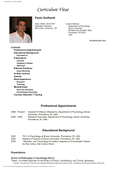
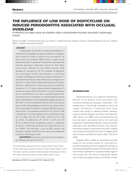
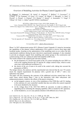
Brazilian Journal of Medical and Biological Research (1998) 31: 1443-1448
Immobilization stress in adult male rats
ISSN 0100-879X
1443
Decreased spermatogenic and
androgenic testicular functions
in adult rats submitted to
immobilization-induced stress
from prepuberty
S.A. Almeida1,
S.O. Petenusci2,
J.A. Anselmo-Franci2,
A.A.M. Rosa-e-Silva1 and
T.L. Lamano-Carvalho2
1Departamento
de Fisiologia, Faculdade de Medicina de Ribeirão Preto, and
de Morfologia, Estomatologia e Fisiologia,
Faculdade de Odontologia de Ribeirão Preto, Universidade de São Paulo,
Ribeirão Preto, SP, Brasil
2Departamento
Abstract
Correspondence
T.L. Lamano-Carvalho
Faculdade de Odontologia de
Ribeirão Preto, USP
Av. Café, s/n
14040-904 Ribeirão Preto, SP
Brasil
Fax: +55-16-633-0999
E-mail: [email protected]
Research supported by FAPESP
(No. 97/2498-1) and CNPq
(No. 520128/96-6). S.A. Almeida
is the recipient of a CNPq
fellowship (No. 141513/95-0).
We investigated whether chronic stress applied from prepuberty to full
sexual maturity interferes with spermatogenic and androgenic testicular functions. Male Wistar rats (40 days old) were immobilized 6 h a
day for 60 days. Following immobilization, plasma concentrations of
corticosterone and prolactin increased 135% and 48%, respectively,
while plasma luteinizing hormone and testosterone presented a significant decrease of 29% and 37%, respectively. Plasma concentration
of follicle-stimulating hormone was not altered in stressed rats. Chronic
stress reduced the amount of mature spermatids in the testis by 16%
and the spermatozoon concentration in the cauda epididymidis by
32%. A 17% reduction in weight and a 42% decrease in DNA content
were observed in the seminal vesicle of immobilized rats but not in its
fructose content. The growth and secretory activity of the ventral
prostate were not altered by chronic stress.
Key words
•
•
•
•
•
•
Immobilization
Spermatogenesis
LH
FSH
Testosterone
Prolactin
Received March 26, 1998
Accepted August 7, 1998
Introduction
Although short- or long-term immobilization is stressful to laboratory animals, the
magnitude of the response depends on the
intensity and duration of the stimulus (1-11).
Experiments are in progress in our laboratory using a model of aversive stimulation –
6 h of daily immobilization – to investigate
the effects of stress on the onset of puberty in
male rats. When applied from prepuberty
(40 days of age) to early puberty (55 days of
age) the stressful stimuli significantly de-
creased plasma luteinizing hormone (LH)
concentration but caused a marked increase
in plasma testosterone; a lower extent of
testicular maturation was also found in pubertal stressed animals (Almeida SA,
Petenusci SO, Anselmo-Franci JA, Rosa e
Silva AAM and Lamano Carvalho TL, unpublished results).
The purpose of the present study was to
investigate the progression of these reproductive disorders by examining the effects
of stress-induced immobilization, applied
from prepuberty, on spermatid production,
Braz J Med Biol Res 31(11) 1998
1444
S.A. Almeida et al.
spermatozoon concentration in the cauda
epididymidis and on the accessory sex glands
of adult rats.
Material and Methods
Male Wistar rats were submitted to immobilization from prepuberty (40 days of
age) to full sexual maturity (100 days of
age). The phases of sexual development were
established according to Zanato et al. (12).
The animals were housed four to a cage in
plastic boxes (40 x 32 x 17 cm) under controlled environmental conditions (12-h light
period starting at 8:00 h, temperature 23 ±
2oC). Laboratory chow and tap water were
given ad libitum.
The animals (N = 8) were immobilized
inside plastic tubes dimensioned to produce
stress without promoting pain (4.5 cm in
diameter x 15.5 cm long up to 55 days of age;
5.0 cm in diameter x 25.5 cm long up to 100
days) for 6 h a day over a period of 60 days.
Control rats (N = 8) were left undisturbed in
their cages. Undesirable stress was avoided
as much as possible by gentle handling and
Table 1 - Plasma hormone concentrations of
chronically immobilized adult rats.
Rats were immobilized for 6 h/day from day 40 to
100 of life. Data were obtained by radioimmunoassay and are reported as means ± SEM for 8
rats in each group. *P<0.05 compared to control
(Mann-Whitney test).
Groups
Braz J Med Biol Res 31(11) 1998
Control
Immobilized
Corticosterone
(ng/ml)
98.8 ± 19.4
231.9 ± 43.9*
Prolactin
(ng/ml)
19.0 ± 2.3
28.1 ± 4.0*
FSH
(ng/ml)
14.1 ± 0.8
12.4 ± 0.3
LH
(ng/ml)
5.19 ± 1.93
3.70 ± 0.40*
Testosterone
(ng/ml)
2.92 ± 0.28
1.83 ± 0.30*
noiselessness throughout the experiment.
Stressed and control rats were killed by
decapitation in the morning after the last
stressing session. Trunk blood was collected
into heparinized tubes and plasma was separated by centrifugation and stored at -20oC
for hormone determinations. Testosterone
(T), prolactin (Prl), LH, and follicle-stimulating hormone (FSH) were measured by
double-antibody radioimmunoassay as described by Zanato et al. (12). A commercial
kit (Coat-A-Count Rat Corticosterone, DPC
- Diagnostic Products Corporation, Los Angeles, CA) was used to measure plasma corticosterone.
The seminal vesicle and ventral prostate
were removed, weighed and stored at -20oC
for colorimetric measurements of DNA and
fructose (13). The sperm suspension was
collected from the cauda epididymidis for
evaluation of spermatozoon concentration,
as described by Kempinas and Lamano
Carvalho (14).
The testes were removed, weighed and
immersed in Alfac fixing solution (85% ethanol 80º, 10% formaldehyde and 5% glacial
acetic acid) for 24 h. Seven-µm thick equatorial paraffin sections were stained with hematoxylin and eosin for histological and
histometric analysis. Spermatid production
was estimated by the amount of spermatids
in the maturation phase (stages 15 to 18 of
spermiogenesis, identified according to Ref.
15) counted in 10 seminiferous tubule crosssections per animal.
The results were analyzed statistically by
the non-parametric Mann-Whitney test.
Results
Sixty days of immobilization markedly
increased plasma corticosterone (135%) and
Prl (48%) concentrations. Plasma LH and T
decreased by 29% and 37%, respectively,
following chronic stress, while no significant change was observed in plasma FSH
(Table 1).
1445
Immobilization stress in adult male rats
Our results support literature data showing no significant change in plasma FSH (7)
and a decrease in plasma LH (7,9,10) after
chronic immobilization of male rats. The
stress-induced decrease in LH has been attributed to inhibition of gonadotropin releasing hormone (GnRH) secretion (10).
In the present study, immobilization of
male rats from prepuberty to sexual maturity
caused a significant decrease in T secretion.
The inhibitory effect of chronic immobilization on plasma T has been a common finding
in adult rats (5-7,10,11) attributed to reduced LH concentration (7,20). The precise
mechanism of the inhibitory effect of prolonged stress on plasma T, however, is not
Discussion
150
Spermatids/tubular section
Stress caused by chronic immobilization
was confirmed by a significant rise in plasma
corticosterone. Literature data have shown
that frequent stimulation with a low-intensity stressor often leads to habituation of the
hypothalamic-pituitary-adrenal axis, while
lower frequency and/or high-intensity stimuli
can promote an exaggerated response (16).
A recent review (17) summarizes the role of
the hippocampus in the control of a variety
of vegetative functions, such as ACTH secretion, and provides evidence that repeated
restraint stress can promote hippocampal
atrophy. Considering the inhibitory role of
the hippocampus in glucocorticoid synthesis, stress-induced dendritic atrophy may have
contributed to high corticosterone levels following prolonged immobilization.
Plasma Prl is also expected to increase in
response to stress (18,19), but the response
is usually of short duration because of the
suppressive effect of the concomitant increase in adrenal glucocorticoids (18). In the
present study, however, Prl remained elevated above control levels even 24 h after
the last episode of repeated exposures to the
stressor.
Stress
Control
250
125
200
*
100
*
150
75
100
50
50
25
0
Spermatozoa (number/ml x 107)
No change was observed in the testicular
weight (1.77 ± 0.03 g in control and 1.69 ±
0.05 g in immobilized rats) or in the gross
histological features. However, a significant
decrease in spermatid production was demonstrable by histometric evaluation, which
revealed a 16% decline in the amount of
maturing spermatids per seminiferous tubule cross-section. A 32% decrease in the
concentration of spermatozoa stored in the
cauda epididymidis was also observed in
stressed rats (Figure 1).
Immobilization caused a significant reduction in seminal vesicle weight (17%) and
DNA content (42%) but not in in its quantity
of fructose. Ventral prostate growth and secretory activity were not altered (Table 2).
Figure 1 - Amount of spermatids
(stages 15 to 18 of spermiogenesis) counted in 10 seminiferous tubular sections per animal
(left) and concentration of spermatozoa in the cauda epididymidis (right), in chronically immobilized adult rats. Rats were immobilized 6 h/day from day 40 to
100 of life. Data are reported as
mean ± SEM for 8 rats in each
group. *P<0.05 compared to
control (Mann-Whitney test).
0
Spermatid
production
Spermatozoon
concentration
Table 2 - Effect of immobilization on rat seminal vesicle and
ventral prostate weight, and DNA and fructose content.
Rats were immobilized 6 h/day from day 40 to 100 of life.
Data are reported as means ± SEM for 8 rats in each group.
*P<0.05 compared to control (Mann-Whitney test).
Group
Weight
(g)
DNA
(mg)
Fructose
(mg)
Seminal vesicle
Control
0.63 ± 0.03 0.33 ± 0.03 0.12 ± 0.01
Immobilized
0.52 ± 0.04* 0.19 ± 0.01* 0.11 ± 0.01
Ventral prostate
Control
0.35 ± 0.03
Immobilized
0.34 ± 0.02
0.14 ± 0.01
0.16 ± 0.02
0.26 ± 0.03
0.27 ± 0.03
Braz J Med Biol Res 31(11) 1998
1446
S.A. Almeida et al.
fully understood since the changes in androgenic response are not always associated
with altered LH levels (8,11,21). It was suggested that restraint stress may induce Leydig
cell hyposensitivity to gonadotropin leading
to a blockade of T biosynthesis at normal LH
levels (21). More recent data have shown
that increased glucocorticoids may act via
glucocorticoid receptors on Leydig cells, thus
suppressing the testicular response to gonadotropins (11). The proopiomelanocortin
(POMC)-derived peptides may also play an
autocrine/paracrine role in mediating the
stress-induced decline in testicular steroidogenesis (8).
In contrast to the considerations discussed
above, studies under way in our laboratory
have shown an increase in T concentration
in early pubertal rats submitted to chronic
immobilization from prepuberty (Almeida
SA, Petenusci SO, Anselmo-Franci JA, Rosa
e Silva AAM and Lamano Carvalho TL,
unpublished results), indicating that the
stressful stimulus probably acts in a different
way on the gonadal axis during distinct phases
of sexual development. Sympathetic innervation seems to modulate androgen biosynthesis, particularly at prepuberty and early
puberty. Previous studies from our laboratory dealing with the effects of chemical
sympathectomy showed decreased steroidogenesis in prepubertal and early pubertal,
but not in adult rats, at normal LH levels, and
suggested that adrenergic stimulation plays
a pivotal role in testicular steroidogenesis
around the onset of puberty (22-24). If so,
and since immobilization induces a significant rise in plasma epinephrine and norepinephrine levels (6), a sympathetic overstimulation might explain the increased T levels
observed in pubertal stressed rats.
No change was observed in testicular
weight or gross histological features after
immobilization from prepuberty to sexual
maturity; however, a significant decrease in
both the production of maturing spermatids
Braz J Med Biol Res 31(11) 1998
and concentration of spermatozoa in the
cauda epididymidis was observed. The decreased androgenic status of stressed rats
may be responsible, at least in part, for depressing spermatogenesis, since the stimulatory action of both FSH and T is needed to
initiate and maintain the process (15). In
addition to the hormonal control, local regulatory interactions occur between all testicular cell types (interstitial, peritubular myoid,
germ and Sertoli cells) and these cell-cell
communications, involving growth factors
and POMC peptides, mediate the cell growth
and differentiation required for the initiation
and maintenance of spermatogenesis (25).
The stressful stimulus may have also interfered with elements of this elaborate paracrine control, impairing the spermatogenic
process.
Although chronic immobilization did not
disturb prostate growth or secretory activity,
the seminal vesicle became atrophied. The
atrophic seminal vesicle of stressed rats might
be a consequence of decreased T secretion,
in addition to high Prl levels, since the growth
and secretory activity of male accessory
glands are under the main control of testicular androgens and Prl (26,27). There are,
however, clear differences between the glandular response to androgens in distinct phases
of sexual development: the prostate of peripubertal rats seems to be particularly responsive to testosterone when compared to
younger or sexually mature animals, while
the seminal vesicles showed decreasing responsiveness with increasing ages (28). Prl
acts both independently and in synergy with
T by intensifying the growth-promoting influence of androgens. However, while a normal circulating Prl level is essential for male
reproductive functions, high levels produce
adverse effects including glandular atrophy
(26,29).
In conclusion, immobilization-induced
stress applied from prepuberty decreased
spermatid production and spermatozoon
1447
Immobilization stress in adult male rats
concentration in the cauda epididymidis
of adult rats. In contrast to the findings in
pubertal rats (Almeida SA, Petenusci SO,
Anselmo-Franci JA, Rosa e Silva AAM
and Lamano Carvalho TL, unpublished
data), however, T secretion decreased in
sexually mature stressed animals. Thus,
besides the intensity and duration of the
stressful stimulus, the phase of sexual development should also be taken into ac-
count in studies of the reproductive responses to stress.
Acknowledgments
The authors thank Dr. C.R. Franci for
providing the facilities for hormone measurements, and Sonia A.Z. Baptista, Mauro F.
Silva, Antonio de Campos and Edna A.S.
Moraes for technical assistance.
References
1. Zemjanis R, Gondos B, Adey WR &
Cockett ATK (1970). Testicular degeneration in Macaca nemestrina induced by
immobilisation. Fertility and Sterility, 21:
335-340.
2. Cockett ATK, Elbadawi A, Zemjanis R &
Adey WR (1970). The effects of immobilisation on spermatogenesis in subhuman
primates. Fertility and Sterility, 21: 610614.
3. Cockett ATK, Zemjanis R, Elbadawi A &
Adey WR (1971). Male infertility: histochemical changes in the subhuman primate testis after prolonged immobilisation. Fertility and Sterility, 22: 565-572.
4. Meitner ER (1976). Einfluss der Immobilisation auf die Spermiogenese. Acta
Anatomica, 95: 300-308.
5. Charpenet G, Taché Y, Forest MG, Haour
F, Saez JM, Bernier M, Ducharme JR &
Collu R (1981). Effects of chronic intermittent immobilisation stress on rat testicular androgenic function. Endocrinology,
109: 1254-1258.
6. Collu R, Gibb W & Ducharme JR (1984).
Role of catecholamines in the inhibitory
effect of immobilisation stress on testosterone secretion in rats. Biology of Reproduction, 30: 416-422.
7. Demura R, Suzuki T, Nakamura S,
Komatsu H, Odagiri E & Demura H (1989).
Effect of immobilisation stress on testosterone and inhibin in male rats. Journal of
Andrology, 10: 210-213.
8. Mann DR & Orr TE (1990). Effect of restraint stress on gonadal proopiomelanocortin peptides and the pituitary-testicular
axis in rats. Life Sciences, 46: 1602-1609.
9. López-Calderón A, González-Quijano MI,
Tresguerres JAF & Ariznavarreta C (1990).
Role of LHRH in the gonadotropin response to restraint stress in intact male
10.
11.
12.
13.
14.
15.
16.
17.
rats. Journal of Endocrinology, 124: 241246.
López-Calderón A, Ariznavarreta C,
González-Quijano MI, Tresguerres JAF &
Calderón MD (1991). Stress induced
changes in testis function. Journal of Steroid Biochemistry and Molecular Biology,
40: 473-479.
Orr TE & Mann DR (1992). Role of glucocorticoids in the stress-induced suppression of testicular steroidogenesis in adult
male rats. Hormones and Behavior, 26:
350-363.
Zanato VF, Martins MP, Anselmo-Franci
JAA, Petenusci SO & Lamano Carvalho
TL (1994). Sexual development of male
Wistar rats. Brazilian Journal of Medical
and Biological Research, 27: 1273-1280.
Kempinas WG, Lamano Carvalho TL,
Petenusci SO, Favaretto ALV, Lopes R &
Azoubel R (1988). Functional disturbance
of rat sexual accessory glands in an early
phase of lead intoxication. Gegenbaurs
Morphologisches Jahrbuch, 134: 791-798.
Kempinas WG & Lamano Carvalho TL
(1988). A method for estimating the concentration of spermatozoa in the rat cauda
epididymidis. Laboratory Animals, 22:
154-156.
Steinberger E & Steinberger A (1975).
Spermatogenic function of the testis. In:
Greep RO & Astwood EB (Editors), Handbook of Physiology. Vol. 5. Williams &
Wilkins, Baltimore, 1-19.
Pitman DL, Ottenweller JE & Natelson
BH (1990). Effect of stressor intensity on
habituation and sensitization of glucocorticoid response in rats. Behavioral Neuroscience, 104: 28-36.
McEwen BS & Magarinos AM (1997).
Stress effects on morphology and function of the hippocampus. Annals of the
18.
19.
20.
21.
22.
23.
24.
New York Academy of Sciences, 821:
271-284.
Gala RR (1990). The physiology and
mechanisms of the stress-induced
changes in prolactin secretion in the rat.
Life Sciences, 46: 1407-1420.
Nonaka KO, Midlej M, Cobas C, Ramalho
MJ, Fregoneze JB, Machado A, AntunesRodrigues J & Castro e Silva E (1991).
Effects of central epinephrine synthesis
inhibition on stress-induced prolactin secretion in male rats. Brazilian Journal of
Medical and Biological Research, 24:
1071-1079.
Sapolsky RM (1985). Stress-induced suppression of testicular function in the wild
baboon: role of glucocorticoids. Endocrinology, 116: 2273-2278.
Charpenet G, Taché Y, Bernier M,
Ducharme JR & Collu R (1982). Stressinduced testicular hyposensitivity to gonadotropins in rats. Role of the pituitary
gland. Biology of Reproduction, 27: 616623.
Lamano Carvalho TL, Guimarães MA,
Kempinas WG, Petenusci SO & Rosa e
Silva AAM (1996). Effects of guanethidine-induced sympathectomy on the spermatogenic and steroidogenic testicular
functions of prepubertal to mature rats.
Andrologia, 28: 117-122.
Kempinas WG, Petenusci SO, Rosa e
Silva AAM, Favaretto ALV & Lamano
Carvalho TL (1995). The hypophyseal-testicular axis and sex accessory glands following chemical sympathectomy with
guanethidine of pre-pubertal to mature
rats. Andrologia, 27: 121-125.
Rosa e Silva AAM, Guimarães MA,
Lamano Carvalho TL & Kempinas WG
(1995). Chemical sympathectomy blocks
androgen biosynthesis during prepuberty.
Braz J Med Biol Res 31(11) 1998
1448
Brazilian Journal of Medical and Biological
Research, 28: 1109-1112.
25. Skinner MK, Norton JN, Mullaney BP,
Rosselli M, Whaley PD & Anthony CT
(1991). Cell-cell interactions and the regulation of testis function. In: Robaire B (Editor), The Male Germ Cell: Spermatogonium to Fertilization. Annals of the New
York Academy of Sciences, 637: 354-363.
26. Mann T & Lutwak-Mann C (1981). Secre-
Braz J Med Biol Res 31(11) 1998
S.A. Almeida et al.
tory function of the prostate, seminal
vesicle, Cowper’s gland and other accessory organs of reproduction. In: Mann T &
Lutwak-Mann C (Editors), Male Reproductive Function and Semen. Springer-Verlag,
New York, 171-193.
27. Coffey DS (1988). Androgen action and
the sex accessory tissues. In: Knobil E &
Neill LJ (Editors), The Physiology of Reproduction. Raven Press, New York, 1081-
1119.
28. Odell WD (1990). Sexual maturation in
the rat. In: Gumbach MM, Sizonenko PC
& Aubert ML (Editors), Control of the Onset of Puberty. Williams & Wilkins, London, 183-210.
29. Bartke A (1980). Role of prolactin in reproduction in male mammals. Federation Proceedings, 39: 2577-2581.
Download