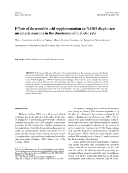
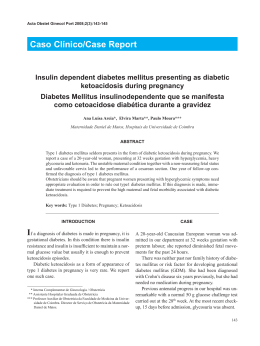
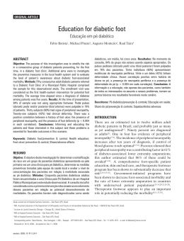
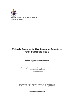
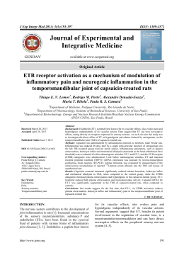
Brazilian
Journalcontrol
of Medical
and Biological Research (2002) 35: 1091-1100
Cardiovascular
in diabetes
ISSN 0100-879X
Review
1091
Cardiovascular control in
experimental diabetes
K. De Angelis1,5,
B.D. Schaan3,
C.Y. Maeda2,
P. Dall’Ago2,6,
R.B. Wichi4,5 and
M.C. Irigoyen2,4,5
1Laboratório
de Cardiovascular, UNIPESQ, Universidade de Santo Amaro,
São Paulo, SP, Brasil
2Laboratório de Fisiologia Cardiovascular, Departamento de Fisiologia,
Instituto de Ciências Básicas da Saúde, Universidade Federal do Rio Grande do Sul,
Porto Alegre, RS, Brasil
3Fundação Universitária de Cardiologia, Instituto de Cardiologia do Rio Grande do Sul,
Porto Alegre, RS, Brasil
4Departamento de Nefrologia, Escola Paulista de Medicina,
Universidade Federal de São Paulo, São Paulo, SP, Brasil
5Unidade de Hipertensão, Instituto do Coração, Hospital das Clínicas,
Faculdade de Medicina, Universidade de São Paulo, São Paulo, SP, Brasil
6Curso de Fisioterapia, FAENFI, Pontifícia Universidade Católica do Rio Grande do Sul,
Porto Alegre, RS, Brasil
Abstract
Correspondence
M.C. Irigoyen
Unidade de Hipertensão
Incor, HC, FMUSP
Av. Enéas C. Aguiar, 44
05403-000 São Paulo, SP
Brasil
Fax: +55-11-3085-7887
E-mail: [email protected]
Presented at the IV International
Symposium on Vasoactive Peptides,
Belo Horizonte, MG, Brazil,
October 19-21, 2001.
Research supported by CNPq,
FAPESP, CAPES, FAPERGS, and
Fundação E.J. Zerbini.
Received February 15, 2002
Several studies have reported impairment in cardiovascular function
and control in diabetes. The studies cited in this review were carried
out from a few days up to 3 months after streptozotocin administration
and were concerned with the control of the circulation. We observed
that early changes (5 days) in blood pressure control by different
peripheral receptors were maintained for several months. Moreover,
the impairment of reflex responses observed after baroreceptor and
chemoreceptor stimulation was probably related to changes in the
efferent limb of the reflex arc (sympathetic and parasympathetic), but
changes also in the central nervous system could not be excluded.
Changes in renal sympathetic nerve activity during volume expansion
were blunted in streptozotocin-treated rats, indicating an adaptive
natriuretic and diuretic response in the diabetic state. The improvement of diabetic cardiovascular dysfunction induced by exercise
training seems to be related to changes in the autonomic nervous
system. Complementary studies about the complex interaction between circulation control systems are clearly needed to adequately
address the management of pathophysiological changes associated
with diabetes.
Key words
·
·
·
·
·
·
·
Experimental diabetes
Arterial pressure
Autonomic control
Baroreflex
Chemoreflex
Cardiopulmonary reflex
Exercise training
Accepted August 5, 2002
Introduction
Diabetes mellitus is one of the most important world health problems, especially in
developing countries, where prevalence and
incidence rates are highest. Diabetic patients
are particularly prone to cardiovascular diseases including hypertension, atherosclerosis, diabetic cardiomyopathy, congestive heart
failure, and cardiac autonomic neuropathy
(1).
Autonomic neuropathy is a frequent complication of diabetes mellitus associated with
high morbidity and mortality in symptomatic
patients (2), which affects the autonomic
modulation of the sinus node, reducing heart
rate variability (HRV) (3). The increased
mortality rate may be related to disorders in
Braz J Med Biol Res 35(9) 2002
1092
K. De Angelis et al.
cardiovascular control, including impairment
of autonomic reflex control leading to orthostatic hypotension, painless myocardial infarction and sudden death (4), the last possibly determined by nocturnal desaturation
episodes (5) or arrhythmias. Recently, it was
demonstrated that cardiac sympathetic dysfunction in diabetes involves areas of dysinnervation (distal), as well as areas of hyperinnervation (proximal) in the left ventricle, which facilitate malignant arrhythmias by altering electrical stability and/or
impairing myocardial blood flow (6).
Abnormalities in the renin-angiotensinaldosterone-kinin cascade have been implicated in the pathogenesis and clinical expression of these cardiovascular-renal sequelae. Angiotensin II, through its effects on
contractility, growth, and the sympathetic
nervous system, may play a key role in this
pathologic process. Angiotensin-converting
enzyme inhibitors and some direct renin inhibitors prevent or reduce the progression of
some of these complications (7). In fact,
angiotensin-converting enzyme inhibitors
have been reported to improve kidney, heart
and, to a lesser extent, eye and peripheral
nerve function of patients with diabetes mellitus (8).
Experimental diabetes induced by streptozotocin (STZ) has been used by several
investigators to study disorders of the autonomic control of the cardiovascular system.
Rats treated with STZ display many of the
features seen in human subjects with uncontrolled diabetes mellitus, including hyperTable 1. Time course of changes in sedentary STZ-induced diabetic rats.
Control
HR (bpm)
MAP (mmHg)
IHR (bpm)
Serum glucose (mg/dl)
332
117
398
106
±
±
±
±
2
3
6
15
STZ (5 days)
291
102
302
306
±
±
±
±
4*
2*
10*
19*
STZ (15 days)
296 ± 11*
99 ± 3*
447 ± 49*
STZ (80 days)
279
91
284
479
±
±
±
±
9*
4*
11*
8*
Values are reported as means ± SEM.
*P<0.05 vs sedentary control group (ANOVA). STZ: streptozotocin, HR: heart rate,
MAP: mean arterial pressure, IHR: intrinsic heart rate (data taken from Refs. 10,14,16).
Braz J Med Biol Res 35(9) 2002
glycemia, hypoinsulinemia, increased urinary glucose levels and consequently polyuria, as well as weight loss (9-11).
Degenerative changes in autonomic neurons were observed from 3 days (12) to
several weeks after STZ administration in
rats (13). We observed that rats presented
lower arterial pressure and heart rate (HR) 5
to 80 days after diabetes induction with STZ
(10,14-16) (Table 1). The mechanisms involved in these alterations are not completely
understood. Some investigators have associated bradycardia and hypotension with reduction in intrinsic HR, in vagal tonus and in
cardiovascular reflex control (10,14-16), suggesting early autonomic dysfunction. Furthermore, impairment of heart contractility
and vascular responsiveness, as well as
changes in blood volume caused by osmotic
diuresis could be involved in the pathogenesis of these alterations.
In this paper we review the effects of
experimental diabetes on the autonomic control of HR and cardiovascular reflexes, as
well as the benefits of exercise training for
the control of these disorders.
Autonomic control of heart rate in
experimental diabetes
Studies from our laboratory have shown
reduction in vagal tonus and maintenance of
sympathetic tonus to the heart evaluated by
pharmacological blockade with propranolol
and methylatropine, respectively (14,16)
(Figure 1), as previously demonstrated by
Wegner et al. (17), suggesting the presence
of cardiac vagal neuropathy. Resting bradycardia in STZ-diabetic rats has been attributed to changes in the sinoatrial node with a
consequent reduction in intrinsic HR (14,16,
18), although functional alterations in the
cholinergic mechanism cannot be excluded
as a causal factor. These data indicate that
vagal function and intrinsic HR are decreased
in short- and long-term STZ-induced diabetes in rats.
1093
Cardiovascular control in diabetes
HRV in STZ-diabetic rats, changes which
were reversed, at least in part, by insulin
administration.
Functional changes in cholinergic or adrenergic mechanisms cannot exclude morphological changes at this early stage of the
diabetic state, since Monckton and Pehowich
(12) reported degenerative changes in autonomic neurons of STZ-diabetic rats. These
investigators found changes in axons from
the sympathetic paravertebral chain within
24 h of STZ injection. Schmidt et al. (13), for
instance, found that mesenteric axonopathy
was apparent in Sprague-Dawley rats treated
with STZ 1.5 to 3 months earlier. Kriel et al.
(24) showed degeneration of sympathetic
neurons in Wistar rats that had been treated
with STZ 1 year earlier. These changes are
consistent with decreases in neuronal activ-
Control
STZ (5 days)
STZ (80 days)
HR: 315 ± 11
HR: 291 ± 4*
HR: 279 ± 9*
Trained
STZ (80 days)
HR: 305 ± 7
450
436 ± 12
425
ST
IHR
390 ± 8
400
Heart rate (bpm)
The bradycardic response to methacholine injection was similar in control and
diabetic rats 5 days after STZ administration
(14). However, in 15-day STZ-treated rats
this response was higher than in normal rats
(10), suggesting that the early impairment in
vagal tonus may be leading to an adaptive
change in muscarinic receptors. Kuntscherova and Vlk (19) found decreased acetylcholine concentrations in isolated auricles of
diabetic rats and Tomlinson et al. (11) observed functional defects in cardiac cholinergic nerves in diabetic rats with vagal dysfunction. Indeed, it was demonstrated that
coupling of cholinergic receptors to adenylate cyclase is altered in STZ-diabetic rats
because the content of Gi proteins in cardiac
tissue increased after STZ administration
(20). Moreover, the interactions between the
sympathetic and parasympathetic system are
complex, suggesting a different vagal action
at different levels of sympathetic function
(21).
HRV may be reduced in diabetic autonomic neuropathy through injury to the parasympathetic fibers. Thus, HRV is a valuable
index of cardiac parasympathetic nerve functional integrity (22). Evaluating rats injected
with STZ 12-18 weeks before, Fazan et al.
(3) observed that the standard deviation of
the lengths of adjacent pulse pressures, an
index of HRV, was reduced in these animals
in comparison to their controls, indicating
the presence of functional cardiac parasympathetic neuropathy. Since we studied the
same HRV index in 45-90-day STZ-diabetic
rats treated with insulin, and did not find
differences between them and their controls,
we suggest a definitive role of metabolic
decompensation in the genesis and maintenance of these autonomic changes. Indeed,
the negative correlation between HRV indices and plasma glucose (r = -0.26, P = 0.012)
supports this idea (Schaan BD, Ferlin E and
Irigoyen MC, unpublished data). Using biotelemetry techniques, Hicks et al. (23) also
demonstrated time-dependent reductions in
383 ± 10
375
350
357 ± 12
VT
ST
346 ± 13
IHR
342 ± 14*+
ST
325
315 ± 15
IHR
302 ± 10*
300
IHR
284 ± 11*
VT
275
VT
ST
307 ± 4*
VT
263 ± 7
259 ± 6*
250
Figure 1. Intrinsic heart rate (IHR), sympathetic tonus (ST) and vagal tonus (VT) of sedentary
control rats (control), 5-day-sedentary diabetic rats (STZ - 5 days), 80-day-sedentary diabetic
rats (STZ - 80 days), and 80-day-trained diabetic rats (trained STZ - 80 days). IHR was
evaluated after simultaneous blockade with propranolol and methylatropine. Sympathetic
tonus was determined as the difference between maximum heart rate after methylatropine
injection and IHR. Vagal tonus was obtained by the difference between the lowest heart
rate after propranolol injection and IHR. Data are reported as means ± SEM. *P<0.05
compared to sedentary control rats (ANOVA); +P<0.05 compared to 80-day-sedentary
diabetic rats (Student t-test). HR: basal heart rate, STZ: streptozotocin (data taken from
Refs. 14,16).
Braz J Med Biol Res 35(9) 2002
1094
K. De Angelis et al.
ity. However, Felten et al. (25) found no
change in the pattern of noradrenergic innervation in the hearts of rats with 8 months of
STZ-induced diabetes. Sharma and Thomas
(26) also could not demonstrate morphological changes in the peripheral nerves of
this animal model even after extended periods of diabetes. Probably the pattern of development of autonomic neuropathy depends
on the rat strain used, animal age when STZ
was given, the use or not of insulin during
the experiments, etc. Finally, it cannot be
ruled out that STZ per se is directly toxic to
the nervous system, since it is certainly toxic
to pancreatic beta cells (27) and the lesions
described in the study of Monckton and
Pehowich (12) occurred so soon after STZ
use (24 h) that they could not have been
determined by diabetes itself.
On the other hand, biochemical changes
have been frequently described in STZdiabetic rats. Schmidt and Plurad (28) found
a reduction in dopamine ß-hydroxylase activity in the superior cervical ganglia from
STZ-treated rats, although the activity of this
enzyme was unaltered in the superior mesenteric ganglia. However, Jobidon et al. (29)
found no change in plasma or cardiac noradrenaline levels after treatment with STZ.
Obviously, tissue or plasma noradrenaline
levels alone provide little information about
sympathetic nervous system activity because
they depend on a number of factors, including rate of synthesis, uptake, and release of
noradrenaline by the tissues. Indeed, Sato et
al. (30) reported a decreased contractile response to noradrenaline in left atria isolated
from STZ-diabetic rats. Carrier and Aronstam
(31) demonstrated that acetylcholinesterase
levels were lower in right atria from STZdiabetic rats. This might increase the effective concentrations of acetylcholine acting
on myocardial receptors, therefore contributing to supersensitivity to acetylcholine.
These alterations in cardiac cholinergic and
noradrenergic function may be involved in
the bradycardia presented by diabetic rats.
Braz J Med Biol Res 35(9) 2002
Baroreflex control in experimental
diabetes
The arterial baroreflex system is one of
the most influential and rapidly acting
mechanisms for blood pressure control. Indeed, the minimization of blood pressure
variability by the baroreflex mechanisms is
important since a reduced baroreflex is an
independent risk factor for sudden death
after myocardial infarction (32). Diabetic
patients with normal cardiovascular reflexes
have a lower incidence of mortality than
diabetic individuals with abnormal autonomic reflex function (2). Some studies have
shown that the arterial baroreceptor reflex
exerts a major influence on both the sympathetic and parasympathetic systems for cardiovascular control. Hence, the disorders in
autonomic efferent and afferent neuronal
systems could have important consequences
for the control of cardiovascular function.
Several studies using experimental models
have been conducted to investigate the
mechanisms of reflex dysfunction of diabetes (10,14-16,33,34).
We have demonstrated that early in the
course of experimental diabetes there is an
impairment of baroreflex control in STZ rats
characterized by reduction of baroreflexmediated tachycardia, while baroreflexmediated bradycardia is still maintained
(14,15). Later (15 and 30 days after STZ),
the baroreflex-mediated bradycardia was also
lost in diabetic rats (10), and these changes
persisted even 80 days after STZ injection
(35) (Figure 2). On the other hand, McDowell
et al. (34) observed maintenance of the response to the increase in blood pressure
induced by infusion of a vasoconstrictor agent
2 weeks after STZ treatment, but their study
was conducted on rabbits. Also in a different
animal model, the spontaneously diabetic rat
(Bio-Breeding), Krizsan-Agbas and Buñag
(36) demonstrated exacerbation of baroreflex-mediated tachycardia, while Eckberg et
al. (37) reported a normal tachycardia re-
1095
Cardiovascular control in diabetes
represent hypersensitivity of receptors linked
to the characteristic reduction in parasympathetic activity shown by these animals. The
up-regulation of muscarinic receptors could
be related to reduced vagal function, probably contributing to the reduction of the
tachycardic response to arterial pressure falls
as described previously in diabetic (14,15)
and aged rats (44). These responses characterized by reflex parasympathetic withdrawal and cardiac sympathetic activation suggest that the interactions between the two
branches of the autonomic nervous system
are complex, with different degrees of modulation at different levels of activity of these
systems (21).
Arterial chemoreceptors in
experimental diabetes
The arterial chemoreceptors represent an
Control
STZ (5 days)
STZ (15 days)
STZ (80 days)
4
3
DHR/DMAP (bpm/mmHg)
sponse to decreases of blood pressure in
diabetic humans.
Different mechanisms may be acting in
the baroreflex control of HR in diabetes
(34,37). Moreover, the different data obtained in the cited studies may be attributed
to time-dependent changes in HR control.
Also, metabolic factors could contribute to
the discrepant results, since our data were
obtained in rats receiving no insulin 5, 15
and 80 days after STZ administration (10,
14,15,35), and other investigators observed
correction of previously abnormal cardiovascular reflexes by treating diabetic rats
with insulin (38).
The impaired ability to perform adequate
HR regulation during changes in arterial pressure has been attributed to some alterations in
cardiac parasympathetic activity (14), although
changes in the receptor function or in the
central mediation of the baroreceptor reflex
cannot be excluded (39). The parasympathetic
dysfunction could be due to alterations in
cardiac muscarinic receptors (31). Williams et
al. (40) reported that the density of cardiac
muscarinic receptors was unaltered in STZdiabetic rats. However, these investigators did
not measure the density of atrial muscarinic
receptors separately. Carrier et al. (41) demonstrated that there was no difference in muscarinic receptor density in ventricles from STZdiabetic and age-matched control rats, but the
density of muscarinic receptors was reduced
in the right and left atria from diabetic rats
(31).
Li et al. (42) demonstrated a higher negative chronotropic effect induced by methacholine injection in 6-week STZ-induced
diabetic rats. In fact, our studies demonstrated that electric vagal stimulation, as well
as myocardial muscarinic receptor stimulation by methacholine injections determined
an increase in the bradycardic response of
short-term STZ-induced diabetic animals,
suggesting efferent pathway impairment in
the reflex arc (43). The increased bradycardia induced by methacholine injection could
Tachycardic response to sodium nitroprusside injection
*
2
*
*
1
0
-1
*
*
-2
Bradycardic response to phenylephrine injection
Figure 2. Tachycardic and bradycardic responses to arterial pressure decreases and increases, respectively, in sedentary control rats (control), 5-day-sedentary diabetic rats
(STZ - 5 days), 15-day-sedentary diabetic rats (STZ - 15 days), and 80-day-sedentary diabetic
rats (STZ - 80 days) *P<0.05 compared to sedentary control rats (ANOVA) (data taken from
Refs. 10,14,39). STZ: streptozotocin, HR: heart rate, MAP: mean arterial pressure.
Braz J Med Biol Res 35(9) 2002
1096
K. De Angelis et al.
tion) in rats. Using the same methodology in
diabetic rats injected 15 days before with STZ,
the concomitant evaluation of baro- and
chemoreflexes showed that both cardiovascular responses were impaired in this model
(10). Indeed, diabetes was associated with
hyporesponsiveness of vagal cardiac activation evoked by the chemoreflex, as demonstrated by the reduction of the bradycardic
responses produced by stimulation of the ca-
important group of afferences that participate
in the control of autonomic function. However, they have been less studied than baroreceptors. Infusion of intravenous potassium
cyanide (KCN) stimulates the parasympathetic
and sympathetic chemoreceptor pathways (45).
Franchini and Krieger (45) showed that stimulation of the chemoreceptors with KCN resulted in bradycardia (cardiac vagal stimulation) and hypertension (sympathetic stimula250
A
1:
250.00 mmHg
1:
125.00 mmHg
mmHg
Figure 3. Arterial pressure during KCN injection into a 15-day
STZ-induced diabetic rat (A) and
a control rat (B). Line graphs
showing the effects of STZ-induced diabetes on mean arterial
pressure (MAP) responses (C)
and heart rate (HR) responses
(D) of control and 15-day STZdiabetic rats to increasing doses
of KCN. *P<0.05 compared to
control (ANOVA) (data taken
from Ref. 10). STZ: streptozotocin.
1s
KCN injection
0
B
1:
2:
0.00 mmHg
250.00 mmHg
2:
125.00 mmHg
mmHg
250
1s
KCN injection
2:
0
.0.00 mmHg
C
D
60
70
*
Control
-50
STZ
50
*
*
40
30
20
DHR (bpm)
DMAP (mmHg)
180
0
60
*
-100
*
-150
-200
*
10
0
60
100
140
KCN (µg/kg)
Control
STZ
-250
Braz J Med Biol Res 35(9) 2002
KCN (µg/kg)
100
140
180
1097
Cardiovascular control in diabetes
rotid body (Figure 3). At the same time the
pressor response induced by vascular sympathetic activation in response to the chemoreflex stimulation was impaired in STZ-induced
diabetic rats (10) (Figure 3).
The impairment of bradycardic responses
elicited by chemo- or baroreceptor stimulation may indicate changes in the efferent
branch of these reflexes, since pharmacological blockade showed reduced heart parasympathetic function in diabetic rats (10,
14,15). Moreover, baro- and chemoreflexes
are integrated by pathways that converge to
the same site in the central nervous system
(46) that could be involved in the changes
demonstrated in both responses in this model of diabetes. Finally, we cannot exclude
that changes in the afferent limb of reflex
arches may be participating in the depressed
responses.
In fact, histological studies performed on
spontaneously diabetic rats showed structural changes in carotid body (47) such as
axonal swelling and intramyelinic edema
suggesting diabetic neuropathy. These histopathological findings could be responsible
for the reduced arterial chemoreceptor drive
through impairment of nerve conduction.
This finding supports data from our laboratory indicating a neurogenic origin of the
reduction of chemoreceptor response after
intravenous injection of KCN.
Cardiopulmonary reflexes in
experimental diabetes
Among the different mechanisms of cardiovascular control, the cardiopulmonary
reflex is activated by stimuli located in different structures of the thorax, including
atria, ventricles, veins and pulmonary parenchyma, which represent an important
source of information about volume and
blood pressure variations in the cardiopulmonary region.
The cardiopulmonary receptors have an
important participation in the homeostasis
of fluid balance, both by modulating the
activity of the sympathetic nervous system
on the cardiovascular system, and by acting
on the kidney (48-50). Intravenous infusion
of chemical substances like serotonin or stimulation of cardiopulmonary receptors promoting bradycardia and hypotension are
methods used to evaluate the cardiopulmonary receptors (Bezold-Jarisch reflex). The
renal response is a reduction of vascular
resistance and efferent sympathetic activity
(50). The reflex response obtained in our
laboratory after the stimulation of cardiopulmonary receptors by injecting serotonin was
similar for diabetic and control rats (48),
suggesting that the cardiovascular response
to stimulation of chemosensitive cardiac receptors is preserved in the STZ-diabetic
model. However, the reflex response induced
by a similar plasma volume expansion and
associated changes in left ventricle end diastolic pressure produced lower bradycardia
and hypotension in diabetic than in control
rats. Furthermore, the modulation of renal
sympathetic activity was abolished in STZ
animals. The physiological role of this altered response in diabetes could be associated with renal dysfunction in the balance
between sodium and water intake and uptake, changing the natriuretic and diuretic
responses in this condition. The reduction in
renal sodium excretion associated with a
decrease in renal sympathetic activation was
also described by Patel and Zhang (49).
Indeed, the examination of the various components of the volume reflex in different
models of diabetic rats indicated an altered
neural component associated with a humoral
component of the effector limb probably
related to atrial natriuretic factor (50).
Exercise training in experimental
diabetes
Cardiovascular, metabolic and autonomic
improvement induced by acute and chronic
exercise have led many investigators to sugBraz J Med Biol Res 35(9) 2002
1098
K. De Angelis et al.
gest exercise training as an important nonpharmacological treatment for different pathologies like diabetes, hypertension and
coronary artery disease (16,51-54). Exercise
influences several aspects of diabetes, including blood glucose concentration, insulin action and cardiovascular risk factors
(35,51). Beyond the acute impact of physical activity, long-term exercise behaviors
have been repeatedly associated with decreased rates of type 2 diabetes (51,52).
In our laboratory, exercise training applied to young and aged normotensive rats
and young hypertensive rats improved the
autonomic control of cardiovascular function (53,55,56). Indeed, we have recently
demonstrated the benefits of exercise training in diabetes-induced dysfunction (16).
The hypotension presented by sedentary STZ
rats was not observed in trained diabetic rats
(11 weeks of training on a treadmill). This
improvement could be related to an increase
in cardiac output in diabetic rats (57), since
Jackson and Carrier (33) have suggested
previously that the decrease in arterial pressure should be the result of a decreased
cardiac output in sedentary diabetic rats due
to hypovolemia caused by hyperglycemic
osmotic diuresis. Moreover, exercise training reverses the changes in the contractile
properties of the heart induced by STZ diabetes in rats such as reduced cardiac contractility and relaxation (16,57), suggesting that
the improvement in cardiac function could
be due to a decrease in the severity of the
diabetic state (58). Besides these changes,
exercise also improves glucose homeostasis, reducing the glucose/insulin ratio and
increasing insulin sensitivity (54,55). We
have also demonstrated that the increase in
body weight observed in trained diabetic
rats seems to indicate an improvement of the
metabolic state. Other changes such as reduction in ultrastructural glomerular lesions
Braz J Med Biol Res 35(9) 2002
and albumin excretion observed in a rat model of type 2 diabetes mellitus submitted to
aerobic training may be an improved metabolic control and delayed diabetic complications (59).
Moreover, we observed an increase in
resting HR in trained diabetic rats that was
correlated with changes in intrinsic HR, confirming the important role of the sinoatrial
node in HR changes in experimental diabetes (16). In contrast, previous studies have
demonstrated that exercise training decreases
resting HR in young (55) and aged (56)
normotensive rats and humans (60). The
decreased intrinsic HR previously observed
in our laboratory in trained control rats (55)
as well as a decreased sympathetic tonus in
spontaneously hypertensive rats (53) after
training may be the mechanisms involved in
exercise bradycardia. In STZ-induced diabetic rats we did not observe changes in
sympathetic tonus between sedentary and
trained groups, suggesting that the increase
in resting HR in trained diabetic rats may be
related to the improvement of intrinsic pacemaker regulation (16) (Figure 1). In contrast
to the impairment of vagal function observed
in normal rats after exercise training by the
reduced bradycardia in response to electrical
vagal stimulation (55), exercise training did
not modify the reduced parasympathetic
function observed in diabetic rats (16,17)
(Figure 1). Therefore, changes in reflex control of the circulation related to parasympathetic function may persist after exercise
training, as demonstrated previously in
trained nitric oxide blockade hypertensive
rats (54).
These data show that physical activity
delays and improves the hemodynamic and
metabolic dysfunction observed in diabetes,
and should be considered in the prevention
and treatment of this disease.
1099
Cardiovascular control in diabetes
References
1. Jarret RJ (1989). Cardiovascular disease
and hypertension in diabetes mellitus.
Diabetes/Metabolism Reviews, 5: 547558.
2. Ewing D, Campbell I & Clarke B (1980).
The natural history of diabetic autonomic
neuropathy. Quarterly Journal of Medicine, 49: 95-108.
3. Fazan R, Ballejo G, Salgado MC, Moraes
M & Salgado HC (1997). Heart rate variability and baroreceptor function in
chronic diabetic rats. Hypertension, 30
(Part 2): 632-635.
4. Page M & Watkins P (1978). Cardiorespiratory arrest and diabetic autonomic neuropathy. Lancet, i: 14-16.
5. Neumann C, Martinez D & Schmid H
(1995). Nocturnal oxygen desaturation in
diabetic patients with severe autonomic
neuropathy. Diabetes Research and Clinical Practice, 28: 97-102.
6. Stevens MJ, Raffel DM, Allman KC,
Dayanikli F, Ficaro E & Sandford T (1998).
Cardiac sympathetic dysinnervation in diabetes: implications for enhanced cardiovascular risk. Circulation, 98: 961-968.
7. Hsueh WA (1992). Effects of renin-angiotensin system in vascular disease of type
II diabetes mellitus. American Journal of
Medicine, 92: 13S-19S.
8. Cordonnier DJ, Zaoui P & Halimi S (2001).
Role of ACE inhibitors in patients with
diabetes mellitus. Drugs, 61: 1883-1892.
9. Schaan BD, Maeda CY, Timm H, Medeiros S, Moraes R, Ferlin E, Fernandes TG,
Ribeiro JP, Schmid H & Irigoyen MC
(1997). Time course of changes in heart
rate and blood pressure variability in streptozotocin-induced diabetic rats treated
with insulin. Brazilian Journal of Medical
and Biological Research, 30: 1081-1086.
10. Dall’Ago P, Fernandes TG, Machado UF,
Belló AA & Irigoyen MC (1997). Baroreflex and chemoreflex dysfunction in streptozotocin-diabetic rats. Brazilian Journal
of Medical and Biological Research, 30:
119-124.
11. Tomlinson KC, Sheila MG, Anhony Hebden
R & Bennett T (1981). Functional consequences of streptozotocin-induced diabetes mellitus, with particular reference to
the cardiovascular system. Pharmacological Reviews, 44: 103-148.
12. Monckton G & Pehowich E (1980). Autonomic neuropathy in the streptozotocin
diabetic rat. Canadian Journal of Neurological Sciences, 7: 135-142.
13. Schmidt RE, Plurad SB & Modert CW
(1983). Experimental diabetic autonomic
14.
15.
16.
17.
18.
19.
20.
21.
22.
23.
24.
neuropathy characterization in streptozotocin-diabetic Sprague-Dawley rats. Laboratory Investigation, 49: 538-552.
Maeda CY, Fernandes TG, Timm HB &
Irigoyen MC (1995). Autonomic dysfunction in short-term experimental diabetes.
Hypertension, 26 (Part 2): 1000-1004.
Maeda CY, Fernandes TG, Lulhier F &
Irigoyen MC (1995). Streptozotocin diabetes modifies arterial pressure and baroreflex sensitivity in rats. Brazilian Journal of
Medical and Biological Research, 28: 497501.
De Angelis KLD, Oliveira AR, Dall’Ago P,
Peixoto LRA, Gadonski G, Fernandes TG
& Irigoyen MC (2000). Effects of exercise
training in autonomic and myocardial dysfunction in streptozotocin-diabetic rats.
Brazilian Journal of Medical and Biological
Research, 33: 635-641.
Wegner JA, Lund DD, Overton JM, Edwards JG, Oda RP & Tipton CM (1987).
Select cardiovascular and metabolic responses of diabetic rats to moderate exercise training. Medicine and Science in
Sports and Exercise, 19: 497-503.
Senges J, Brachmann J, Pelzer D, Hasslacher C, Weihe E & Kübler W (1980).
Altered cardiac automaticity and conduction in experimental diabetes mellitus.
Journal of Molecular and Cellular Cardiology, 12: 1341-1351.
Kuntscherova J & Vlk J (1970). Influence
of alloxan diabetes on acetylcholine synthesis in tissues of the albino rat. Physiologia Bohemoslovaca, 19: 431-434.
Nishio Y, Kashiwagi A, Kida Y, Kodama M,
Abe N, Saeki Y & Shigeta Y (1988). Deficiency of cardiac B-adrenergic receptor in
streptozotocin-induced diabetic rats. Diabetes, 37: 1181-1187.
Chassaing C, Duchene-Marullaz P &
Veyrac J (1983). Effects of catecholamines on cardiac chronotropic response
to vagal stimulation in the dog. American
Journal of Physiology, 245: H721-H724.
Van Ravenswaaij-Arts CMA, Kollé LAA,
Hopman JCW, Stoelinga GB & Van Geijn
HP (1993). Heart rate variability. Annals of
Internal Medicine, 118: 436-447.
Hicks KK, Seifen E, Stimers JR & Kennedy
RH (1998). Effects of streptozotocininduced diabetes on heart rate, blood
pressure and cardiac autonomic nervous
control. Journal of the Autonomic Nervous System, 69: 21-30.
Kriel PC, Juncker U, Perrin IV, Bestetti GE
& Rossi GL (1986). Varied effect of experimental diabetes on the autonomic ner-
25.
26.
27.
28.
29.
30.
31.
32.
33.
vous system of the rat. Laboratory Investigation, 54: 523-530.
Felten SY, Peterson RG, Shea PA, Besch
JR & Felten DL (1982). Effects of streptozotocin diabetes on the noradrenergic innervation of the rat heart: A longitudinal
histofluorescence and neurochemical
study. Brain Research Bulletim, 8: 593607.
Sharma AK & Thomas PK (1987). Animal
models: pathology and pathophysiology.
In: Dick PJ, Thomas PK, Asbury AK,
Winegrad AI & Porte D (Editors), Diabetic
Neuropathy. Section IV. Pathology and
Pathophysiology - Human and Experimental. W.B. Saunders, Philadelphia, PA, USA,
237-252.
Junod A, Lambert AE, Stauffacher W &
Renold AE (1969). Diabetogenic action of
streptozotocin: relationship of dose to
metabolic response. Journal of Clinical Investigation, 48: 2129-2139.
Schmidt RE & Plurad SB (1986). Ultrastructural and biochemical characterization of autonomic neuropathy in rats with
chronic streptozotocin diabetes. Journal
of Neuropathology and Experimental Neurology, 45: 525-544.
Jobidon C, Nadeu A, Tancrede G, Nguyen
MH & Rousseau-Migneron S (1985).
Plasma, adrenal, and heart catecholamines in physically trained normal and
diabetic rats. Diabetes, 34: 532-535.
Sato N, Hashimoto H, Takigughi Y & Nakashima M (1989). Altered responsiveness to sympathetic nerve stimulation and
agonists of isolated left atria of diabetic
rats: no evidence for involvement of hypothyroidism. Journal of Pharmacology
and Experimental Therapeutics, 248: 367371.
Carrier GO & Aronstam RS (1987). Altered muscarinic receptor properties and
function in the heart in diabetes. Journal
of Pharmacology and Experimental Therapeutics, 242: 531-535.
Farrel TG, Bashir Y, Cripps T, Malik M,
Poloniecki J, Bennett ED, Ward DE &
Camm AJ (1991). Risk stratification for
arrhythmic events in postinfarction patients based on heart rate variability, ambulatory electrocardiographic variables
and the signal-averaged electrocardiogram. Journal of the American College of
Cardiology, 18: 667-697.
Jackson CV & Carrier GO (1983). Influence of short-term experimental diabetes
on blood pressure and heart rate in response to norepinephrine and angiotensin
Braz J Med Biol Res 35(9) 2002
1100
34.
35.
36.
37.
38.
39.
40.
41.
42.
K. De Angelis et al.
II in the conscious rat. Journal of Cardiovascular Pharmacology, 5: 260-265.
McDowell TS, Chapleau MW, Hajduczok
G & Abboud FM (1994). Baroreflex dysfunction in diabetes mellitus. I. Selective
impairment of parasympathetic control of
heart rate. American Journal of Physiology, 266: H235-H243.
De Angelis KLD, Dall’Ago P, Gadonski G,
Peixoto LRA, Fernandes TG & Irigoyen
MC (1999). Exercise training improves cardiovascular and metabolic status in diabetic rats. Hypertension, 33: 1073 (Abstract).
Krizsan-Agbas D & Buñag RD (1991). Normotensive diabetic BB/W rats show enhanced reflex tachycardia. Diabetes, 40:
1504-1510.
Eckberg DL, Harkins SW, Fritsch JM,
Musgrave GE & Gardner DF (1986). Baroreflex control of plasma norepinephrine
and heart period in healthy subjects and
diabetic patients. Journal of Clinical Investigation, 78: 366-374.
Chang KSK & Lund DD (1986). Alterations
in the baroreceptor reflex control of heart
rate in streptozotocin diabetic rats. Journal of Molecular and Cellular Cardiology,
18: 617-624.
Fazan R, Irigoyen MC, Moraes MFD,
Maeda CY & Salgado HC (1995). Baroreceptor function in short-term streptozotocin diabetic rats. Hypertension, 25: 1398
(Abstract).
Williams RS, Schaible TF, Scheuer J &
Kennedy R (1983). Effects of experimental diabetes on adrenergic and cholinergic
receptors of rat myocardium. Diabetes,
32: 881-886.
Carrier GO, Edwards AD & Aronstam RS
(1984). Cholinergic supersensitivity and
decreased number of muscarinic receptors in atria from short term diabetic rats.
Journal of Molecular and Cellular Cardiology, 16: 963-965.
Li X, Tanz RD & Chang KSK (1989). Effect
of age and methacholine on the rate and
Braz J Med Biol Res 35(9) 2002
43.
44.
45.
46.
47.
48.
49.
50.
51.
52.
coronary flow of isolated hearts of diabetic rats. British Journal of Pharmacology, 97: 1209-1217.
Dall’Ago P, Silva VOK, De Angelis KLD,
Irigoyen MC, Fazan Jr R & Salgado HC
(2002). Reflex control of arterial pressure
and heart rate in short term streptozotocin diabetic rats. Brazilian Journal of Medical and Biological Research (in press).
Werner A, Rosa NRS, Oliveira AR, Fernandes TG, Belló AA & Irigoyen MC
(1995). Changes in blood pressure control
in aged rats. Brazilian Journal of Medical
and Biological Research, 28: 603-607.
Franchini KG & Krieger EM (1993). Cardiovascular response of conscious rats to
body chemoreceptor stimulation by intravenous KCN. Journal of the Autonomic
Nervous System, 42: 63-69.
Heistad DD, Abboud AL, Mark AL &
Schimid PG (1974). Interaction of baroreceptor and chemoreceptor reflexes:
modulation of the chemoreceptor reflex
by changes in baroreceptor activity. Journal of Clinical Investigation, 53: 12261236.
Clarke JA, Daly M de B, Ead HW &
Hennessy EM (1999). The carotid body of
the spontaneous insulin-dependent diabetic rat. Brazilian Journal of Medical and
Biological Research, 32: 85-91.
Oliveira VLL, Moreira ED, Farah V,
Consolim-Colombo F, Krieger EM &
Irigoyen MC (1999). Cardiopulmonary reflex impairment in experimental diabetes
in rats. Hypertension, 34: 813-817.
Patel KP & Zhang PL (1994). Reduced
renal sympathoinhibition in response to
acute volume expansion in diabetic rats.
American Journal of Physiology, 267:
R372-R379.
Patel KP (1997). Volume reflex in diabetes. Cardiovascular Research, 34: 81-90.
Chipkin SR, Klugh AS & Chasan-Taber L
(2001). Exercise and diabetes. Cardiology
Clinics, 19: 489-505.
Hardman AE (1996). Exercise in the pre-
53.
54.
55.
56.
57.
58.
59.
60.
vention of atherosclerotic, metabolic and
hypertensive diseases: a review. Journal
of Sports Sciences, 14: 201-218.
Gava NS, Véras-Silva AS, Negrão CE &
Krieger EM (1995). Low-intensity exercise
training attenuates cardiac beta-adrenergic tone during exercise in spontaneously
hypertensive rats. Hypertension, 26:
1129-1133.
De Angelis KLD, Gadonski G, Fang J,
Dall’Ago P, Albuquerque VL, Peixoto LRA,
Fernandes TG & Irigoyen MC (1999). Exercise reverses peripheral insulin resistance in trained L-NAME-hypertensive
rats. Hypertension, 34: 768-772.
Negrão CE, Moreira ED, Santos MCLM,
Farah VMA & Krieger EM (1992). Vagal
function impairment after exercise training. Journal of Applied Physiology, 72:
1749-1753.
De Angelis KLD, Oliveira AR, Werner A,
Bock P, Belló-Klein A & Irigoyen MC
(1997). Exercise training in aging: hemodynamic, metabolic, and oxidative stress
evaluations. Hypertension, 30: 767-771.
De Blieux PM, Barbee RW, McDonough
KH & Shepherd RE (1993). Exercise training improves cardiac performance in diabetic rats. Proceedings of the Society for
Experimental Biology and Medicine, 203:
209-213.
Paulson DJ, Mathews R, Bowman J &
Zhao J (1992). Metabolic effects of treadmill exercise training on the diabetic heart.
Journal of Applied Physiology, 73: 265271.
Chiasera JM, Ward-Cokk KM, McCune AS
& Waldlaw GM (2000). Effects of aerobic
training on diabetic nephropathy in a rat
model of type 2 diabetes mellitus. Annals
of Clinical and Laboratory Science, 30:
346-353.
Katona PG, McLean M, Dighton DH &
Guz A (1982). Sympathetic and parasympathetic cardiac control in athletes and
non-athletes at rest. Journal of Applied
Physiology, 52: 1652-1657.
Download