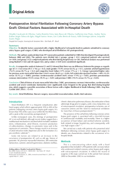
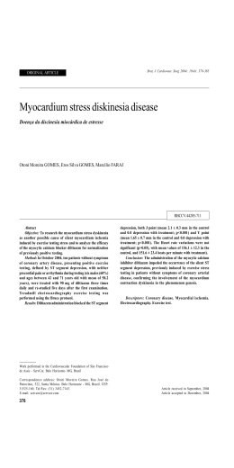
CASE REPORT
Rev Bras Cir Cardiovasc 2007; 22(4): 505-508
Myocardial revascularization in anomalous origin of
the right coronary artery: case report
Revascularização do miocárdio em origem anômala da artéria coronária direita: relato de caso
Marcelo Sávio da Silva MARTINS1, Eduardo Sérgio BASTOS2, Jorge Viana ANNIBAL3, Alvaro Barde BEZERRA4
RBCCV 44205-937
Abstract
A 21-year-old man with angina-like chest pain and
syncope related to ischemic ECG changes due to an
anomalous origin of the right coronary artery. The patient
was submitted to surgical correction with myocardial
revascularization with internal thoracic artery. A literature
review of this rare congenital heart disease is presented.
Resumo
O objetivo deste artigo é mostrar o caso de um paciente de
21 anos com episódio de angina e lipotímia relacionados com
alteração isquêmica do miocárdio decorrente de origem
anômala da artéria coronária direita a partir do seio da aorta
(seio de Valsalva) esquerdo. O paciente foi submetido à cirurgia
de revascularização do miocárdio, com artéria torácica interna
direita para artéria coronária direita. É realizada também uma
revisão da literatura desta rara doença cardíaca congênita.
Descriptors: Coronary vessel anomalies. Sinus of
Valsalva. Coronary disease.
Descritores: Anomalias dos vasos coronários. Seio aórtico.
Coronariopatia.
1. Cardiovascular Surgery Specialist; Heart Surgeon, Hospital de
Força Aérea do Galeão (HFAG). Postgraduate student,
Departament of Surgery, Faculdade de Medicina da Universidade
Federal do Rio de Janeiro.
2. Associate Professor, Departament of Surgery, Discipline of Heart
Surgery, Faculdade de Medicina da Universidade Federal do Rio de
Janeiro; Heart Surgeon, Heart Surgery Service, Universidade
Federal do Rio de Janeiro, HFAG.
3. Head Clinical Surgeries, HFAG; Heart Surgeon, HFAG.
4. Director of Hospital de Aeronáutica de São Paulo; Heart Surgeon.
This study was carried out at Hospital de Força Aérea do Galeão, RJ.
Correspondence address:
Marcelo Sávio da Silva Martins. Rua Ary Parreiras, 65/904, Icaraí,
Niterói, RJ. CEP 24230-320.
E-mail address: [email protected]
Article received in 19 May 2007
Article accepted in 20 Jul 2007
505
MARTINS, MSS ET AL - Myocardial revascularization in anomalous
origin of the right coronary artery: case report
INTRODUCTION
Right and left coronary arteries arise from the aortic
sinus behind their respective semilunar cups. Usually, the
ostia are localized in the superior third of aortic sinus (sinus
of Valsalva). Because the oblique plane of aortic valve, left
coronary (LC) artery ostium is superior to the right coronary
(RC) artery ostium.
Coronary artery abnormalities result from disorders
occurring during fetal development and may present
varying anatomic aspects according to their route and
origin. These abnormalities without another congenital
defect associated are rare and are identified only in 0.25%
to 0.9% of the cineangioicoronariographies. Gender
prevalence was not observed [1].
Among these abnormalities, the most frequent one is the
anomalous origin of the circumflex branch of right coronary
artery. It is described for the first time in the literature. Yet
most unusual is the anomalous origin of the left coronary
(LC) artery from the right coronary (RC) artery, i.e., the isolated
origin of the posterior interventricular branch (posterior
descending branch) of the left coronary artery [2].
The origin of the right coronary artery from the left aortic
sinus corresponds to 16% of these anatomic variations. It
was firstly described in 1984 by White et al.. It is a potential
cause of sudden death in children and athletes due to
myocardial ischemia, acute myocardial infarction (AMI) or
sudden death [2].
The treatment is surgical and Bett performed the first
surgical correction in 1985.
Rev Bras Cir Cardiovasc 2007; 22(4): 505-508
The transesophageal echocardiogram was normal, with
left ventricular global and segmental contractions
preserved. Holter monitor showed ischemic alterations
during maximum exertion.
Transesophageal echocardiogram showed the anomalous
right coronary artery pathway between the aorta and
pulmonary trunk. Nuclear magnetic resonance (NMR) was
not conclusive. Cineangiocoronariography showed a normal
left coronary artery and the anomalous origin of the left
coronary artery from the left aortic sinus (Figure 1), with a
possible pathway between the aorta and pulmonary trunk.
CASE REPORT
Fig. 1 - Cineangiocoronariografia da CD
A 21-year-old male athlete was asymptomatic until
February 2003, when presented syncope as a result of
physical activity. One month later, the patient presented a
sharp precordial pain and lipothymia when walking. Patient
was referred to the Hospital de Força Aé rea do Galeão
(HFAG) emergency service, where on physical examination,
he presented blood pressure of 190 x 120 mmHg. The patient
was discharged from hospital after being treated with
calcium channel blocker and once blood pressure was under
control.
A family history of sudden death was already
established as a consequence of his 13-year-old brother’s
sudden death with reports of precordial pain and cardiac
murmur. In this case, the presence of cardiac murmur
associated to precordial pain raises the possibility of the
patient to present with a coronary fistula.
After 30 days, the patient presented with the same
symptoms and he received medical care at HFAG. At the
time, he reported typical precordial pain and the
electrocardiogram showed ischemic alterations.
506
Patient underwent myocardial revascularization with the
right internal thoracic artery grafting the right coronary
artery proximal segment and proximal ligation of RCA with
a good postoperative outcome.
In a new exercise test performed six months
postoperatively, signs of myocardial ischemia were not
noted even under extreme exertion.
DISCUSSION
This is an unusual anomaly and a potential cause of
sudden death in children and athletes with the majority of
the cases being reported by both pediatrician and mainly
pathologist authors. In reviews of the current literature, it
is more common to find reports of coronary fistula correction
surgery due to cardiac murmur, what draws surgical attention
leading to the performance of complementary tests that
diagnose the anomaly [1].
MARTINS, MSS ET AL - Myocardial revascularization in anomalous
origin of the right coronary artery: case report
Rev Bras Cir Cardiovasc 2007; 22(4): 505-508
In the literature, the first case was published, in 1948,
by White et al. in an autopsy finding [2]. In 1974, Cheitlin
published a catheterization finding. In 1980, Benge et al. [2]
presented the first publication addressing the morbidity of
this coronary artery anomaly; however, only in 1985, the
first surgical correction of the anomalous origin of right
coronary artery from the left aortic sinus was performed by
Bett, who used a saphenous vein graft [3].
In an analysis of 12,595 patients undergoing
catheterization, coronary artery anomalies were found in
1,686 patients (1.3%). Of the 1,686 patients, 1,461 (87%) had
anomalies of origin and distribution, and 225 (13%) had
coronary artery fistulae. Among patients with anomalies of
origin, 16% had right coronary origin from the left aortic
sinus [4]. Regarding anomalies of origin, they can arise from
abnormal origin in aortic sinus, from pulmonary trunk origin,
or present a single ostium. These anomalies were classified
as benign and malignant according to the symptoms
presented. The benign anomalies do not result in signs,
symptoms, or complications. The malignant anomalies carry
a high risk of angina pectoris, syncope, acute myocardial
infarction, congestive heart failure, and sudden death and
require surgical correction. Among the so-called malignant
anomalies, we can include ectopic coronary origin from the
contralateral aortic sinus [4], similar to the case herein
presented.
The pathophysiology of myocardial ischemia (Figure 2)
provoked by this anomaly is due to three causes resulting
from right coronary artery pathway between the aorta and
pulmonary trunk: 1. Slit-like ostium: because of the pressure
put on this artery by the two large vessels, a hypertrophy
of the tunica intima and a deformity of this coronary artery
diameter occur; 2. Output angle, rather than perpendicular
to the aorta, nearly forms a right angle with the aorta wall;
3. Systolic compression that is the most leading explanation
for the etiology of sudden death among athletes occurring
when there is an increased arterial pressure, what increases
the aorta diameter and the pulmonary trunk as well, then
occurring the right coronary artery segment collapse
between both structures [3].
Many patients with anomalous coronaries are
asymptomatic and signs of myocardial ischemia in youths
are not valuable making difficult the diagnosis of this
anomaly. Clinical complications occur usually during
physical exertion evoked by the anomalous pathway of
these arteries, especially in the pathway between the aorta
and pulmonary trunk. Myocardial perfusion changes can
occur as a consequence of the movements of the vessels
during cardiac cycle. It is due to this physical exertion that
acute myocardial infarction, or even ischemia-induced
arrhythmias may occur [3].
Repair surgery can be performed by right coronary
artery reimplantation, or using a graft for this coronary. On
reimplantation, the artery is moved to its proximal portion
and anastomosed to the right aortic sinus. However, in these
cases, the coronary artery proximal segment, which is
compressed between the aorta and pulmonary trunk, can
present intimal hyperplasia, thus favoring thrombosis and
the occurrence of local atherosclerosis [5,6].
The choice of surgical procedure was the use of right
internal thoracic artery (RITA) grafted into the right coronary
artery just after its emergence in the pathway between the
aorta and pulmonary trunk, with ligation of proximal vessel.
Fig. 2 - Compressão da CD entre aorta e o tronco pulmonar
CONCLUSION
In the anomalous origin of the right coronary artery
from left aortic sinus, with a route between the aorta and
pulmonary trunk, surgical anastomosis of right internal
thoracic artery to the anomalous origin of the vessel and
proximal ligation can be considered as a safe and easily
reproducible technical option.
507
MARTINS, MSS ET AL - Myocardial revascularization in anomalous
origin of the right coronary artery: case report
REFERENCES
1. Cieslinski G, Rapprich B, Kober G. Coronary anomalies:
incidence and importance. Clin Cardiol. 1993;16(10):711-5.
Rev Bras Cir Cardiovasc 2007; 22(4): 505-508
4. Giroux SK, Cartier R. Atypical presentation of an anomalous
origin of the right coronary artery with severe compression
between the great vessels. Ann Thorac Surg. 2002;73(5):1636-8.
2. Benge W, Martins JB, Funk DC. Morbidity associated with
anomalous origin of the right coronary artery from the left
sinus of Valsalva. Am Heart J. 1980;99(1):96-100.
5. Di Lello F, Mnuk JF, Flemma RJ, Mullen DC. Successful
coronary reimplantation for anomalous origin of the right
coronary artery from the left sinus of Valsalva. J Thorac
Cardiovasc Surg. 1991;102(3):455-6.
3. Yamanaka O, Hobbs RE. Coronary artery anomalies in 126,595
patients undergoing coronary arteriography. Cathet Cardiovasc
Diagn. 1990;21(1):28-40.
6. Nelson-Piercy C, Rickards AF, Yacoub MH. Aberrant origin of
the right coronary artery as a potencial cause of sudden death:
successful anatomical correction. Br Heart J. 1990;64(3):208-10.
508
Download