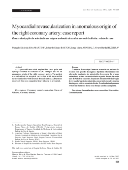
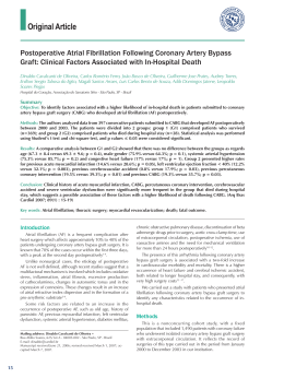
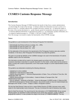
Braz J Cardiovasc Surg 2004; 19(4): 378-385
ORIGINAL ARTICLE
Myocardium stress diskinesia disease
Doença da discinesia miocárdica de estresse
Otoni Moreira GOMES, Eros Silva GOMES, Marcílio FARAJ
RBCCV 44205-711
Abstract
Objective: To research the myocardium stress dyskinesia
as another possible cause of silent myocardium ischemia
induced by exercise testing stress and to analyze the efficacy
of the myocytic calcium blocker diltiazem for normalization
of previously positive testing.
Method: In October 2004, ten patients without symptoms
of coronary artery disease, presenting positive exercise
testing, defined by ST segment depression, with neither
precordial pain or arrhythmia during testing (six males (60%)
and ages between 42 and 71 years old with mean of 58.2
years), were treated with 90 mg of diltiazem three times
daily and re-studied five days after the first examination.
Treadmill electrocardiography exercise testing was
performed using the Bruce protocol.
Results: Diltiazem administration blocked the ST segment
depression, both J point (mean 2.1 ± 0.3 mm in the control
and 0.0 depression with treatment; p<0.001) and Y point
(mean 1.65 ± 0.7 mm in the control and 0.0 depression with
treatment; p<0.001). The Heart rate variations were not
significant (p>0.05), with mean values of 156.1 ± 12.3 in the
control, and 151.6 ± 23.4 beats per minute with treatment).
Conclusion: The administration of the myocytic calcium
inhibitor diltiazem impeded the occurrence of the silent ST
segment depression, previously induced by exercise stress
testing in patients without symptoms of coronary arterial
disease, confirming the involvement of the myocardium
contraction dyskinesia in the phenomenon genesis.
Descriptors: Coronary disease. Myocardial ischemia.
Electrocardiography. Exercise test.
Work performed in the Cardiovascular Foundation of São Francisco
de Assis - ServCor, Belo Horizonte- MG, Brazil
Correspondence address: Otoni Moreira Gomes. Rua José do
Patrocínio, 522, Santa Mônica. Belo Horizonte - MG, Brazil. CEP:
31525-160. Tel/Fax: (31) 3452-7143.
E-mail: [email protected]
378
Article received in September, 2004
Article accepted in December, 2004
GOMES, OM ET AL - Myocardium stress diskinesia disease
Resumo
Objetivo: Pesquisando a discinesia de contração miocárdica
como outra possível causa da isquemia silenciosa de estresse,
analisar a eficácia do cálcio inibidor miocítico diltiazem na
normalização de testes ergométricos previamente positivos.
Método: Em outubro de 2004, dez pacientes, sem sintomas
de doença arterial coronária, com teste ergométrico positivo
definido por infradesnível de segmento ST, sem dor precordial
nem arritmia durante os exames (Seis --60%-- do sexo
masculino; idades entre 42 e 71 anos, média de 58,2 anos),
foram tratados com três doses diárias de 90mg de diltiazem e
reestudados após período de cinco dias. Os exames foram
realizados em esteira ergométrica, no protocolo de Bruce.
Resultados: O diltiazem impediu a depressão do segmento
ST, tanto do ponto J (infradesnível médio e 2,1 ± 0,3mm, no
controle, e 0,0 desnível com tratamento; p<0,001), quanto do
INTRODUCTION
Since the studies of PARDEE in 1920 [1], alterations in
electrocardiograms (ECG) of myocardial ischemia patients
have been well defined, characterizing the relationship
between the anomalous Q waves with inactive zones due to
necrosis and consequent fibrosis, the ST segment deviated
by the pre-necrotic ischemic lesion and T wave alterations
by the establishment of ischemia in its earliest phases.
The advent and evolution of the diagnosis of coronary
insufficiency by induced stress increased even more the
importance of the analysis of ST segment deviations, not
only because of the diagnostic specificity in itself, but also
because of evidence of its almost immediate reversibility
with the cessation of ischemia-inducing stress. Individual
differences in the electrocardiographic responses to stress,
with varying patterns of the ST deviation in individuals
with normal coronary arteries, motivated the need of
improvement in the criteria and the introduction of analysis
of the J and Y points, which among other things facilitated
the interpretation of descending and ascending ST designs
(concave or convex). Even so there are still cases of induced
ST variations diagnosed as false positive or false negative,
that is, imprecise sensitivity and specificity.
It is also well known that individuals presenting with
partial obstructive coronary disease, ventricular
hypertrophy or significant overloads during stress, preserve
the normal ST pattern, whilst others, without diagnosed
heart disease, evolve with significant changes of the ST
segment, specifically at point J, with maximum responses at
higher heart rates. These patients, even with normal coronary
arteries or with non-occlusive parietal alterations evidenced
by scintigraphic or angiographic studies, generally present
with a syndromic profile defined by atypical precordial or
Braz J Cardiovasc Surg 2004; 19(4): 378-385
ponto Y (infradesnível de 1,65 ± 0,7 mm, no controle, e 0,0
desnível com tratamento; p<0,001). A freqüência cardíaca não
apresentou variação estatisticamente significante (média de
156,1 ±12,3 bpm, no controle, e 151,6 ± 23,4 bpm com
tratamento/ p>0,05).
Conclusão: A administração do cálcio inibidor miocítico,
diltiazem, impediu a depressão silenciosa do segmento ST
previamente induzida por teste ergométrico em pacientes
sem sintomas de doença arterial coronária, confirmando o
envolvimento da discinesia da contração miocárdica na gênese
do fenômeno.
Descritores: Coronariopatia. Isquemia miocárdica.
Eletrocardiografia. Teste de esforço.
retrosternal pain reducing their physical capability and a
horizontal depression of the ST segment, marked by a 2millimeter depression or greater at point J and a normal point
Y or one with less than one-millimeter of depression.
The horizontal depression of the ST segment has been
interpreted (not rarely as a physiological response of tachycardia)
as dependent on the heart beat [2], with DETRANO et al. [3] in
1986 and LACHTERMAN et al. [4] in 1990 studying the ST/FC
stress induced index, as a diagnostic element in exercise stress
testing. Even so, this interpretation has been contested,
demonstrating that it does not have a greater diagnostic
significance than the simple ST variations [5-8].
In 1984, PIC & BROUSTED [9] observed that the ST
depression in isolation presents a sensitivity rate of 72.5%
and a specificity rate of 62.5% in patients without previous
infarction, an increase in the R-wave sensitivity 58.5% and
specificity 67.5% (or of the QRS with 78% and 57.5%,
respectively) and the pain 63% and 75%. The three together
positively diagnosed in 100% of cases. The positivity of
only one signal corresponds to normal coronary arteries in
most cases. This difficulty in guaranteeing the
electrocardiographic diagnosis to guide patients is
continuously being highlighted in recent studies [10-12].
KURL et al. [13] in 2003 demonstrated a significant
increase in cardiovascular death rate (3.5 times greater) and
death due to brain strokes (2.2 times greater) in asymptomatic
patients with a horizontal depression of the ST segment
during exercise stress testing.
These results prove to be risk free only for patients where
the basal ECG is normal and the TE normal. In all other
circumstances, they seem to indicate the need of
complementary angiographic, angiokinetic or scintigraphic
studies with cineangiography being the most reliable for a
definitive diagnosis.
379
GOMES, OM ET AL - Myocardium stress diskinesia disease
Definitive studies that diagnose patients without
coronary artery obstructions and alterations of the ST
segment seen during exercise stress tests with sufficient
precision over the long-term are still rare and relatively
incomplete. However, there is already sufficient evidence to
justify preventative treatment in these cases, so that the
risks to which patients are exposed are not ignored [13].
The challenge includes a search for other etiopathogenic
factors, as well as arriving at a consensus that silent myocardial
ischemia depends only on coronary vascular disease.
Taking the classic pattern of ST segment with deviations
of less than 1 mm as normal and considering additional
deviations of the ST segment as ischemic, the possibility of
an asymmetric response between the myocardial oxygen
demand and the dynamics of the coronary reserve in these
patients is acceptable, (a) by the intensity in the contraction
produced being greater than that necessary for systolic
outflow and for the existent peripheral demand, (b) by the
inferior reflex vasodilation necessary for the existent
contractile demand or (c) due to the atypical and
heterogeneous pattern of the myocardial sectorial
myofibrillar contraction. Under these conditions, the areas
of less spastic contraction may act with a steal effect from
the areas of more intense contraction.
An interpretation of the asymmetric intramural
contractions corroborates with the results published by Lins
[14], demonstrating the appearance of an intraventricular
pressure gradient in the echocardiographic study using
induced stress in a group of patients with positive exercise
stress tests.
Accepting abnormal myocardial contractions as the main
generator of the ischemic condition, highlights the role of
adrenergic inotropic stimulants and Calcium++ dependents.
Evidence that patients being treated for hypertension using
beta-blockers also presented silent depression of the ST
segment, suggests that mechanisms dependent on
transmembrane calcium flow are involved in the genesis of
the disorder.
It is been demonstrated that in the first phases of
myocardial ischemia contraction bands can occur together
with the reduction of cytoplasmatic glycogen, impeding
normal myofibrillar relaxation and making ventricular diastole
difficult, a fundamental factor in the increased calcium++
flow into the myocell. These cases support the use of calcium
antagonists as the first therapeutic option, not only because
of the specific physiopathologic and pharmacokinetic
implications, but also due to experience based on their use,
biocompatibility and multiple cardiovascular benefits.
The aim of this present investigation is to analyze the
results of a study of patients with atypical precordial pain,
without obstructive coronary disease, submitted to exercise
stress testing with or without treatment using diltiazem
myocytic calcium-inhibitor.
380
Braz J Cardiovasc Surg 2004; 19(4): 378-385
METHOD
With the approval of the Bio-ethics Commission of the
Institution, ten consecutive patients were studied in the
Laboratory of stress exercise tests (FCSFAServcor). The
exercise stress test was defined as positive when a horizontal
depression of the ST segment occurred without precordial
pain or arrhythmia during the examinations. Six (60%)
patients were men and the ages of the patients ranged from
42 to 71 years (mean age 58.2 years). All the patients of the
present series were restudied five days after being treatment
with three daily doses of 90 mg of diltiazem.
The examinations were performed on a computerized
treadmill (model Digistress/ MG), following the Bruce protocol.
The J (STj) and Y (Sty) point variations of the ST segment, the
maximum VO2 reached (mL/kg/min), heart rate (bpm), the double
product (DP), the METs reached and the functional class (AHA)
were analyzed, using the following criteria: 1- excellent, 2- good,
3-regular (medium), 4- weak and 5- very weak.
The Student t-test was employed to evaluate the
statistical significance of differences between two parallel
samples, with the level of significance was fixed with a pvalue < 0.05. The EPIINFO version 6.04 computer program
of the World Health Organization was used.
RESULTS
Table 1 presents the obtained results. It was confirmed
that the administration of calcium-inhibitors impedes
depression of the ST segment, both at point J (mean
horizontal depression 2.1 ± 0.3 mm for controls, and 0.0
depression with treatment - p<0.001) and at point Y
(horizontal depression of 1.65 ± 0.7 mm for controls, and 0.0
depression with treatment - p< 0.001). The heart rate did not
present with statistically significant variations (average 156.1
± 12.3 bpm in the controls and 151.6 ± 23.4 bpm with treatment
– p-value > 0.05). There was a statistically significant
improvement in the functioning (p-value < 0.05) with the
treatment (mean 2.6 ± 0.7 in controls and 1.9 ± 0.7 after
treatment), without significante differences in the VO2 or in
the double product (p>0.05).
In two patients, the horizontal depression of the ST
segment did not appear homogeneous in all complexes in
each of the derivations analyzed, but the alterations were
manifested progressively, with an appearance of claudication
and pathologic complexes in with alternate anachronic and
normal complexes on the same derivation (Figure 1).
COMMENTS
The consensus that deviations of the ST segment are
more reliable to confirm myocardial ischemia during exercise
stress testing compared to other hemodynamic or
GOMES, OM ET AL - Myocardium stress diskinesia disease
Braz J Cardiovasc Surg 2004; 19(4): 378-385
Table 1. Re-synchronism of the myocardial contraction-perfusion by diltiazem
CONTROL
NB nº
Age
Gender
METZ
VO 2
FC
DP
AHA
STJ
STY
1
2
3
4
5
6
7
8
9
10
Mean
SD+/
42
70
69
71
61
52
67
54
44
52
F
M
M
M
M
F
M
M
F
F
10
8
8
12
8
8
10
12
9
8
9.3
1.6
25
29
28
49
28
30
34
44
36
38
34.1
7.7
169
137
166
153
140
154
169
171
148
154
156.1
12.3
30420
30277
31540
26010
25910
24640
30420
33345
13458
24640
27066
5692.6
3
3
3
1
3
3
2
2
3
3
2.6
0.7
-2
-2
-2
-2
-2
-2
-3
-2
-2
-2
-2.1
0.3
-2
-2
-1.5
-2
-1
-1
-3
-1
-2
-1
-1.65
0.7
DILTIAZEM 90-120mg, 8/8h
NB nº
Age
Gender
METZ
VO 2
FC
DP
AHA
STJ
STY
1
2
3
4
5
6
7
8
9
10
Mean
SD+/
42
70
69
71
61
52
67
54
44
52
F
M
M
M
M
F
M
M
F
F
10
8
8
14
11
9
12
11
10
9
10.2
1.9
36
26
28
46
4
32
44
39
51
32
33.8
13.2
171
121
112
136
137
170
153
171
175
170
151.6
23.4
29070
19200
19040
21080
26715
30600
27540
30780
31500
30600
26613
4976.2
2
3
3
1
2
2
1
2
1
2
1.9
0.7
0
0
0
0
0
0
0
0
0
0
0
0
0
0
0
0
0
0
0
0
0
0
0
0
Fig. 1 – Ischemic claudication (arrows) presenting with normal
between pathologic complexes
electrocardiographic variables
(arrhythmia, Q wave or R wave
alterations) is well known [15].
The current universal consensus
of a relationship between induced
stress ischemia, evidenced by silent
depression of the ST segment, with
coronary artery perviousness does
not adequately explain cases of
normal coronary arteries, nor does it
offer perspectives of therapeutic
options for the patients involved.
When considering another
possible cardiovascular nosologic
entity responsible for the ST
segment depression in patients
without organic coronary lesions
and without defined heart disease,
the following physiopathologic
factors are especially interesting:
the physiologic mechanism of
supply-demand in the myocardial
metabolism and interactive
physiopathology of the ischemic
degenerative alterations (with
electrocardiographic
and
pharmacokinetic signs) of the
transmembrane
activation/
inhibition of myocardial contraction
and angiokinesis.
Mechanism of supply-demand in the myocardial
metabolism
The preservation of the myocardial integrity depends
basically on the relation between the supply and demand of
essential metabolic substrates, with the coronary arterial
blood flow as the factor responsible for cellular supply. In
adverse circumstances, the cellular mechanisms of the
coronary reserve and ischemic adaptation are determinants
which respond to the myocardial survival capacity.
The phenomenon of coronary reserve can be
demonstrated in flowmetry by the reactive hyperemia that
follows occlusion of a coronary artery at an interval of ten
seconds. Even occlusions of only one second cause
reperfusion at an increased flow rate, estimated as normal
hyperflow when levels are five times the basal rate, that is,
from 250 mL to 1250 mL/min, or from 0.9 mL/g of myocardium
to 4.5 mL/g [16]. Coronary artery diseases, including
endothelial dysfunction and alterations in the extra-vascular
resistance factor, can significantly alter the coronary reserve.
Ischemic adaptation is the resource by which the
myocardium is able to present sensitive changes in its
381
GOMES, OM ET AL - Myocardium stress diskinesia disease
oxygen needs to protect itself from ischemia, whether
reducing its inotropism and myofilamentar tension, or
changing enzymatic channels to reduce energy use. The
phenomenon of ischemic adaptation is well characterized in
ischemic preconditioning, where it has been consistently
demonstrated that a brief period of ischemia, succeeded by
another period of reperfusion, increases the tolerance of
the myocardium to anoxia and prolongs the time necessary
for degeneration to necrosis [17], with the following
beneficial effects demonstrated: reduction of the intensity
and frequency of anginal events – warm up phenomenon,
horizontal depression of the ST segment, depletion of PTA,
enzyme release (CK-MB, troponin), production of lactic acid
and free radicals, apoptosis, ultra-structural injury, infarction
area, the incidence of arrhythmia, noxious remodeling,
activation of leukocytes, hospitalization, with reduction of
mortality and protection of the endothelium during
reperfusion.
Although a simple episode of transitory ischemia can
produce heart protection, repetitive episodes of brief periods
of occlusion, if they are frequent, can cause trachyphilaxy
resulting in myocardial injury.
Any expression of the proprieties of adaptation/ischemic
resistance will depend on the evolution of the myocardial
ischemia for the conditions of integral recovery, necrosis,
stunning or hibernation. Stunning, which is the maximum
reversible ultrastructural lesion and hibernation representing
the condition of tissue survival with optimized minimum
flow, by the maximum ischemic adaptation capacity or
minimum sustainable metabolic reserve?
Electrocardiographic signal of myocardial ischemia
The correlation between the evolution of acute myocardial
ischemia and the pattern of electrocardiographic register is
sufficiently defined, with the T wave altered at the start of
ischemia, the ST segment showing the progression of cellular
degeneration and the Q wave diagnosing the definitive
establishment of necrosis. All these signs are intimately linked
to variations in the ionic flow of the cellular membrane and
modifications resulting from the transmembrane potential. It is
notable that the ECG can remain unaltered even in situations of
complete inactivation of the myocardial contraction, even in
mechanical uncoupling, which is reproducible in the laboratory
using drugs. With this limitation always in view, is it possible
to make a correlation between the biochemical and
histopathologic phases of ischemia and alterations on the ECG.
Basically, the following phases can be considered for
myocardial ischemia: 1 - cytoplasmatic hypoxia with a
reduction in the pH, reduction of the ionic pumping activity
and the start of the formation of oxygen free radicals; 2 Edema and vacuolization of mitochondrias; 3 - Rupture of
mitochondrias and of the myofibrils; 4 - karyolysis and 5 382
Braz J Cardiovasc Surg 2004; 19(4): 378-385
Fibrosis of substitution. The first and the second phases
are rapidly induced and reversible; the second generally
appears after 5-10 minutes of ischemia and the third phase
marks the start of necrosis, which generally occurs after 1520 minutes of normothermic ischemia, with reversibility
depending of the extension of the injured area and capacity
of viable sustentation of the myocardium [18-22]. The
myocardial ischemic stunning can be between phases 2 and
3 with most lesions reversible.
Basically, it is known that the variations of the T wave
happen in the first phase and those of the ST segment
correspond to lesions in the second and third phases and
the Q wave marks the necrotic areas of Phases 4 and 5.
This interpretation valorizes the appearance of the
horizontal depression of the ST segment as representative
of regional myocardial involvement always accompanied
by histologic myocardial aggression, however little, whose
intensity will define the velocity of the tissue recovery and
the normalization of the ECG, which is also influenced by
the velocity and efficiency of reperfusion.
Physiopathology of ischemia with silent depression of
the ST segment
The etiopathological correlation of the transmembrane
disorder of calcium flow, as a marker of silent stress ischemia
without coronary arterial disease, has clinical proof of cause
and effect in electrocardiographic alterations present in the
surgical post-cardioplegia myocardial reperfusion, in patients
presenting with hyperpotassemia (K+ > 7 mEq/L) and
hypocalcaemia (Ca++ < 7 mg/dL): the electrocardiogram
presents ST horizontal depression with the T wave negative,
similar to the “Pardee Complex” of acute infarction,
progressively corrected until complete normalization is
achieved by the endovenous use of calcium gluconate and
renal depuration of the potassium [23, 24].
Such solid physiopathologic evidence justified the
hypothesis of treatment of the disorder using a calcium
inhibitor.
The initial results so far (shown in Table 1), confirmed
this physiopathologic and pharmacokinetic evidence. Note
that treatment with diltiazem normalized the response to
exercise stress test.
It is notable, in respect to the data, that maintenance of
heart frequency was possible without the ischemic signs
seen in the control group and without therapeutic
stabilization.
On the other hand, myocardial disease with diskinetic
contraction-perfusion, as it is related to the progressive
functional response of the heart muscle, presupposes the
possibility of installation and progressive regional extension
of the contraction-perfusion asymmetry. This is a fact not
infrequently observed in these patients, with the initial
GOMES, OM ET AL - Myocardium stress diskinesia disease
appearance of alternate or claudicant horizontal depression
of the ST segment, to the establishment of a uniform
definition of the regional involvement with the continuation
of effort. Figure 1 demonstrates an example of this
phenomenon.
The ischemic stress marker does not have a fixed pattern
in the ventricular anatomy and can occur only in the inferior,
lateral or anterior walls or be generalized and is diagnosed
as a diskinetic condition of the myocardial contraction with
regions showing different patterns of contraction/
relaxation. The ischemic claudication phenomenon, with
complexes of normal ST segments with intermittent
ischemic patterns, similar to focal zones of tetany/spasms
that do not affect the basic hemodynamic performance is
not rare. This is similar to what happens in the skeletal
musculature, with the manifestation of myalgia and local
contractures (only precursory of cramps and contractures)
which do not impede the performance but induce fatigue
quicker. Figuratively, myocardial dyskinesia can function
as an opening and closing of manual pumping, as if one or
more fingers contract at different levels of tension, without
prejudicing the total compression force, enabling the
ventricles to maintain the systolic outflow and the
hemodynamic stability.
Etymologically speaking, disease is “the lack or any
disorder of the health [25]” and, thus, depression of the ST
segment identifies existent heart diseases, such as
obstructive coronary disease and myocardial hypertrophies.
Innumerous lethal organic diseases also present silent
evolution with totally asymptomatic phases and of extremely
difficult diagnosis, with lethal results as one of their first
clinical manifestations for examples the rupture of brain
aneurysms and thromboembolic states and the always feared
tumoral metastases.
Thus, the myocardial condition responsible for the
appearance of effort-induced depression of the ST segment,
even without organic obstructive coronary lesions is an
important disease, which over the long-term causes higher
morbid-mortality rates compared to other diseases, even
congenital or acquired heart diseases.
The social aspect of the problem, in some studies, is
similar to incidence of hypertension, with possible significant
regional variations [26-28], as the positive exercise stress
test in asymptomatic patients includes from 0.6 to 15% of
the studied population (eight studies). In a maximum of 0.06
to l.6% cases this is associated with significant arterial
disease (narrowing of 50% or more of the epicardial coronary
artery), suggesting that stress myocardial dyskinesia is a
disease occurring in 0.54 to l3.4% of the asymptomatic
population [29]. KURL et al.[13], in 2003, demonstrated a
significant increase in cardiovascular death (3.5 times
greater) and of strokes (2.2 times greater) in asymptomatic
Braz J Cardiovasc Surg 2004; 19(4): 378-385
individuals with horizontal depression of the ST segment
evidenced during exercise stress testing.
Evidence that silent depression of the ST segment can
be cured with drugs highlights the importance of adhesion
of the patient to treatment. The classification of the problem
as a reversible disease offers highly favorable psychological
support, in contrast with the anxiety caused by the insecurity
and lack of definition of its pleomorphic symptomatology,
with a reduction of the physical aptness and pseudo-anginal
algic sensations. The constructive emotion of the safety
and satisfaction of patients is clearly seen when they are
informed that the ECG has normalized after treatment.
The ideal situation is that all patients with stress
myocardial dyskinesia without coronary arterial disease, a
condition which defines the nosology, are completely studied
in respect to the normality of the coronary arteries. However,
the condition of public health system is complex, because the
number of positive cases is relatively small and the negative
cases high. On the other hand the evidence shows that the
disease increases considerably the morbid-mortality of
individuals, thus it requires special preventive treatment.
The initial experience of our institution, supporting the
use of 90-120 mg diltiazem three times daily, highlights
another significant aspect of the study. Re-synchronization
of the myocardial contraction/perfusion by the drug appears
to be a diagnostic test option to improve the criterion of the
indication of complementary studies of coronary
perviousness. As Antônio Felipe Simão and Maurício Nunes
assure us there are no drugs, in acceptable clinical doses,
able to normalize exercise stress testing in patients with
significant coronary obstructions. Moreover, the use of
diltiazem increases the safety in the initial period of
rehabilitation, until effective myocardial conditioning,
allowing a faster improvement of the functional performance,
as occurred in previously treated patients, providing
psychological and functional benefits of motivation and
adherence.
Recent research suggests, without sufficient proof of
cause, the occurrence of myocardial perfusion disorders
of the microcirculation in the postoperative period of
coronary revascularization [30]. From this it can be inferred
that dyskinesia of myocardial contraction may also
influence the direction of intramyocardial flow patterns,
determining postoperative ventricular functional
alterations. There are no studies yet comparing the
evolution of these patients.
In a general analysis, the results obtained in the current
investigation, without similar results reported in the literature,
indicate a disorder of the myocardial contraction
synchronism is another factor of silent ischemia induced by
exercise stress, efficiently treated using diltiazem calcium
inhibitor.
383
GOMES, OM ET AL - Myocardium stress diskinesia disease
As frequently in preliminary clinical research, the small
sample size, notwithstanding its absolute statistical
significance and the restricted number of parameters
analyzed, suggest the necessity of further investigations to
provide an uncontestable scientific definition of the
phenomenon observed.
Braz J Cardiovasc Surg 2004; 19(4): 378-385
9. Pic A, Broustet JP. Diagnostic value of amplitude variations of
the QRS complex during in computerized exercise testing. Arch
Mal Coeur Vaiss 1984;77:54-63.
10. Severi S, Orsini E, Marracini P, Michelassi C, L'Abbate A.
The basal electrocardiogram and the exercise stress test in
assessing prognosis in patients with unstable angina. Eur
Heart J 1988;9:441-6.
11. Fruegaard P, Launbjerg J, Jacobsen HL, Madsen JK. Sevenyear prognostic value of the electrocardiogram at rest and an
exercise test in patients admitted for, but without, confirmed
myocardial infarction. Eur Heart J 1993;14:499-504.
12. Grundy D, Gibler WB, Bassan R. O eletrocardiograma e o
monitor de tendência do segmento ST na avaliação diagnóstica
e no prognóstico da dor torácica. In: Bassan R, editor. Síndrome
coronariana aguda nas unidades de dor torácica. Rio de
Janeiro:Atheneu;2000. p.71-95.
BIBLIOGRAPHIC REFERENCES
1. Pardee HEB. An electrocardiographic sign of coronary artery
obstruction. Arch Intern Med 1920;26:244-57.
2. Okin PM, Kligfield P. Computer-based implementation of the
ST-segment/heart rate slope. Am J Cardiol 1989;64:926-30.
3. Detrano R, Salcedo E, Passalacqua M, Friis R. Exercise
electrocardiographic variables: a critical appraisal. J Am Coll
Cardiol 1986;8:836-47.
4. Lachterman B, Lehmann KG, Detrano R, Neutel J, Froelicher
VF. Comparison of ST segment/heart rate index to standard
ST criteria for analysis of exercise electrocardiogram. Circulation
1990;82:44-50.
13. Kurl S, Laukkanen JA, Tuomainen TP, Rauramaa R, Lakka
TA, Salonen R et al. Association of exercise-induced, silent
ST-segment depression with the risk of stroke and
cardiovascular disease in men. Stroke 2003;34:1760-5.
14. Lins RHC. Gradiente intraventricular de estresse. In: Simpósio
do Departamento de Fisiologia Cardiovascular e Cardiologia
Experimental. 59o Congresso da Sociedade Brasileira de Cardiologia.
Rio de Janeiro:Sociedade Brasileira de Cardiologia;2004.
15. Uchida AH, Canabrava MVF. Teste ergométrico em
assintomáticos: uma análise baseada em evidências. Revista
DERC 2004;10:18-9.
16. Gould KL. Coronary artery stenosis. New York:Elsevier
Science Publishers;1991.
17. Murry CE, Jennings RB, Reimer KA. Preconditioning with
ischemia: a delay of lethal cell injury in ischemic myocardium.
Circulation 1986;74:1124-36.
5. Froelicher VF, Lehmann KG, Thomas R, Goldman R, Morrison
D, Edson R et al. The electrocardiographic exercise test in a
population with reduced workup bias: diagnostic performance,
computerized interpretation, and multivariable prediction. Ann
Intern Med 1998;128:965-74.
18. Gomes OM. Análise comparativa das alterações ultraestruturais e bioquímicas determinadas no miocárdio, pelas
paradas cardíacas anóxicas normotérmica e hipotérmica (20ºC):
estudo experimental. [Tese de doutorado]. São Paulo:Faculdade
de Medicina da Universidade de São Paulo,1975.
6. Okin PM, Grandits G, Rautaharju PM, Prineas RP, Cohen JD,
Crow RS et al. Prognostic Value of heart rate adjustment of
exercise-induced ST segment depression in the multiple risk
factor intervention trial. J Am Coll Cardiol 1996;27:1437-43.
19. Gomes OM, Weigl DR, Pedroso FI, Gomes ES, Faraj M, Carvalho
JI et al. Cardioplegia diastólica: o fator endotelial e o paradoxo do
ATP na parada cardíaca hipotérmica. Coração 1995;5:9-18.
7. Okin PM, Anderson KM, Levy D, Kligfield P. Heart rate
adjustment of exercise-induced ST segment depression:
improved risk stratification in the Framingham Offspring
Study. Circulation 1991;83: 866-74.
8. ACC/AHA 2002 Guideline Update for Exercise Testing:
Summary Article. Circulation 2002;106:1883-903.
384
20. Gomes OM, Weigl DR, Pedroso FI, Pitchon M, Caetano MC,
Oliveira NA et al. Classificação das lesões anóxicas
ultramicroscópicas do miocárdio. Coração 1991;2:5-9.
21. Braile DM. Fisiopatologia da proteção miocárdica. In: Martins
AS, editor. Proteção miocárdica e função ventricular.
Botucatu:Editora Cultura Acadêmica;2004. p.13-44.
GOMES, OM ET AL - Myocardium stress diskinesia disease
Braz J Cardiovasc Surg 2004; 19(4): 378-385
22. Martins AS, Matsubara BB, Braile DM, Gomes OM. Proteção
miocárdica e função ventricular. Botucatu:Editora Cultura
Acadêmica;2004.
27. Gomes OM, Crizola R, Kazzaz NM, Anjos MLM, Caetano
MC, Pitchon M et al. Censo pressórico: estudo Belo Horizonte.
Coração 1989;1:20-5.
23. Gomes OM. Síndromes da proteção miocárdica. Coração
1992;2:3-5.
28. Gomes OM, Caetano MC, Pitchon M, Teixeira RMB, Barros
MVL, Gomes ES et al. Censo pressórico: estudo Belo
Horizonte II/Seguimento tardio. Coração 1990;2:13-6.
24. Gomes OM. Myocardial protection distress. CN NetWork
2002;1:19.
25. Ferreira ABH. Novo dicionário da Língua Portuguesa. Rio de
Janeiro:Editora Nova Fronteira;1975.
26. Organização Panamericana da Saúde. La hipertensión arterial
como problema de salud comunitario. Serie Paltex para
ejecutores de Programa de Salud, N.3;1984.
29. Fowler-Brown A, Pignone M, Pletcher M, Tice JA, Sutton SF,
Lohr KN. Exercise tolerance testing to screen for coronary heart
disease: a systematic review for the technical support for the US
Preventive Services Task Force. Ann Inter Med 2004;140:W9-24.
30. Spyrou N, Khan MA, Rosen SD, Foale R, Davies DW, Sogliani
F et al. Persistent but reversible coronary microvascular
dysfunction after bypass grafting. Am J Physiol Heart Circ
Physiol 2000;279:H2634-40.
385
Download