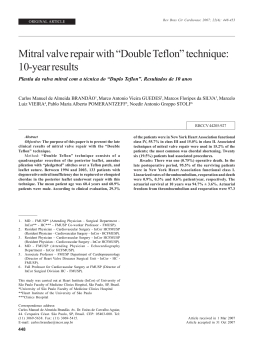
Double Orifice Mitral Valve in an
Asymptomatic Adult with an Unusual
Combination of Congenital Malformations:
A Case Report [16]
GONÇALO PROENÇA, ANTÓNIO FREITAS, SÉRGIO BAPTISTA, BOBAN THOMAS,
JOSÉ FRAGATA, RAFAEL FERREIRA
Serviço de Cardiologia, Hospital Fernando da Fonseca
Amadora, Portugal
Rev Port Cardiol 2004; 23 (2) : 233-236
ABSTRACT
We report a case of an asymptomatic adult
patient, with several congenital
malformations including an infrequent
variant of double orifice mitral valve, postductal aortic coarctation, bicuspid aortic
valve and an aneurysm of the right Valsalva
sinus. The loss of support of the right
coronary cusp of the aortic valve caused
major aortic regurgitation. With the
exception of the mitral valve, which was left
untouched because it was neither stenotic
nor regurgitant, all the other abnormalities
were successfully corrected, in a two-step
surgical approach.
Key words
Double orifice mitral valve; Aortic coarctation;
Bicuspid aortic valve; Aneurysm of the Valsalva sinus;
Aortic regurgitation
CASE REPORT
W
e present the case of a 32-year-old man
who was referred to our department due
to a systolic murmur detected on a routine medical examination. He was asymptomatic, performing heavy duties and practicing sports.
RESUMO
Duplo Orifício Valvular Mitral em
Adulto Assintomático com Combinação
Pouco Usual de Malformações
Congénitas: A Propósito de um Caso
Clínico
Os autores apresentam o caso clínico de um
adulto assintomático, referenciado para
esclarecimento de um sopro sistólico, em
que se diagnosticou um conjunto de
malformações congénitas incluindo duplo
orifício valvular mitral, coarctação da aorta,
bicuspidia aórtica e aneurisma do seio de
valsalva direito. Este último condicionava
ainda perda de suporte da cúspide coronária
direita e regurgitação aórtica major. Com
excepção da válvula mitral que, por não
apresentar alterações funcionais, não foi
corrigida, todas as outras anomalias foram
objecto de correcção cirúrgica, em duas
etapas.
Palavras-Chave
Cardiopatia congénita; Duplo orifício valvular mitral;
Coarctação da aorta; Bicuspidia aórtica;
Aneurisma de seio de Valsalva; Insuficiência aórtica
On physical examination his blood pressure
was 150/70 mmHg in the right arm and 110/70
mmHg in the left leg. The radial pulse was
regular, rhythmic, and had normal amplitude.
By contrast, the femoral arterial pulses were
difficult to detect and clearly delayed relative
to the radial pulse. Breath sounds were normal
Recebido para publicação: Julho de 2003 • Aceite para publicação: Dezembro de 2003
Received for publication: July 2003 • Accepted for publication: December 2003
233
but on cardiac auscultation an S4 was heard at
the apex as well as a systolic and protodiastolic grade II/VI aortic murmur. The upper and
lower extremities were equally developed and
no abnormal collateral circulation was seen on
the chest wall.
The ECG showed AQRS left axis deviation
with left anterior fascicular block. On the chest
X-ray there was extensive bilateral and
symmetrical rib notching, an increased cardiothoracic index, and the classic inverted 3 sign
(Fig. 1). The transthoracic and transesophageal
echocardiograms revealed a bicuspid aortic
valve, an outlet defect of the interventricular
septum (aneurysm of the right Valsalva sinus)
causing loss of support to the right coronary
cusp, and major aortic regurgitation. The left
ventricle was slightly enlarged. We could also
see a markedly dilated aortic root and supra-
aortic branches. In suprasternal view the descending thoracic aorta seemed to be cut off.
Surprisingly, there were two normally functioning mitral valve orifices (Figs. 2, 3, 4),
roughly the same size, with a central fibrous
subdivision. No regurgitation or stenosis was
detected. There was neither atrial nor ventricular septal defect. Magnetic resonance imaging
confirmed extensive aortic coarctation (Fig. 5).
Finally, prior to surgery, cardiac catheterisation
confirmed the diagnosis, excluding coronary
artery malformation and demonstrating a transcoarctation gradient of 55 mmHg.
The patient successfully underwent surgical
correction of the aortic coarctation with a patch
of bovine pericardium. Four months later, he
started complaining of fatigue in his daily activities and the echocardiogram revealed further
left ventricular enlargement. The aortic valve
Fig. 1-A Enlarged left ventricle, frontal view and inverted 3 sign (arrow); B - Frontal view – zoom, with rib notching (arrow).
234
Fig. 2. Transesophageal transgastric short-axis view (A), at the level of the mitral valve showing two separate mitral valve orifices (asterisks). The posterior orifice is larger than the anterior. (B) Transgastric longitudinal view showing two mitral valve orifices (arrow).
LA = left atrium; LV = left ventricle.
Fig. 3 Transesophageal longitudinal two-chamber view in systole (A) and diastole (B) showing two separate mitral valve orifices, with
a central bridge that connects the two orifices. LA = left atrium; LV = left ventricle.
Fig. 4 Transesophageal aortic valve short-axis view demonstrating a bicuspid aortic valve with aneurysm of the anterior Valsalva sinus (arrow). Ao = aorta; RA = right atrium; LA = left
atrium; PA = pulmonary artery.
Fig. 5 Contrast-enhanced MRI showing post-ductal aortic
coarctation and numerous collaterals.
was then replaced by a St Jude 25 ® mechanical prosthesis and the Valsalva sinus aneurysm
was closed by suturing its isthmus. Three
months after the second operation, he had resumed his job and was again asymptomatic.
two major types of DOMV. The most common
is called double parachute or hole type MV
(85 % of cases), because there is a smaller accessory orifice at either the anteroseptal or
posteromedial commissure and a larger main
orifice, and both insert exclusively in one papillary muscle. It is usually associated with
other anomalies of the MV apparatus. Only
five (15 %) of these cases resembled our patient’s with two separate mitral orifices approximately equal in size, with separate leaflet
structures, due to the presence of a central fibrous subdivision (central type).
Frequently, this abnormality is associated
with other cardiac malformations such as ventricular septal defect, bicuspid aortic valve,
coarctation of the aorta and, most commonly,
atrio-ventricular septal defects (2, 3). The morphology as well as the presence of significant
DISCUSSION
Double orifice mitral valve (DOMV) is a
rare congenital malformation, first described
by Greenfield in 1876. It is characterized by a
mitral valve (MV) with a single fibrous annulus
and two orifices opening into the left ventricle
(LV). Subvalvular structures, especially the
tensor apparatus, may show varying degrees of
abnormalities.
In the largest published series, Baño-Rodrigo and co-workers reported on twenty-seven
post-mortem pediatric cases (1). They described
235
regurgitation or stenosis can usually be clearly
delineated by transthoracic or transesophageal
echocardiography (4). However, diagnosis is not
always straightforward. In a series of 549 patients who had surgical repair of atrioventricular septal defects (21 patients had associated
DOMV), a high percentage of missed diagnosis
on preoperative transthoracic echocardiography
was reported, stressing the need for careful observation in parasternal short axis view. In this
report, DOMV was the most frequently missed
lesion (14 of 21 cases) (5).
REFERENCES
1. Baño-Rodrigo A, van Praagh S, Growitzsch E, van Praagh
R. Double orifice mitral valve: a study of 27 postmortem cases with developmental, diagnostic and surgical considerations. Am J Cardiol 1988;61:152-60.
2. Purnode P, Rombaut E, Alkhori M, Marchandise B. Double orifice mitral valve with flail leaflet: a transesophageal
echocardiographic examination. Eur J Echocardiography
2000;1:144-16.
3. Sousa R, Correia D. Válvula mitral com duplo orifício. A
propósito de um caso clínico. Rev Port Cardiol 1995;14(3):
233-8.
4. Solorio S, Badui E, Yanez M, Enciso R, Rodriguez L,
Quintero LR. Double mitral valve orifice. Two dimensional
and Doppler echocardiographic diagnoses. Arch Med Res
1996;27(4):491-4 (Abs.)
5. Sittiwangkul R, Ma R., McCrindle B, Coles J. Smallhorn
J. Echocardiographic assessment of obstructive lesions in
atrioventricular septal defects. JACC 2001;38:253-61.
Address for reprints:
Pedidos de separatas para:
GONÇALO MIRANDA PROENÇA
Hospital Fernando da Fonseca
Serviço de Cardiologia (Piso 4 – Torre Amadora)
IC 19
2720-276 AMADORA, PORTUGAL
e-mail: [email protected]
Fax: 21-4348466
236
Download