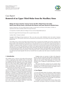
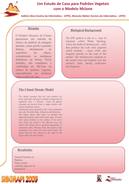
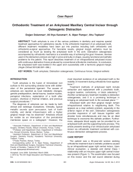
RSBO Revista Sul-Brasileira de Odontologia
ISSN: 1806-7727
[email protected]
Universidade da Região de Joinville
Brasil
Maris LOSSO, Estela; PIZZATTO, Eduardo; Miranda ULBRICH, Lucienne
Complex odontoma associated to a primary maxillary canine: case report
RSBO Revista Sul-Brasileira de Odontologia, vol. 6, núm. 2, 2009, pp. 204-207
Universidade da Região de Joinville
Joinville, Brasil
Available in: http://www.redalyc.org/articulo.oa?id=153013734013
How to cite
Complete issue
More information about this article
Journal's homepage in redalyc.org
Scientific Information System
Network of Scientific Journals from Latin America, the Caribbean, Spain and Portugal
Non-profit academic project, developed under the open access initiative
ISSN:
Versão impressa: 1806-7727
Versão eletrônica: 1984-5685
Case Report Article
Artigo de Caso Clínico
Complex odontoma associated to a primary
maxillary canine: case report
Odontoma complexo associado ao canino
decíduo superior: relato de caso
Estela Maris LOSSO*
Eduardo PIZZATTO**
Lucienne Miranda ULBRICH***
Address for correspondence:
Endereço para correspondência:
Estela Maris Losso
Universidade Positivo – Mestrado Profissional em Odontologia Clínica
Rua Professor Pedro Viriato Parigot de Souza, 5.300 – Campo Comprido
CEP 81280-330 – Curitiba – PR
E-mail: [email protected]
* PhD, Professor of Pediatric Dentistry – Positivo University (UP-PR).
** PhD, Professor of Collective Health – Positivo University (UP-PR).
*** MSc, Professor of Dental Surgery – Positivo University (UP-PR).
Received on October 1st, 2008. Accepted on November 3, 2008.
Recebido em 1.º/10/08. Aceito em 3/11/08.
Keywords:
odontoma; impacted
tooth; child.
Abstract
Introduction: Odontomas are malformations of the dental tissues and
may interfere with the eruption of the associated tooth. The early
diagnosis, followed by a proper treatment at the right time, will result
in a favorable prognosis and a desirable occlusion development.
Complex odontomas associated to primary teeth are rare. Case report
and conclusion: This article describes a case of a complex odontoma
in a four-year-old girl that prevented eruption of the left primary canine.
The treatment choice was enucleation of the odontoma and the
maintenance of the left primary canine. In this case, complete removal
of the complex odontoma was successfully conducted, since after one
year of follow-up the primary maxillary canine restarted its eruption
process.
RSBO v. 6, n. 2, 2009 –
Palavras-chave:
odontoma; dente
impactado; criança.
205
Resumo
Introdução: Odontomas são malformações dos tecidos dentais e
podem interferir no processo eruptivo de um dente associado a eles.
O diagnóstico precoce, seguido de um tratamento adequado e feito no
momento certo, resultará em um prognóstico favorável e em um melhor
desenvolvimento da oclusão. Odontomas complexos associados a
dentes decíduos são raros. Relato do caso e conclusão: Este artigo
descreve um caso de odontoma complexo em uma criança de 4 anos
de idade, o qual impediu a erupção do canino decíduo superior
esquerdo. O tratamento deu-se pela remoção cirúrgica do odontoma
complexo e pela manutenção do referido dente. Nesse caso a completa
remoção do odontoma foi considerada um sucesso, uma vez que após
um ano de acompanhamento o canino decíduo superior esquerdo
irrompeu na cavidade bucal.
Introduction
An odontoma is a benign tumor of mixed tissue origin
that is located within the facial bones. It consists of
masses of enamel, dentin, cementum and pulpal tissues.
Radiographically, the lesion presents a wellcircumscribed radiolucent image and generally shows
a radiopaque border at its periphery. Compound
odontomas have a similarity to normal teeth, while
complex odontomas are irregular masses. Both
odontoma occur predominantly in the second decade
of life [5].
The treatment choice for compound odontomas
associated to the primary dentition is described in the
literature as the surgical removal of the lesion and the
maintenance of the arch space until the eruption of
the deciduous tooth. This process is estimated to take
from 3 months to 2 years. Afterward, the patients
could be submitted to the surgical exposure of the
deciduous tooth and orthodontic treatment, when
needed [2, 7, 8].
There are only a few case reports of complex
odontomas associated to the primary dentition in
children below five years old [4, 8]. However, there is no
case report of spontaneous eruption of the primary tooth
after the surgical removal of the complex odontoma.
border of the canine (figure 1B), thus preventing its
eruption process. The other teeth were present and
showed normal development. Based on the clinical and
radiographic evaluation, the diagnosis of a complex
odontoma associated with this tooth was established.
Case report
A four-year-old white girl was referred to the
pediatric dentist’s office due to the delayed eruption of
the maxillary left primary canine. Extra-oral
examination showed no asymmetry. Intra-oral
examination revealed normal colored mucosa,
increased in volume of the alveolar ridge and absence
of the canine (figure 1A). No other abnormalities were
found in this examination. The radiograph exam
exhibited an irregular radiopaque mass at the incisal
Figure 1 — Clinical evaluation before surgery: (1A)
Absence of right primary canine with volume increase
of the alveolar ridge; (1B) Radiographic exam showing
an irregular radiopaque mass associated to the incisal
border of the right primary canine
Losso et al.
206 – Complex odontoma associated to a primary maxillary canine: case report
The patient underwent enucleation of the lesion under general anesthesia (figure 2A and 2B). Since the
impacted primary canine was clearly separated from the capsule of the lesion, after the entire lesion had
been removed it was decided to keep the primary canine in place and wait its eruption (figure 2C and 2D).
During the enucleation of the odontoma, the mobility of the canine revealed no ankylosis of this tooth.
Figure 2 — Surgical procedure: (2A) View of left canine primary region; (2B) View of the complex odontoma before
its removal; (2C) View of surgical area after complex odontoma removal; (2D) Surgically removed odontoma
macroscopically shows irregular hard tissues
Clinical follow-up was conducted monthly. Four months after surgery, the radiographic examination
showed a reduction of the alveolar ridge, although there was no eruption of the canine (figure 3A). After one
year of follow-up, the tooth restarted its eruption process (figure 3B and 3C) without any complication.
Figure 3 — Clinical follow-up: (3A) 4 months, (3B and 3C) 1 year after the surgery
Discussion and conclusion
A complex odontoma associated to the maxillary left second molar that delayed its eruption process
in a three-year-old child was reported by Motokawa et al. [4]. Eight months after the surgical removal
of the lesion, surgical exposure of the tooth and orthodontic treatment were administered.
RSBO v. 6, n. 2, 2009 –
Since the canine of this case report was not
inside the odontoma, special care was taken while
performing minimum osteotomy in order to remove
the lesion and keep enough amount of bone to allow
the eruption process of the retained tooth. The child
had Baume’s arch type I and had no loss of space.
Other authors described spontaneous eruption
of primary teeth associated to compound
odontomas after the enucleation of the lesion [1, 3,
6, 9]. The time ranged from 3 weeks to 2 years.
There was no description of spontaneous eruption
of primary teeth associated to complex odontoma
in the literature reviewed.
Early diagnosis of odontomas is important for
preventing craniofacial and tooth developmental
problems. The early diagnosis accompanied by a
proper treatment at the right time will result in a
favorable prognosis. In order to diagnose
developmental abnormalities as soon as possible,
a professional team of pediatric dentists should be
aware of the importance of clinical and radiographic
examinations.
In the present case, there was no need for a
second surgery or an orthodontic intervention, so
the clinician could preserve the deciduous tooth,
avoiding the use of dental appliance as well as
aesthetic and emotional problems due to the early
loss of deciduous tooth. Thus, this appears to be a
suitable treatment strategy for complex odontomas
in primary dentition.
References
1. Bacetti T. Interceptive approach to tooth eruption
abnormalities: 10-year follow-up of a case. J Clin
Pediatr Dent. 1995;19(4):297-300.
207
2. Chen YK, Lin LM, Huang HC, Lin CC, Yan YH. A
retrospective study of oral and maxillofacial biopsy
lesions in a pediatric population from southern
Taiwan. Pediatric Dent. 1998;20(7):404-10.
3. Levine N, Stoneman DW. Compound odontoma
associated with a primary dentition. Ont Dent.
1977;54(5):12-4.
4. Motokawa W, Breaham RL, Morris ME, Tanaka
M. Surgical exposure and orthodontic alignment of
an unerupted primary maxillary second molar
impacted by an odontoma and a dentigerous cyst: a
case report. Quintessence Int. 1990;21(2):159-62.
5. Neville BW, Damm DD, Allen CM, Bouquot JE.
Oral & maxillofacial pathology. 2ª ed. Rio de
Janeiro: Guanabara Koogan; 2004. 605 p.
6. Noonan RG. A compound odontoma associated
with a deciduous tooth. J Oral Surg.
1971;32(5):740-2.
7. Sato M, Tanaka N, Sato T, Amagasa T. Oral
and maxillofacial tumours in children: a review.
Br J Oral Maxillofacial Surg. 1997;35(2):92-5.
8. Sheehy EC, Odeel EW, Al-Jaddir G. Odontomas
in primary dentition: literature review and case
report. J Dent Child. 2004;71(1):73-6.
9. Yassim OM. Delayed eruption of maxillary primary
cuspid associated with compound odontoma. J Clin
Pediatr Dent. 1999;23(2):147-9.
Download