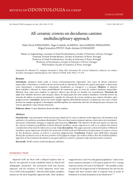
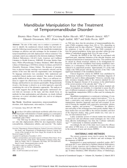
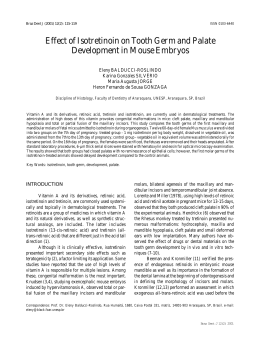
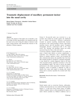
Braz Dent J (2009) 20(1): 84-86
84
F.R. Victorino et al.
ISSN 0103-6440
Bilateral Mandibular Canines with Two Roots
and Two Separate Canals - Case Report
Fausto Rodrigo VICTORINO
Ricardo Affonso BERNARDES
Jarcio Victorio BALDI
Ivaldo Gomes de MORAES
Norberti BERNARDINELLI
Roberto Brandão GARCIA
Clovis Monteiro BRAMANTE
Department of Restorative Dentistry, Endodontics and Dental Materials,
Bauru Dental School, University of São Paulo, Bauru, SP, Brazil
The mandibular canine is usually considered a single-rooted tooth with a single root canal. However, two canals and more rarely two
roots may also occur. This paper reports the case of a patient with bilateral mandibular canines with two roots and two root canals.
The initial periapical radiographs of the mandibular right and left canines for endodontic treatment revealed the presence of two roots
in each tooth. After coronal opening, the cervical third was prepared with a SX file of the ProTaper® system and root canal length was
confirmed using Root ZX electronic apex locator. Root canal preparation was completed with the series of ProTaper® instruments and
the root canal was filled with gutta-percha and an epoxy resin-based endodontic sealer according to Tagger’s hybrid technique. The
final radiographs showed two well-obturated canals ending at the electronically located apexes. The 6-month posttreatment follow-up
showed apparent clinical and radiographic success. Clinicians should always consider the presence of anatomical variations in the teeth
during endodontic treatments. Despite the low prevalence, variations may occur in the number of roots and root canals of mandibular
canines, as demonstrated in this case report.
Key Words: mandibular canine, internal anatomy, root canal.
INTRODUCTION
Knowledge of root canal anatomy is mandatory
for the success of endodontic treatment. In most cases,
the mandibular canines present only one root (1-5).
The occurrence of two roots and even more two root
canals is rare, ranging from 1% (4) to 5% (5). Despite
the low prevalence, clinicians should consider the possible variations in the number of roots and root canals
of mandibular canines. This paper reports the case of a
patient with bilateral mandibular canines with two roots
and two root canals.
CASE REPORT
A healthy 40-year-old Caucasian female was
referred to the Clinic of Endodontics of Bauru Dental
School, University of São Paulo for endodontic treatment of the mandibular canines and a mandibular lateral
incisor for prosthetic rehabilitation. Initial periapical
radiographic examination revealed the presence of two
roots on both mandibular canines (Fig. 1).
Endodontic treatment was performed in a single
session because the teeth presented pulp vitality on
clinical examination. After local anesthesia bilaterally, a
rubber dam was placed and endodontic access was performed with a #1014 round diamond bur (KG Sorensen,
São Paulo, SP, Brazil) and an Endo Z tapered safe-end
bur (Dentsply/Maillefer, Ballaigues, Switzerland). Location and negotiation of root canals were done with
a size 10 K-file (Maillefer, Ballaigues, Switzerland).
The cervical and middle thirds were prepared with a
Correspondence: Dr. Fausto Rodrigo Victorino, Rua Nossa Senhora do Rocio, 981, 86990-000 Marialva, PR. Tel: +55-44-3015-4534. e-mail: [email protected]
Braz Dent J 20(1) 2009
1136.indd 84
26/4/2009 18:51:10
Canines with two roots
SX file of the ProTaper® system (Dentsply/Maillefer).
Root canal length was determined radiographically and
confirmed with an electronic apex locator (Root ZX; J
Morita Co, Kyoto, Japan). Chemomechanical preparation was performed with hand Flexofile files (Maillefer)
and irrigation with 1% sodium hypochlorite at each
change of file. The instrumented root canals were filled
with gutta-percha cones and an epoxy resin-based root
canal sealer (Sealer 26; Dentsply/Maillefer) according
to Tagger’s hybrid technique (Figs. 2a and 2b). The
final radiographs showed two well-obturated canals
ending at the electronically located apexes.The 6-month
posttreatment follow-up showed apparent clinical and
radiographic success.
DISCUSSION
Diagnosis and identification of the number of
roots and root canals are key factors for endodontic
treatment. The initial radiograph is extremely important
because it allows for the identification or suspicion of
root and root canal anatomical variations. Bifurcations
in the cervical and middle thirds may be observed ra-
Figure 1. Initial radiograph.
85
diographically when the x-ray incidence angle does not
cause superimposition of images. In mandibular canines,
bifurcation at these sites has been shown to occur in
43.1% of the situations (6). In the present case, identification of the second root was evident, especially due
to absence of some teeth. However, it does not always
occur. Identification of the second root is even more
difficult in the presence of tooth crowding. Therefore,
the radiographic image should be carefully analyzed in
order to interpret and identify details that may suggest
the presence of bifurcations or trifurcations, such as
sudden root canal discontinuity (7).
Endodontists should always search for two
canals in mandibular canines during endodontic treatment, even in single-rooted teeth. Green (2) observed
two canals in a single root in 13 out of 100 mandibular
canines examined. This is consistent with the findings
of Hess (8), who observed two canals in 15% of the
cases. Vertucci (9) reported the presence of two canals
in 18% of the mandibular canines. Mandibular canines
with two separated canals also demonstrated in a recent
case report (10).
However, the presence of two roots in mandibular
canines is rarely observed (11,12). Quellet (5) described
the occurrence of two roots and two canals in mandibular
canines in only 5% of all analyzed teeth. Laurichesse et
al. (4) described the second root of mandibular canines
in only 1% of cases. A previous study that investigated
the internal anatomy, direction and number of roots and
Figure 2. Final radiographs. A: Mandibular right canine; B:
Mandibular left canine.
Braz Dent J 20(1) 2009
1136.indd 85
26/4/2009 18:51:11
86
F.R. Victorino et al.
size of 830 extracted human mandibular canines found
only 1.7% of the teeth with two roots and separate two
canals (13). D´Arcangelo et al. (14) reported two cases
of endodontic treatment of mandibular canines with two
roots. A case report of two roots in mandibular canine
was also presented by Heling et al. (15), yet the tooth
exhibited three root canals. To the best of our knowledge,
the occurrence of bilateral mandibular canines with two
roots and two separate canals, as described in the present
case, has not yet be published.
Even though the most common anatomy of mandibular canines comprises a single root and a single root
canal, clinicians should consider the possible variations
and always search for the second root canal in teeth with
either one or two roots.
RESUMO
O canino inferior é tido como um dente unirradiculado e com
apenas um canal. Todavia, pode ocorrer o aparecimento de dois
canais e, mais raramente, duas raízes. Este artigo apresenta o
caso clínico de uma paciente com ambos os caninos inferiores
apresentando duas raízes e dois canais radiculares. A radiografia
inicial periapical dos caninos mandibulares inferiores direito e
esquerdo para tratamento endodôntico revelou a presença de
duas raízes em cada um deles. Uma vez realizada a abertura
coronária, o preparo do terço cervical foi feito com a lima SX do
sistema ProTaper® e a odontometria foi determinada e confirmada
eletronicamente com o localizador apical eletrônico Root ZX.
O preparo foi finalizado com a série de instrumentos do sistema
ProTaper® e a obturação foi realizada com cones de guta-percha
e cimento obturador resinoso sob a técnica Híbrida de Tagger.
As radiografias finais mostraram dois canais bem obturados
terminando nos limites apicais estabelecidos pelos localizadores
apicais eletrônicos. O controle de 6 meses mostrou sucesso clínico
e radiográfico do tratamento endodôntico. Os clínicos devem
sempre considerar a presença de variações anatômicas em todo
tratamento endodôntico. A despeito da baixa prevalência, pode
haver variações no número de raízes e canais de caninos inferiores,
como demonstrado neste caso clínico.
REFERENCES
1. Pineda F, Kuttler Y. Mesiodistal and buccolingual. Roentgenographic investigation of 7275 root canals. Oral Surg, Oral Med
and Oral Pathol 1972;33:101-110.
2. Green D. Double canal in single roots. Oral Surg, Oral Med and
Oral Pathol 1973;35:689-696.
3. Vertucci FJ. Root canal anatomy of the human permanent teeth.
Oral Surg, Oral Med and Oral Pathol 1984;58:589-599.
4. Laurichesse JM, Maestroni J, Breillat J. Endodontie Clinique. 1st
ed. Paris, France: CdP;1986:64-66.
5. Ouellet R. Mandibular permanent cuspids with two roots. J Can
Dent Assoc 1995;61:159-61.
6. Sharma R, Pécora JD, Lumley PJ, Walmsley AD. The external and
internal anatomy of human mandibular canine teeth with two roots.
Endod Dent Traumatol 1998;14:88-92.
7. Ingle JI, Walton RE, Malamed SF, Coil JM, Khademi JA, Kahn
FH et al. Preparation for endodontic treatment. In: Endodontics.
Ingle JI, Bakland LK (Editors). 5th ed. Hamilton: BC Decker Inc.,
2002:357-404.
8. Hess W. The anatomy of the root canals of teeth of the permanent
dentition. New York: Williams Wood Co.; 1925.
9. Vertucci FJ. Root canal anatomy of the mandibular anterior teeth.
J Am Dent Assoc 1974;89:369-371.
10. Ghoddusi J, Zarei M, Vatanpour M. Mandibular canine with two
separated canals. N Y State Dent J 2007;73:52-53.
11. Green D. Morphology of the pulp cavity of the permanent teeth.
Oral Surg 1955;8:743-759.
12. Kutler Y. Anatomia topografica de la cavidad pulpar. In: Endodoncia Prática. Kutler Y. Mexico: Alpha; 1961, p. 29.
13. Pécora JD, Sousa Neto MD, Saquy PC. Internal anatomy, direction
and number of roots and size of human mandibular canines. Braz
Dent J 1993;4:53-57.
14. D’Arcangelo C, Varvara G, De Fazio P. Root canal treatment in
mandibular canines with two roots: a report of two cases. Int End
J 2001;34:331-334.
15. Heling I, Gottlieb-Dadon I, Chandler NP. Mandibular canine with
two roots and three root canals. Endod Dent Traumatol 1995;11:
301-302.
Accepted February 4, 2009
Braz Dent J 20(1) 2009
1136.indd 86
26/4/2009 18:51:11
Download