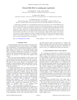
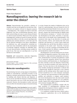
This Accepted Author Manuscript is copyrighted and published by Elsevier. It is posted here by
agreement between Elsevier and University of Brasilia. Changes resulting from the publishing
process - such as editing, corrections, structural formatting, and other quality control
mechanisms - may not be reflected in this version of the text. The definitive version of the text
was subsequently published in [Journal of Magnetism and Magnetic Materials, Volumes 272–
276, Part 3, May 2004, Pages 2400–2401, doi:10.1016/j.jmmm.2003.12.998].You may download,
copy and otherwise use the AAM for non-commercial purposes provided that your license is
limited by the following restrictions:
(1) You may use this AAM for non-commercial purposes only under the terms of the CC-BY-NCND license.
(2) The integrity of the work and identification of the author, copyright owner, and publisher
must be preserved in any copy.
(3) You must attribute this AAM in the following format: [agreed attribution language, including
link to CC BY-NC-ND license + Digital Object Identifier link to the published journal article on
Elsevier’s ScienceDirect® platform].
________________________________________________________________________
Este Manuscrito do Autor Aceito para Publicação (AAM) é protegido por direitos autorais e
publicado pela Elsevier. Ele esta disponível neste Repositório, por acordo entre a Elsevier e a
Universidade de Brasília. As alterações decorrentes do processo de publicação - como a edição,
correção, formatação estrutural, e outros mecanismos de controle de qualidade - não estão
refletidas nesta versão do texto. A versão definitiva do texto foi posteriormente publicado em
[Journal of Magnetism and Magnetic Materials, Volumes 272–276, Parte 3, Maio de 2004,
Paginas 2400–2401, doi:10.1016/j.jmmm.2003.12.998]. Você pode baixar, copiar e utilizar de
outra forma o AAM para fins não comerciais , desde que sua licença seja limitada pelas
seguintes restrições:
(1) Você pode usar este AAM para fins não comerciais apenas sob os termos da licença CC- BYNC-ND.
(2) A integridade do trabalho e identificação do autor, detentor dos direitos autorais e editor
deve ser preservado em qualquer cópia.
(3) Tem de atribuir este AAM no seguinte formato: [acordo na linguagem atribuída, incluindo o
link para CC BY-NC-ND licença Digital + DOI do artigo publicado na revista Elsevier ScienceDirect
® da plataforma].
Magnetic resonance investigation of magnetic-labeled baker's yeast cells
J.P.M. Godoy Morais
R.B. Azevedo
L.P. Silva
Z.G.M. Lacava
S.N. Báo
O. Silva
F. Pelegrini
C. Gansau
N. Buske
I. Safarik
M. Safarikova
P.C. Moraise
Abstract
In this study, the interaction of DMSA-coated magnetite nanoparticles (5 and 10 nm core-size)
with Saccharomyces cerevisae was investigated using magnetic resonance (MR) and
transmission electron microscopy (TEM). The TEM micrographs revealed magnetite
nanoparticles attached externally to the cell wall. The MR data support the strong interaction
among the nanoparticles supported by the cells. A remarkable shift in the resonance field was
used as signature of particle attachment to the cell wall.
PACS
75.50.Mm; 76.50+g; 87.68.+z
Keywords: Magnetic fluid; Magnetic resonance; Transmission electron microscopy; Yeast cell
The use of nanosized magnetic materials in the development of field-gradient
magnetic separation (FGMS) technologies has attracted much attention in recent years.
Wastewater containing processed textile dyes, for instance, is an increasing source of
environmental
contamination.
Recent
experiments
showed
that
the
yeast
cells
(Saccharomyces cerevisiae) efficiently interact with magnetic nanoparticles dispersed as
magnetic fluids (MFs), leading to the formation of magnetic-labeled cells which could be easily
separated from the system using appropriated magnetic separators [1]. Also, yeast cells are
good candidates to promote removal and degradation of dyes from wastewater [2]. In this
study, transmission electron microscopy (TEM) and magnetic resonance (MR) were used to
investigate the interaction of S. cerevisiae cells with magnetic nanoparticles (MNPs) after
incubation with magnetite-based, DMSA-coated MF samples (DMSA-MF).
Baker's yeast (S. cerevisiae) was obtained locally. Magnetite-based (5 and 10 nm
average diameter), DMSA-coated biocompatible MF samples were prepared as described in
the literature [3]. Compressed baker's yeast (2 g) was suspended in saline (6 mL), centrifuged
and resuspended in 6 mL 0.1 M PBS (pH 7.4). After centrifugation the pellet was once again
resuspended in PBS to obtain a 33% yeast suspension (v/v; yeast cells volume determined after
sedimentation for 24 h at 1 g). Three mL of the yeast suspension was added to 1 mL of the
DMSA-MF sample containing 1.2×10¹⁶ particle/cm3. Two distinct DMSA-MF samples (5 and 10
nm average diameter MNP's) were used in the experiments. The suspension was mixed and
then incubated at room temperature for 1 h without mixing. After this time period most of the
yeast cells were magnetically modified by the added DMSA-MF (the cells responded to
external magnetic field gradients). Nonmagnetic yeast cells and residual MF were removed by
repeated static magnetic separation using PBS and saline as washing liquids. After treatment
cells were harvested, fixed, and processed for TEM analysis. Modified cells were also used for
MR measurments. The TEM analysis (see Fig. 1) revealed that the 5 nm-size, DMSA-coated
magnetite nanoparticles, was externally attached to the S. cerevisiae cell wall. Similar result
was observed using the 10 nm-size, magnetite-based DMSA-MF sample. Incubation for 4 h
instead of 1 h leads to similar observations.
Fig. 1. TEM picture of S. cerevisiae incubated with 5 nm DMSA-MF. Observe MNP's attached to the cell
wall (arrows). No particles were found inside the cell.
MR experiments have been successfully used to investigate biodistribution of MNPs
introduced in living beings [4]. Fig. 2 shows the MR spectra of the two modified cells (open
circles for the 5 nm and full circles for the 10 nm MNPs) plus the spectra of two 10 nm-size
(dashed and solid lines), magnetite-based DMSA-MF samples containing 4.8×10¹⁶ (1:1) and
4.8×10¹³ particle/cm3 (1:1000). The huge dilution (from 1:1 to 1:1000) of the 10 nm-size MF
sample causes a relative shift in the resonance field of about 73 G. However, the shift observed
in the resonance field between the concentrated 10 nm-size MF sample (1:1) and the modified
cells using the 10 nm-size, magnetite-based DMSA-MF sample, was about 250 G. This
remarkable resonance field shift is the signature of a strong interaction among the MNP's
when attached to the S. cerevisiae cell wall. The MR spectrum of the modified cells using the 5
nm-core, magnetite-based DMSA-MF sample, is also shown in Fig. 2 (open circles).
Fig. 2. Magnetic resonance spectra of the 5 (open circles) and 10 nm (full circles) nanoparticles in
labeled cells. Solid and dashed lines represent the spectra of the 10 nm magnetite suspended as a stable
magnetic fluid samples at different concentrations (see text).
In conclusion, 5 and 10 nm-sized magnetite particles, DMSA-coated, do attach to the S.
cerevisiae cell wall after incubation of the yeast cells with the corresponding biocompatible
magnetic fluid samples. Magnetic resonance data confirm that both nanoparticles are firmly
attached to the cells as revealed by the TEM micrographs.
The authors acknowledge the financial support of the Brazilian agencies CNPq and FINATEC.
References
[1] I. Safarik, L. Ptackova, M. Safarikova, Eur. Cells Mater. 3 (S2) (2002) 52.
[2] M.A.M. Martins, M.H. Cardoso, M.J. Queiroz, M.T. Ramalho, A.M.O. Campos, Chemosphere
38 (1999) 2455.
[3] T. Goetze, C. Gansau, N. Buske, M. Roeder, P. Gornert, .M. Bahr, J. Magn. Magn. Mater. 252
(2002) 399.
[4] L.M. Lacava, Z.G.M. Lacava, M.F. Da Silva, O. Silva, S.B. Chaves, R.B. Azevedo, F. Pelegrini, C.
Gansau, N. Buske, D. Sabolovic, P.C. Morais, Biophys. J. 80 (2001) 2483.
Download