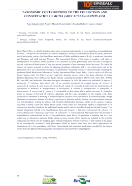
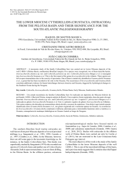
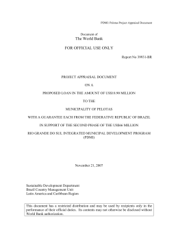
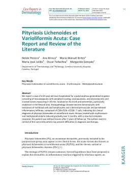
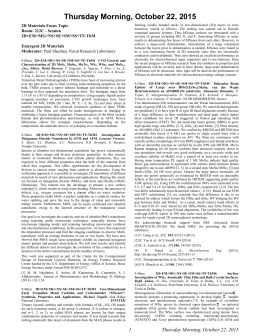
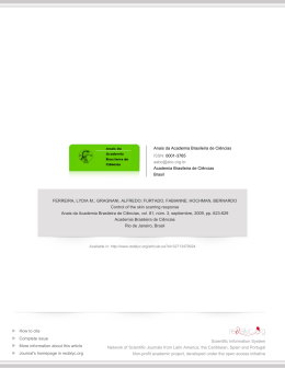
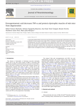
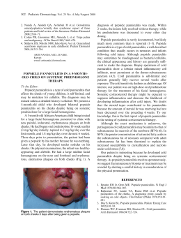
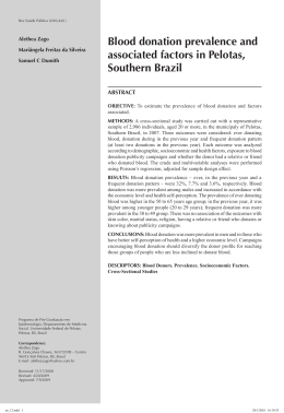
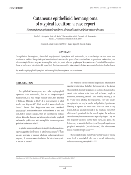
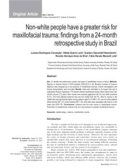
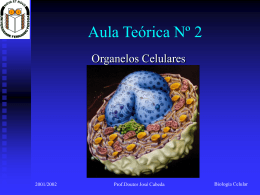
art 927
DERMATOPATHOLOGY
117
▲
Light and transmission electron microscopy of generalized
dystrophic epidermolysis bullosa (Pasini's albopapuloid
subtype) *
Microscopia óptica e eletrônica de transmissão da epidermólise bolhosa
distrófica generalizada (subtipo albo-papulóide de Pasini)
Hiram Larangeira de Almeida Jr1
Nara Moreira Rocha3
Lísia Nudelmann2
Luis Antonio Suita de Castro4
Abstract: Pasini's albopapuloid epidermolysis bullosa is a very rare subtype of generalized dystrophic
dominant epidermolyis bullosa. A 30 year-old white female patient presented since her childhood disseminated small blisters and papules. Light microscopy of a blister showed dermal-epidermal cleavage;
moreover, focal areas of dermal-epidermal splitting were also observed. Transmission electron microscopy also identified focal areas of cleavage, which were seen below the lamina densa. It is important to
recognize this condition as a variant of epidermolysis bullosa, since the most important cutaneous findings are generalized papules and not blisters and erosions as in other forms of epidermolysis bullosa.
Keywords: Epidermolysis bullosa dystrophica; Epidermolysis bullosa, junctional; Epidermolysis bullosa
simplex; Histology; Microscopy, electron, transmission
Resumo: A epidermólise bolhosa albo-papulóide de Pasini é uma variante rara da forma generalizada
de epidermólise bolhosa distrófica dominante. Uma paciente de 30 anos apresenta desde a infância
pápulas e bolhas disseminadas. A microscopia óptica de uma bolha demonstrou clivagem dermo-epidérmica; além disso áreas focais de desprendimento dermo-epidérmico foram encontradas. A microscopia eletrônica de transmissão identificou a clivagem abaixo da lâmina densa. É importante que se
reconheça essa variante de epidermólise bolhosa, já que o aspecto clínico predominante são pápulas
disseminadas e não bolhas como nas outras formas de epidermólise bolhosa.
Palavras-chave: Epidermólise bolhosa distrófica; Epidermólise bolhosa juncional; Epidermólise bolhosa
simples; Histologia; Microscopia eletrônica de transmissão
INTRODUCTION
Pasini’s albopapuloid epidermolysis bullosa
(PAP-EB), described in 1928, is a very rare subtype of
generalized dystrophic dominant epidermolyis bullosa (DDEB). 1-3 This group of EB has a cleavage under
the lamina densa, due to dysfunction of the anchoring filaments. Very few cases were reported in the literature. 4
CASE REPORT
A 30 year-old white female patient was examined in our outpatient clinic. She informed that since
her childhood small blisters and papules appear on
her skin. Some blisters are induced by trauma while
others are spontaneous. Her older sister has similar
cutaneous changes.
Skin examination showed disseminated whitish
Received on 01.12.2010.
Approved by the Advisory Board and accepted for publication on 01.02.2011.
* Study carried out at the Catholic University of Pelotas (Universidade Católica de Pelotas - UCPEL), Federal University of Pelotas (Universidade Federal de Pelotas
- UFPEL) and Dermatology Service of the Mercy Hospital of Porto Alegre (Santa Casa De Misericórdia de Porto Alegre) – Porto Alegre (RS), Brazil.
Conflict of interest: None / Conflito de interesse: Nenhum
Financial funding: None / Suporte financeiro: Nenhum
1
2
3
4
Lecturer – Assistant Professor, Federal University of Pelotas (Universidade Federal de Pelotas - UFPEL) – Pelotas (RS), Brazil.
Dermatologist – enrolled in the Master's degree program, Catholic University of Rio Grande do Sul (Pontifícia Universidade Católica do Rio Grande do Sul PUC-RS) – Porto Alegre (RS), Brazil.
Biologist – Laboratory of Electronic Microscopy – Brazilian Agricultural Research Corporation, Center for Technical Production – Temperate Climate
(EMBRAPA CPT - CT) – Pelotas (RS), Brazil.
Master's degree - Laboratory of Electronic Microscopy – Brazilian Agricultural Research Corporation, Center for Technical Production – Temperate Climate
(EMBRAPA CPT - CT) – Pelotas (RS), Brazil.
©2012 by Anais Brasileiros de Dermatologia
An Bras Dermatol. 2012;87(1):115-8.
118
Almeida Jr HL, Nudelmann L, Rocha NM, Castro LAS
papules, which measured a few millimeters. Parallel to
these papules a few erosions with hemorrhagic crusts
were also observed (Figures 1A and 1B). She presented normal nails, teeth and hairs.
Under light microscopy a dermal-epidermal
cleavage was easily identified (Figure 2A). With higher
magnification it could be seen that the whitish
papules corresponded to superficial dermal elevations, which had higher cellularity and sparse collagen, not arranged in bundles, when compared to the
adjacent dermis (Figures 2B and 2C). Scattered in the
specimen focal areas of dermal-epidermal splitting
were also observed (Figures 3A and 3B). Staining for
elastic fibers revealed their absence in these dermal
elevations.
Transmission electron microscopy also identified focal areas of cleavage, which were seen below
the lamina densa (Figure 4A and B). The dermis
beneath the cleavage was formed by loose collagen
fibers, which were not forming bundles and by an
amorphous material (Figure 4C).
A
B
C
FIGURE 2: Light microscopy: a. dermal-epidermal cleavage (HE x
100). b. detail of the papule with high cellularity and collagen
fibers that do not form bundles (HE x400). c. normal adjacent
dermis with collagen bundles
A
DISCUSSION
DDEB is caused by mutations in the COL7A1
gene leading to dysfunction of the anchoring filaments, differently from the recessive group, in which
the absence of collagen VII leads to severe clinical features. 5-7 Pasini’s albopapuloid subtype belongs to the
Generalized Dystrophic subgroup, and is characterized by disseminated whitish papules associated with
blisters and erosions, as seen in this case. 8 Nail and
mucosal involvement is possible, not found in this
patient.
B
A
FIGURE 1: Whitish papules and hemorrhagic crusts in the
interscapular (a) and periumbilical region (b)
An Bras Dermatol. 2012;87(1):115-8.
B
FIGURE 3: Light microscopy: a. and b. focal dermal-epidermal
cleavage (HE x 400)
Light and transmission electron microscopy of generalized dystrophic epidermolysis bullosa (Pasini's albopapuloid subtype)
A
119
C
B
FIGURE 4: Transmission electron microscopy: a. and b. dermal cleavage (asterisks) under the lamina densa (KF – keratin filaments)
(x 12.000) c. isolated collagen fibers and deposition of amorphous material in the albopapuloid lesion(x 12.000)
With light microscopy we could demonstrate
that the whitish papules were formed by immature
collagen with a higher number of fibroblasts, when
compared to the adjacent area, according to a previous report. 9 Moreover, many focal areas of dermal-epidermal cleavage could be seen.
Transmission electron microscopy also showed
some areas of focal cleavage, which occurred under
the lamina densa. Similarly to what was seen with
light microscopy, the dermal component of the papule
is formed by isolated collagen fibers. In addition, an
amorphous material was also observed, probably a
secondary dermal proliferative phenomenon, as
described in another case of PAP-EB. 9
Although this condition has been reclassified as
generalized DDEB, it is important to recognize it as a
variant of EB, since the most important cutaneous
findings are generalized papules and not blisters and
erosions as in others forms of EB. 8 q
REFERENCES
1.
2.
3.
4.
5.
6.
7.
Spinelli LP, Orofino RR, Kac BK, Sodré C, Azulay RD. Epidermólise bolhosa albopapulóide (variante de Pasini). An Bras Dermatol 2003;78:459- 63.
Alonso AM, Marinho DEA, Leal MPL. Epidermólise bolhosa albopapulóide (variante de Pasini). Med Cut ILA. 1981;9:307-10.
Shenefelt PD, Castellano LM. Successful treatment of albopapuloid epidermolysis
bullosa (Pasini`s variant) with pulse topical corticosteroid therapy. J Am Acad
Dermatol. 1993;29:785-6.
Horn HM, Tidman MJ. The clinical spectrum of dystrophic epidermolysis bullosa.
Br J Dermatol. 2002;146:267-74.
Kon A, Nomura K, Pulkkinen L, Sawamura D, Hashimoto I, Uitto J. Novel glycine
substitution mutations in COL7A1 reveal that the Pasini and Cockayne-Touraine
variants of dominant dystrophic epidermolysis bullosa are allelic. J Invest
Dermatol. 1997;109:684-7.
Jonkman MF, Moreno G, Rouan F, Oranje AP, Pulkkinem L, Uitto J. Dominant
Dystrophic Epidermolysis Bullosa (Pasini) caused by a novel substation mutation
in the type VII collagen gene (COL7A1). J Invest Dermatol. 1999;112:815- 7.
Mellerio JE, Salas-Alanis JC, Talamantes ML, Horn H, Tidman MJ, Ashton GH, et
al. A recurrent glycine substation mutation, G2043R, in the type VII collagen gene
(COL7a1) in dominant dystrophic epidermolysis bullosa. Br J Dermatol.
1998;139:730-7.
8.
9.
Fine JD, Eady RA, Bauer EA, Bauer JW, Bruckner-Tuderman L, Heagerty A, et al.
The classification of inherited epidermolysis bullosa (EB): Report of third international consensus meeting on diagnosis and classification of EB. J Am Acad
Dermatol. 2008;58:931- 50.
Nomura K, Umeki K, Sawamura D, Hashimoto I. Dominant dystrophic epidermolysis bullosa albopapuloidea Pasini -- ultrastructural observations of albopapuloid
lesions and a type VII collagen DNA polymorphism study of a family. Acta Derm
Venereol. 1997;77:277-80.
MAILING ADDRESS / ENDEREÇO PARA CORRESPONDÊNCIA:
Dr. Hiram Larangeira de Almeida Jr.
Rua Barroso 1202
96010-280 Pelotas, RS
Phone: +55 53 3278 7582 Fax: +55 53 3278 3909
E-mail: [email protected]
How to cite this article/Como citar este artigo: Almeida Jr HL, Nudelmann L, Rocha NM, Castro LAS. Light and
transmission electron microscopy of generalized dystrophic epidermolysis bullosa (Pasini's albopapuloid subtype). An Bras Dermatol. 2012;87(2):00-0.
An Bras Dermatol. 2012;87(1):115-8.
Baixar