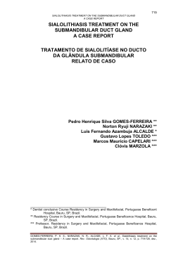
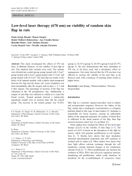
Original Article
Braz J Oral Sci.
October | December 2013 - Volume 12, Number 4
Non-white people have a greater risk for
maxillofacial trauma: findings from a 24-month
retrospective study in Brazil
Luciana Domingues Conceição1, Rafael Guerra Lund1, Gustavo Giacomelli Nascimento1,
Ricardo Henrique Alves da Silva2, Fábio Renato Manzolli Leite3
1
Department of Restorative Dentistry, Dental School, Federal University of Pelotas, Pelotas, RS, Brazil
2
3
Forensic Dentistry, Ribeirão Preto Dental School, University of São Paulo, Ribeirão Preto, SP, Brazil
Department of Semiology and Clinics, Dental School, Federal University of Pelotas, Pelotas, RS, Brazil
Abstract
Aim: To identify the predominant causes and types of maxillofacial trauma in Brazil. Methods:
Reports of corporal trauma (7,536) between 2009-2010 in the Brazilian Institute of Forensic
Medicine were analyzed as to the presence of maxillofacial traumas. Victims’ demographic and
trauma characteristics were recorded. Results: Data were submitted to chi-square test and to
multivariate Poisson regression. 778 reports referred maxillofacial trauma. Most victims were men
(50.8%) around 27.6 years. Main causes were physical aggression (88.1%) and traffic accidents
(6.7%). The most affected extraoral area was the middle third (60.7%). Risk for trauma in the
middle third was significantly higher among patients aged 61-75 (RR 1.32), and non-white patients
(black-skinned RR 1.21; brown-skinned RR 1.18); while falls were associated with trauma in the
lower third (RR1.79). Conclusions: Violence was the main cause of maxillofacial trauma.
Prevention of interpersonal violence may be a key element to prevent maxillofacial trauma.
Keywords: epidemiology, violence, maxillofacial injuries.
Introduction
Received for publication: August 30, 2013
Accepted: November 28, 2013
Correspondence to:
Fábio Renato Manzolli Leite
Faculdade de Odontologia,
Universidade Federal de Pelotas
Rua Gonçalves Chaves, 457, CEP: 96015-560
Centro, Pelotas, RS, Brasil
Phone +55 53 32256741
E-mail: [email protected]
The face is usually the first area to be damaged in case of physical aggression,
car accidents and falls, which makes the maxillofacial region very susceptible to
traumas due to its prominence1. Traumas of the maxillofacial complex represent
one of the most important health problems worldwide, especially because of the
high incidence and the diversity of facial lesions2. Moreover, the face represents
the center of human attention and sometimes lesions may leave marks or
unrepairable sequels that cause physical or psychological damages, burdening
the country economy3. Within the same country and among different countries
the type of maxillofacial trauma is influenced by socio-economic status, cultural
and environmental factors, and the period of investigation 4.
Brazil presents the world’s fifth largest geographical area and population.
Especially after the 2007-2008 crises, the country has strengthened its status as
an economic power, developing more employment opportunities, vehicle sales
and social mobility, which may influence public policies. However, the economic
growth is not being followed by reduction of social inequalities. Dark-skinned
Braz J Oral Sci. 12(4):313-318
314
Non-white people have a greater risk for maxillofacial trauma: findings from a 24-month retrospective study in Brazil
people are still the poorest, consequently, the ones who
concentrate more social and health problems 5. Moreover, as
they usually live far from the central urban areas, they are
more exposed to violent episodes 6.
There are many studies worldwide evaluating traumas in
the oral and maxillofacial region 1,3,7 and some of them
conducted in Brazil4,8. Literature has identified different causes
for traumas in developed and developing countries. There are
controversies regarding the association of traffic accident and
physical aggression with the country’s economic status 3,7,9.
Authors also observed the high prevalence of falls and sports
traumas in both developed and developing countries 10. In
Brazil, the conducted studies presented some limitations: (a)
evaluation of a specific population, like children 11 or rural
population; 8 (b) evaluation of fractures and the required
treatment only;12 (c) analysis of the dental traumas only13.
Although there are some minor studies on maxillofacial
trauma in some parts of Brazil, this is the first report analyzing
data of oral and maxillofacial traumas in the last decade.
The main purpose of this study is to evaluate the
epidemiological characteristics of prevalence, cause and
associated factors of maxillofacial traumas in Southern Brazil
in 2009 and 2010.
Material and methods
This retrospective and cross-sectional study was carried
out on the records of consecutive patients with maxillofacial
traumas who were referred to the Brazilian Institute of
Forensic Medicine, Pelotas, Southern Brazil, from January
2009 to December 2010 (n=7,536). The institute is a
reference for 11 cities with a total of 600,000 inhabitants.
From these records a selection was made according to the
following inclusion criteria: (1) offense to the integrity and/
or health of the victim and (2) presence of maxillofacial
traumas. Maxillofacial lesions were grouped in the following
extraoral regions: lower third (masseter, mandible and mentum
regions), middle third (infraorbital, zygomatic and nasal
regions) and oral (intraoral, lips and perioral soft tissues).
Oral lesions were defined as those involving the following
areas: (a) teeth and surrounding supportive tissues
(periodontium); (b) oral mucosa including gums, alveolar
mucosa in edentulous patient, palate and mucosa; (c) jaw
bones (upper and lower); (d) lips (mucosa and skin); (e)
tongue; (f) perioral soft tissues (extraoral tissues that surround
mouth and cover upper and lower jaw). This study followed
the Declaration of Helsinki on medical protocol and was
approved by the Institutional Review Board of the Federal
University of Pelotas, Dental School (protocol 88/2009).
The selected cases (n=648) were studied for data
regarding the victim’s and offender’s demographic
characteristics, nature and number of inflicted traumas and
their consequences. Personal information such as gender, race,
age and marital status, and lesion’s characteristics (location,
etiology, type) were recorded in an Excel spreadsheet. In
addition, anatomic location and nature of the trauma were
identified to evaluate oral traumas.
Braz J Oral Sci. 12(4):313-318
Data were double typed and analyzed by Stata 12.0 software
(StataCorp, College Station, TX, USA). Descriptive statistics
was performed using frequency analysis for categorical variables
and descriptive analysis for continuous variables. The statistical
significance of the difference in the prevalence of oral and
maxillofacial traumas according to gender, age group, skin color
and cause of trauma was tested using the chi-square test.
Multivariate Poisson regression analysis was conducted by using
traumas in the different parts of face as the dichotomized
dependent variable in order to test the association between the
outcomes and the independent variables, adjusting it for potential
confounders. For variable selection, the stepwise method with
backward selection was used. Variables with p<0.25 were
included in the final model, estimated their Risk Ratio (RR)
and set the interval confidence at 95% .
Results
In this study, out of a total of 7,536 victims only 892
(11.8%) presented maxillofacial traumas. Patients with missing
data were excluded from the study, totalizing 648 (8.6%)
patients presenting 785 traumas. The number of cases was
similar in 2009 (n=306; 47.2%) and 2010 (n=342; 52.8%).
The majority were men (50.1%), single (75.2%), most of them
white (80.6%). The mean age was 27.6 years (SD=7.37), and
victims aged between 16 to 30 years were the most affected
(46.1%), followed by those from 31 to 45 years (24.0%).
The specialized police station for women’s defense (209;
31.9%) referred most of the patients followed by the specialized
police station for children and adolescent defense (124; 19.1%)
and first assistance police station (120; 18.1%). Most of the
maxillofacial traumas were due to physical aggressions (563;
86.8%), traffic accidents (47; 7.2%) and falls (32; 4.9%). About
the damage caused by lesions, 22 patients (3.3%) presented
permanent and irreversible consequences, becoming unable
for daily, social and working activities.
Traumas occurred on all regions of the face in different
proportions, with the middle third concentrating most of the
traumas (475, 73.3%), followed by the lower third (170, 26.2%)
and by the oral region (140, 21.6%). Table 1 describes the
associations between the traumas and independent variables,
which are listed according to the different regions of face.
Traumas occurred in single thirds or more than one third (Figure
1). For dental traumas, tooth fracture was the most prevalent
(12, 33.4%) followed by luxation (8, 22.2%) With respect to
the intraoral soft tissue lesions, oral and gingival mucosa and
tongue were the most affected sites (Table 2).
When the associations between the occurrence of oral
and maxillofacial trauma and explanatory variables were
considered simultaneously, in the multivariate regression
model, (Tables 3 and 4), the risk for trauma in the middle
third was significantly higher among patients aged between
61-75 years (RR, 1.32; 95%CI, 1.07-1.62), and non-white
patients (brown skinned, RR 1.21, 95% CI, 1.09-1.34; black
skinned RR 1.18; 95%CI, 1.01-1.38). The risk for lower third
trauma was significantly higher when falls were the main
causes of trauma (RR 1.79; 95%CI, 1.18-2.70).
Non-white people have a greater risk for maxillofacial trauma: findings from a 24-month retrospective study in Brazil
315
Table 1. Lesions distribution in the different regions of the face according to gender,
age, skin color and cause of trauma (n=785)
Gender
Male
Female
Age
0-15
16-30
31-45
46-60
61-75
>75
Skin color
White
Brown
Black
Cause of trauma
Physical Agression
Trafic Accident
Firearm
Fall
Middle third (%)
p value
0.71
236 (72.4)
239 (74.2)
0.44
74 (68.5)
225 (74.8)
110 (70.8)
46 (74.2)
16 (88.9)
4 (80.0)
0.005
369 (70.5)
29 (82.8)
77 (85.6)
0.85
416 (73.9)
33 (70.2)
4 (66.7)
22 (68.7)
Lower third (%)
p value
0.54
90 (27.3)
80 (25.1)
0.48
28 (25.9)
79 (26.7)
47 (30.3)
13 (20.1)
2 (11.1)
1 (20.0)
0.08
147 (28.1)
7 (20.0)
16 (17.8)
<0.05
139 (24.7)
16 (34.0)
1 (16.7)
14 (43.8)
Oral region (%)
p value
0.30
77 (23.3)
63 (19.8)
0.94
24 (22.2)
66 (22.1)
33 (21.9)
14 (22.6)
2 (11.1)
1 (20.0)
0.48
117 (22.3)
8 (22.9)
15 (16.7)
0.31
122 (21.6)
10 (21.3)
3 (50.0)
5 (15.6)
Fig. 1. Isolated and combined maxillofacial lesions distribution and number of victims.
Discussion
Information on health is important for planning,
monitoring and management of collective and individual
health interventions. In the last years, traumas to the
maxillofacial region are becoming more common both in
the urban and rural areas. 1 Changes in the global socioeconomic scenery are responsible for switches in the pattern
of maxillofacial traumas etiologies.
Brazil has emerged as an economic power in the last
years, resulting in higher employment and immigration rates,
but on the other side growth was accompanied by social
disparities. In this way, more traumas due to physical
aggression are expected. This study shows for the first time
that Brazilian growth is reflecting in increased reports of
trauma due to interpersonal violence. In the present study,
the main cause for traumas in all three analyzed regions was
physical aggression, followed by car accident and falls, as
Braz J Oral Sci. 12(4):313-318
316
Non-white people have a greater risk for maxillofacial trauma: findings from a 24-month retrospective study in Brazil
Table 2. Distribution of intraoral trauma (n = 36)
n
Soft tissue lesions
Tongue
Buccal mucosa
Gingival mucosa
Mouthfloor mucosa
Palate
Dental tissue lesions
Tooth Fracture
Tooth Subluxation
Tooth Luxation
Tooth Avulsion
Total
% of intraoral traumas
% of total traumas
3
2
4
0
0
8.3
5.6
11.1
0
0
0.5
0.3
0.6
0
0
12
0
8
7
36
33.4
0
22.2
19.4
100
1.9
0
1.2
1.1
5.6
Table 3. Poisson regression crude (b) and adjusted (a) analyzes for
occurrence of oral and maxillofacial trauma in the middle third of face.
Pelotas, Brazil (n=75)
Independent Variables
Gender
Female
Male
Age (years)
0-15
16-30
31-45
46-60
61-75
>75
Skin Color
White
Brown
Black
Cause of trauma
Physical Agression
Trafic Accident
Firearm
Fall
RRb (CI 95%)
RRa (CI 95%)
0.021
0.24
1.09
1.03
1.08
1.29
1.16
p-value
-
1.0
0.97 (0.88-1.07)
1.0
(0.94-1.26)
(0.88-1.21)
(0.89-1.31)
(1.05-1.59)
(0.73-1.84)
1.10
1.03
1.10
1.32
1.15
1.0
(0.95-1.27)
(0.87-1.21)
(0.90-1.33)
(1.07-1.62)
(0.74-1.78)
<0.001
<0.001
1.0
1.21 (1.09-1.34)
1.18 (1.01-1.38)
1.0
1.21 (1.09-1.34)
1.17 (1.01-1.37)
0.41
1.0
0.95 (0.78-1.15)
0.90 (0.51-1.59)
0.93 (0.73-1.18)
seen in many urban centers in Germany14 and the United States15.
The explanation for the increase if interpersonal violence
is higher alcohol consumption, drug abuse and social
disparities due to unequal wealth distribution 1,16,17. It was
found that skin color, a marker of social inequality, 5,6
represents a risk factor for facial traumas, since black and
brown victims tended to have more lesions in the middle
third of the face. It is important to emphasize that no
previously published paper has reported a social marker as a
risk factor for oral and maxillofacial traumas. According to
Minayo18 (1990), non-white people are the most vulnerable
to violence in urban areas with low quality of life, since
they live along with violence on a daily basis. As seen in
other reports, non-white people at greatest risk of being
victims of violence are men, young, single and belonging to
low-income families. 6 On the other hand, the stiffening of
road traffic laws and safety norms such as obligatory use of
Braz J Oral Sci. 12(4):313-318
p-value
0.61
-
seat belts, air bags, helmet wearing for motorized two-wheelers
and speed surveillance reduced maxillofacial traumas due
to traffic accidents 16,17,19.
According to the age groups, maxillofacial traumas were
more frequent in people between 16 and 30 years followed
by ages between 31 to 45 years which concurs with previous
studies1,17,20-22. People in these age groups have more social
interaction than other age groups, with higher alcohol and
other drugs consumption1,20. Despite of it, an increase of oral
and maxillofacial traumas in the elderly is being observed.
According to Al-Khateeb and Abdullah 9 (2007) this fact is
due to an increase in average life expectancy, a more active
lifestyle and higher percentage of elderly people in the
population. The main cause of traumas in the elderly
population was due to falls, which has been related to reduced
physical agility, presence of systemic pathologies and use
of psychotropic medications. Our data corroborate other studies
Non-white people have a greater risk for maxillofacial trauma: findings from a 24-month retrospective study in Brazil
317
Table 4. Poisson regression crude (b) and adjusted (a) analyzes for
occurrence of oral and maxillofacial trauma in the lower third of face.
Pelotas, Brazil (n=170)
Independent Variables
Gender
Female
Male
Age (years)
0-15
16-30
31-45
46-60
61-75
>75
Skin Color
White
Brown
Black
Cause of trauma
Physical Agression
Trafic Accident
Firearm
Fall
RRb (CI 95%)
p-value
0.54
RRa (CI 95%)
1.0
1.08 (0.83-1.40)
p-value
0.38
1.02
1.16
0.80
0.42
0.77
1.0
(0.70-1.48)
(0.78-1.74)
(0.45-1.44)
(0.11-1.64)
(0.13-4.58)
-
0.16
1.0
0.63 (0.39-1.00)
0.71 (0.36-1.40)
0.07
1.0
0.61 (0.38-0.98)
0.72 (0.36-1.42)
1.0
1.37 (0.90-2.10)
0.67 (0.11-4.06)
1.77 (1.16-2.70)
0.02
1.0
1.39 (0.91-2.12)
0.66 (0.11-3.94)
1.79 (1.18-2.70)
where the middle third facial area is more affected in elderly
victims, with special regards to the orbital-zygomatic region23,24.
Another finding was a greater risk for trauma on the lower
third associated with falls after adjustment in the final regression
model. This fact may be explained by the chin prominence
trauma when the victims fall. In addition, Iida et al.7,14 (2001,
2003) reported that fall is usually observed as a chin impact
leading to condyle fracture, and in less cases, multiple fractures
when the impact occurs in the lateral sides of mandible.
In contrast to other studies that reported mandible as the
most commonly affected site,22,25,26 in the results of this study
middle third was more affected (73.3%) than the lower third
(26.2%), which agrees with the studies conducted by Gandhi
et al1 (2011). Among the lower third maxillofacial lesions,
dentoalveolar traumas presented a low prevalence (3.6%)
concurring with previous studies 27-30. In this report, crown
fracture was the most common (1.9%) followed by luxation
(1.2%). As expected, most of these lesions correlate with lowimpact traumas due to interpersonal violence that are usually
observed as soft tissue abrasion, hematoma, and dentoalveolar
fractures 8. It is supposed that the importance, number and
severity of the perioral and intraoral lesions would change
with the presence of a forensic dentist at the Institutes of
Forensic Medicine and their prevalence would increase.
An example of specific professional care that has
increased the number of notified lesions was the creation of
specialized police stations for women’s defense in Brazil.
These units stimulated the notification of aggression against
women and reduced the male-to-female ratio of reported
traumas to 1.03:1. Some countries have ratios of up to 8:1,
but recent studies show a trend towards an equal male-tofemale ratio 17,22 . The increase in the number of women
0.02
presenting maxillofacial traumas was attributed to an increase
in the women’s working force and many of them working
outdoors in more high-risk occupations, thus becoming more
exposed to traumas9.
In the last decade, changes in global economy reflected
in different aspects of the worldwide development. New
economies are emerging with consequences to their
population. Specific preventive public policy must respect
the differences of each country, especially in countries with
different social and economic realities aggravated due to
social inequalities. As seen in many countries, there is a
worldwide trend of decreasing traffic-related traumas and
increasing violence-related traumas. Thus, appropriate
strategies at both community and individual levels should
be implemented to prevent and reduce overall trauma.
References
1.
2.
3.
4.
5.
Gandhi S, Ranganathan LK, Solanki M, Mathew GC, Singh I, Bither S.
Pattern of maxillofacial fractures at a tertiary hospital in northern India: a 4year retrospective study of 718 patients. Dent Traumatol. 2011; 27: 257-62.
Haug RH, Prather J, Indresano AT. An epidemiologic survey of facial fractures
and concomitant injuries. J Oral Maxillofac Surg. 1990; 48: 926-32.
Kostakis G, Stathopoulos P, Dais P, Gkinis G, Igoumenakis D, Mezitis
M, et al. An epidemiologic analysis of 1,142 maxillofacial fractures and
concomitant injuries. Oral Surg Oral Med Oral Pathol Oral Radiol. 2012;
114(Suppl 5): 69-73.
Brasileiro BF, Passeri LA. Epidemiological analysis of maxillofacial fractures
in Brazil: a 5-year prospective study. Oral Surg Oral Med Oral Pathol Oral
Radiol Endod. 2006; 102: 28-34.
Silva A, Faleiros HH, Shimizu WAL, Nogueira L de M, Nhãn LL, Silva,
BMF da. et al. [The prevalence of falls and associated factors among the
elderly according to ethnicity]. Cien Saude Colet. 2012; 17: 2181-90.
Braz J Oral Sci. 12(4):313-318
318
6.
7.
8.
9.
10.
11.
12.
13.
14.
15.
16.
17.
18.
19.
20.
21.
22.
23.
24.
25.
26.
27.
Non-white people have a greater risk for maxillofacial trauma: findings from a 24-month retrospective study in Brazil
Soares Filho AM. Homicide victimization according to racial characteristics
in Brazil. Rev Saude Publica. 2011; 45: 745-455.
Iida S, Kogo M, Sugiura T, Mima T, Matsuya T. Retrospective analysis of
1,502 patients with facial fractures. Int J Oral Maxillofac Surg. 2001; 30:
286-90.
Batista AM, Marques LS, Batista AE, Falci SG, Ramos-Jorge ML. Urbanrural differences in oral and maxillofacial trauma. Braz Oral Res. 2012; 26:
132-8.
Al-Khateeb T, Abdullah FM. Craniomaxillofacial injuries in the United Arab
Emirates: a retrospective study. J Oral Maxillofac Surg. 2007; 65: 1094101.
Van den Bergh B, Karagozoglu KH, Heymans MW, Forouzanfar T.
Aetiology and incidence of maxillofacial trauma in Amsterdam: a
retrospective analysis of 579 patients. J Craniomaxillofac Surg. 2012; 40:
e165-9.
Munante-Cardenas JL, Olate S, Asprino L, de Albergaria Barbosa JR, de
Moraes M, Moreira RW. Pattern and treatment of facial trauma in pediatric
and adolescent patients. J Craniofac Surg. 2011; 22: 1251-5.
Martini MZ, Takahashi A, de Oliveira Neto HG, de Carvalho Junior JP,
Curcio R, Shinohara EH. Epidemiology of mandibular fractures treated in
a Brazilian level I trauma public hospital in the city of São Paulo, Brazil.
Braz Dent J. 2006; 17: 243-8.
Gulinelli JL, Saito CTMH, Garcia-Júnior IR, Panzarini SR, Poi WR,
Sonoda CK, et al. Occurrence of tooth injuries in patients treated in hospital
environment in the region of Aracatuba, Brazil during a 6-year period. Dent
Traumatol. 2008; 24: 640-4.
lida S, Hassfeld S, Reuther T, Schweigert Hans-Gert, Haag C, Klein J,
et al. Maxillofacial fractures resulting from falls. J Craniomaxillofac Surg.
2003; 31: 278-83.
Laski R, Ziccardi VB, Broder HL, Janal M. Facial trauma: a recurrent
disease? The potential role of disease prevention. J Oral Maxillofac Surg.
2004; 62: 685-8.
Bacchieri G, Barros AJ. Traffic accidents in Brazil from 1998 to 2010:
many changes and few effects. Rev Saude Publica. 2011; 45: 949-63.
Van Beek GJ, Merkx CA. Changes in the pattern of fractures of the
maxillofacial skeleton. Int J Oral Maxillofac Surg. 1999; 28: 424-8.
Minayo MCS. Violence in adolescence: a public health problem. Cad
Saude Publica. 1990; 6: 278-92.
De Matos FP, Arnez MF, Sverzut CE, Trivellato AE. A retrospective
study of mandibular fracture in a 40-month period. Int J Oral Maxillofac
Surg. 2010; 39: 10-5.
Lee JH, Cho BK, Park WJ. A 4-year retrospective study of facial fractures
on Jeju, Korea. J Craniomaxillofac Surg. 2010; 38: 192-6.
Cheema SA, Amin F. Incidence and causes of maxillofacial skeletal
injuries at the Mayo Hospital in Lahore, Pakistan. Br J Oral Maxillofac
Surg. 2006; 44: 232-4.
Bakardjiev A, Pechalova P. Maxillofacial fractures in Southern Bulgaria a retrospective study of 1,706 cases. J Craniomaxillofac Surg. 2007; 35:
147-50.
Gerbino G, Roccia F, De Gioanni PP, Berrone S. Maxillofacial trauma in
the elderly. J Oral Maxillofac Surg. 1999; 57: 777-82; discussion 82-3.
Falcone PA, Haedicke GJ, Brooks G, Sullivan PK. Maxillofacial fractures
in the elderly: a comparative study. Plast Reconstr Surg. 1990; 86: 443-8.
Kieser J, Stephenson S, Liston PN, Tong DC, Langley JD. Serious facial
fractures in New Zealand from 1979 to 1998. Int J Oral Maxillofac Surg.
2002; 31: 206-9.
Erol B, Tanrikulu R, Gorgun B. Maxillofacial fractures. Analysis of
demographic distribution and treatment in 2,901 patients (25-year experience).
J Craniomaxillofac Surg. 2004; 32: 308-13.
Motamedi MH, Sagafinia M, Famouri-Hosseinizadeh M. Oral and
maxillofacial injuries in civilians during training at military garrisons:
prevalence and causes. Oral Surg Oral Med Oral Pathol Oral Radiol.
2012; 114: 49-51.
Braz J Oral Sci. 12(4):313-318
28. Castro JC, Poi WR, Manfrin TM, Zina LG. Analysis of the crown fractures
and crown-root fractures due to dental trauma assisted by the Integrated
Clinic from 1992 to 2002. Dent Traumatol. 2005; 21: 121-6.
29. Santos SE, Marchiori EC, Soares AJ, Asprino L, de Souza Filho FJ, de
Moraes M, et al. A 9-year retrospective study of dental trauma in Piracicaba
and neighboring regions in the State of São Paulo, Brazil. J Oral Maxillofac
Surg. 2010; 68: 1826-32.
30. Caldas IM, Magalhães T, Afonso A, Matos E. The consequences of
orofacial trauma resulting from violence: a study in Porto Dent Traumatol.
2010; 26: 484-9.
Download