ISSUE 02 / 2009
A SINGHEALTH NEWSLETTER FOR GENERAL PRACTITIONER
EMPOWERMENT PROGRAMME (GPEP) MEMBERS
Visit our website at www.singhealth.com.sg
Hyperbaric Oxygen Therapy (HBOT) in Problem Wound
Management
Dr Kim Soo Joang, Principal Resident Physician, Hyperbaric and Diving Medicine Centre, Singapore General Hospital
Introduction
Hyperbaric Oxygen Therapy (HBOT) is not a new treatment modality and has
been around for more than half a century. In Singapore, the first hyperbaric
chamber can be traced back to the Singapore General Hospital, in the 70’s, where
it was used by the Orthopaedic Department. However, this was transferred to
the Singapore Navy where the bulk of hyperbaric medical work was carried out
by the Republic of Singapore Navy until recently. Whilst the application of HBOT
in the management of decompression illness is well established, its application
for other clinical conditions is less well known. Currently, there are 13 accepted
indications for Hyperbaric Oxygen Therapy supported by good evidence.
Hyperbaric and Diving Medicine Centre
Indications for Hyperbaric Oxygen Therapy
... Hyperbaric
Oxygen
Therapy can
be an effective
adjunctive
therapy for
problem wounds.
Approved uses as recommended by the Undersea and Hyperbaric Medical Society.
(Hyperbaric Oxygen 2003, Indications and Results. The Hyperbaric Oxygen Therapy
Committee Report)
1) Air or gas embolism
2) Carbon monoxide poisoning
3) 0 Clostridial myositis and myonecrosis (Gas gangrene)
4) 0 Crush injury, compartment syndrome and other acute ischaemias
5) 0 Decompression sickness
6) 0 Enhancement of healing in selected problem wounds
7) 0 Exceptional anaemia
8) 0 Intracranial abscess
9) 0 Necrotising soft tissue infections
10) 0Osteomyelitis (Refractory)
11) 0Delayed radiation injury (soft tissue and bony necrosis)
12) 0Skin grafts and flaps
13)0 Thermal burns
One of the most common indications in clinical practice is enhancement of wound
healing in selected problem wounds.
Effective problem wound management requires an integrated multidisciplinary team
approach. Hyperbaric Medicine is one important component of this team. When used
01
05
07
Hyperbaric Oxygen Therapy (HBOT)
in Problem Wound Management
Updates on Retinal Disease
The Management of Head and
Neck Cancers
Medical Update
Medical Update
Medical Update
13
14
15
New Brace Offers Better Treatment
Option For Sciolosis
General Practitioners Seminar On
Laser Vision Correction
Contact Lens Update For
Family Physicians
Service Packages & Updates
KKH Sleep Disorders Centre 1st in Asia
to be accredited by regional bodies
Continuing Medical Education
11
Service Packages & Updates
KKH Inpatient Screening Highlights
Poor Osteoporosis Awareness
16
Continuing Medical Education / Continuing Medical Education
1st SGH Obesity & Metabolic Unit Symposium
Hotline Numbers
01
Apr-Jun 2009
MICA(P) 094/01/2009
MEDIC AL UPDATE
appropriately, Hyperbaric Oxygen Therapy can be an effective adjunctive therapy for
problem wounds.
What is Hyperbaric Oxygen Therapy?
It is a form of therapy where patients breathe 100% oxygen intermittently at pressures
greater than sea level. In order to receive oxygen at these pressures, patients will need to
be pressurized in a specially designed and built vessel. These vessels can accommodate
one or more patients. The vessel is usually compressed with air and the patient breathes
100% oxygen via a transparent hood or mask. Some one-man chambers are compressed
with 100% oxygen. By breathing oxygen at high pressures, a large amount of oxygen is
carried dissolved in the plasma.
Topical Hyperbaric Oxygen Therapy where oxygen is delivered directly to a wound is not
considered Hyperbaric Oxygen Therapy.
How Does Hyperbaric Oxygen Help in Wound Healing?
Wounds without adequate tissue oxygen levels will not heal. Hyperbaric Oxygen Therapy
when used appropriately can reverse tissue hypoxia so that wound healing can proceed.
Hyperbaric oxygen therapy has been shown to have the following effects:
•
•
•
•
•
•
•
•
•
Stimulation of angiogenesis
Enhancement of fibroblast replication and collagen synthesis
Enhancement of epithelialization
Reduction of local oedema
Improved leukocyte killing
Direct toxic effects on anaerobic bacteria
Suppression of bacterial toxin production
Synergism with certain antibiotics
Prevention of leukocyte mediated post-ischaemic reperfusion injury
For Hyperbaric Oxygen Therapy to be effective there must be adequate blood flow to the
wound. Without adequate blood flow, the oxygen carried in the blood and plasma will
not be able to reach the site where it is needed most and in some cases revascularization
procedures may be required first.
Hyperbaric Oxygen Therapy
15 July 2008
17 July 2008
Who Can Benefit From Hyperbaric Oxygen Therapy?
Patients with the following types of wounds may benefit from Hyperbaric Oxygen Therapy:
1) Diabetic lower extremity wounds - failure to heal or improve with conventional
management.
2) Venous stasis ulcers – failure to heal or improve with adequate control of oedema.
3) Late radiation injury – wounds that fail to heal or improve with conventional
management.
02
18 Aug 2008
27 Aug 2008
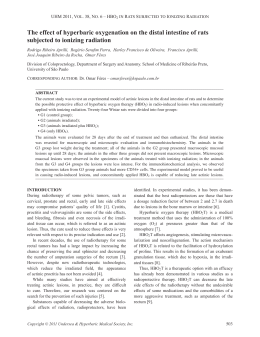
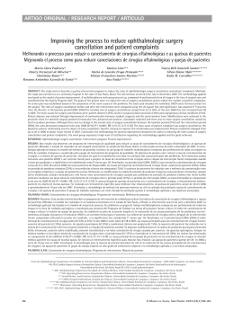
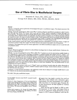
49 year old lady with a history of Diabetes Mellitus.
Presented with poor healing wound (Right big
toe). Received 30 sessions of HBOT.
MEDIC AL UPDATE
4) Arterial insufficiency ulcers – failure to heal or improve despite maximum
revascularisation.
5) Any wound that has failed to heal with conventional management where hypoxia is
a contributing factor.
Patients with problem wounds will undergo a non-invasive test called Transcutaneous
Oximetry. This test measures the transcutaneous oxygen tension at the level of the skin
and is an indirect measurement of microcirculatory blood flow. A baseline measurement
is taken on room air and another while breathing 100% oxygen at sea level.
Patients who have reversible periwound hypoxia are suitable candidates for HBOT. If
perfusion is too poor, referral to vascular team is warranted. Patients can be reassessed
after vascular intervention if any.
Before a patient is selected for Hyperbaric Oxygen Therapy, an assessment is made
to determine the patient’s fitness for exposure to high ambient pressure and high
oxygen concentration.
Malignant ulcers are generally not accepted for Hyperbaric Oxygen Therapy.
When skin grafts and flaps are used to cover problem wounds, Hyperbaric Oxygen
Therapy can be beneficial in support of compromised skin grafts and flaps.
What is Treatment Like?
Problem wounds usually require 20 to 30 treatment sessions or more depending on
response. Each treatment session lasts about 2 hours and the treatment pressure is
between 2 to 3 ATA (equivalent to 10 to 20 metres underwater).
The actual treatment can be divided into 3 phases – compression, maintenance of
pressure and decompression.
Compression
29 July 2008
01 Aug 2008
During this phase, the pressure in the chamber is increased slowly to the treatment
depth. There will be a sensation of fullness in the ears similar to that felt during take off
and landing in an airplane. Equalization techniques are taught. Patients will feel warm
during this phase.
Maintenance of pressure
Once the depth is reached, patients can relax and read a book, or watch a program on
the in chamber entertainment system, while breathing oxygen in a transparent hood
or mask.
17 Sept 2008
19 Nov 2008
Decompression
Once treatment is completed, patients will be decompressed back to sea level. Again,
there will be a sensation of fullness in the ears. It is a normal sensation which will resolve
spontaneously. Patients will feel cold during this phase.
03
MEDIC AL UPDATE
Contraindications
The only true absolute contraindication to HBOT is an
untreated pneumothorax. Once treated, the patient
can proceed with HBOT. Prior exposure to Bleomycin is
not compatible with HBOT.
Complications associated with HBOT
• Barotrauma
- Middle ear
- Tooth
- Sinus
- Pulmonary
• Oxygen toxicity
• Temporary worsening of myopia
Serious complications are extremely rare. The most
common complaint is ear pain which can easily be
managed with no serious consequences.
29 Aug 2008
01 Sept 2008
19 Sept 2008
29 Sept 2008
13 Oct 2008
05 Nov 2008
10 Sept 2008
03 Oct 2008
10 Nov 2008
19 Dec 2008
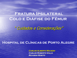
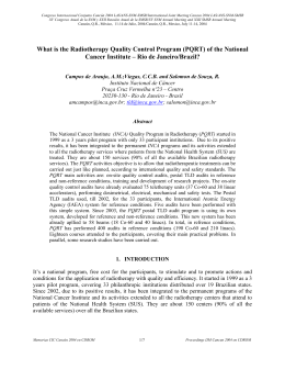
58 year old gentleman with a history of Diabetes Mellitus. Presented with poor healing wound (Left leg).
Received 30 sessions of HBOT.
For more information, please visit our website:
http://www.sgh.com.sg/MedicalSpecialtiesnServices/SpecialistCentres/HDM/
References
1) Hyperbaric Oxygen 2003. Indications and Results. The Hyperbaric Oxygen Therapy
Committee Report. Pg 41 – 55. Enhancement of Healing in selected problem wounds.
Robert A. Warriner III and Harriet W. Hopf.
2) Wound Care Practice. Paul J. Sheffield, Ph.D., Adrianne P.S. Smith, M.D., Caroline E. Fife, M.D.
Pg 661-684. Hyperbaric Oxygen Therapy Applications in Wound Care. Caroline E. Fife.
Problem wounds usually require
20 to 30 treatment sessions or
more depending on response. Each
treatment session lasts about 2
hours and the treatment pressure is
between 2 to 3 ATA (equivalent to
10 to 20 metres underwater).
04
MEDIC AL UPDATE
Updates on Retinal Disease
By Dr Chan Choi Mun, Associate Consultant, Vitreo-Retinal Service, Singapore National Eye Centre
In Ophthalmology, the subspecialty managing
vitreo-retinal diseases has classically been divided
into Surgical Retina and Medical Retina. Surgical
retinal cases include retinal detachments, trauma
and intraocular foreign bodies. The field of Medical
Retina is increasingly gaining importance as it
encompasses several significant eye diseases, all
of which can have a devastating impact on one’s
vision. Age-related macular degeneration, which
includes choroidal neovascularisation and polypoidal
choroidal vasculopathy, is one of these conditions.
Also falling within the purview of Medical Retina are
the conditions of diabetic retinopathy, retinal vein
occlusions, central serous retinopathy and hereditary
retinal diseases.
Age-related Macular Degeneration
Age-related macular degeneration (AMD) is the
leading cause of irreversible vision loss in the
industrialised world. With Singapore’s ageing
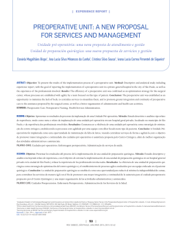
population, we are already noticing an increase in age- The colour fundus photograph shows an epiretinal membrane over the macula. The high resolution 3D
related macular degeneration. As its name implies, it Optical Coherence Tomograph (OCT) illustrates the pathology beautifully.
is a condition of older adults which affects the macula
- the centre of the retina, and the area responsible for clear
Management of AMD
central vision. In the wet, or exudative, form of AMD, pathological
The traditional treatment of AMD has been to employ thermal
choroidal neovascular membranes (CNV) develop under the
laser to destroy the CNV. However, laser photocoagulation
macula, leading to leakage and accumulation of fluid and blood
of juxta or subfoveal CNV, that is CNV which lies within 200
over the macula. Ultimately, a central disciform scar develops
micrometers of the fovea, induces an immediate iatrogenic
over the macula if AMD is left untreated. As such, a patient with
central scotoma. It was only recently that antiangiogenic agents,
AMD notices blurred central vision, disturbances of colour vision
injected directly into the vitreous humour of the eye, were
or metamorphopsia (where straight lines appear wavy). This
employed in the arsenal to treat AMD. The rationale for this is
translates to problems with recognising faces, reading, driving
that animal and clinical studies have establied vasular endothelial
and all activities requiring good central vision.
growth factor or VEGF as a key mediator in ocular angiogenesis.
Investigations
Before treatment can be instituted, specialised investigations
need to be carried out. These tests are employed not just
for AMD, but also for all retinal pathologies. Colour fundus
photography, optical coherence tomography (OCT), rapid
sequence fundal fluorescein angiography (FFA) and indocyanine
green angriography (ICG) are essential.
Latest Developments
SNEC now has the latest 3D OCT, which provides a highly
detailed three-dimensional view of the macula. There is also
the Heidelberg Reina Angiograph (HRA) confocal laser scanning
system which delivers crisp, minutely detailed, high-speed realtime digial angiographic images of the retinal vasculature. These
greatly contribute to accurate and precise diagnoses.
Anti-VEGF agents can cause regression of the abnormal blood
vessels and improvement of vision when injected directly into the
vitreous humor of the eye. Examples of these anti-VEGF agents
include Ranibizumab (Lucentis, Genentech) and Bevacizumab
(Avastin, Genentech), and they are usually injected on a monthly
basis for the initial 3 months after diagnosis. Studies have
demonstrated that treatment with these agents improves or
stabilises vision.
In some situations, photodynamic therapy (PDT) is used in
tandem with anti-VEGF agents. Also available at SNEC, PDT first
involves the intravenous injection of verteporfin before directing
a “cold laser”, which activates the verteporfin, at the affected
area. Verteporfin is a photosensitizing dye, which reacts with
water to create oxygen and hydroxyl free radicals. These radicals
05
MEDIC AL UPDATE
induce occlusion of the pathologic vasculature by means of
massive platelet activation and thrombosis while preserving
the normal choroidal vasculature and nonvascular tissue. The
advantage conferred by PDT is that it avoids creating a central
blinding scotoma when treating subfoveal CNV.
Diabetic Retinopathy
This is a condition very familiar to us. The hallmark of treatment
for severe non-proliferative of proliferative diabetic retinopathy
is panretinal photocoagulation laser treatment. That is
unchanged. However, there is now the additional option of
employing intravitreal injections of antiVEGF agents, similar to
that employed in the management of AMD, for the treatment
of vitreous haemorrhage secondary to proliferative diabetic
retinopathy or for persistent macula oedema which is resistant
to focal laser treatment.
Low Vision Clinic
Sometimes, in spite of the best treatment, or due to the inherent
nature of the eye disease, some patients end up with permanent
poor vision. These patients can be reviewed at the Low Vision
Clinic at SNEC. This is a special service where patients are
counseled on how to optimise their vision; are given certain
optical aids, such as magnifiers, to facilitate their reading vision;
and are refracted by an optometrist familiar with dealing with
low vision patients. Hope and optimism, which is so important
for patients with low vision, is upheld.
Central Retinal Vein Occlusion
Akin to a stroke in the eye, the ischaemic form or central
retinal vein occlusion (CRVO) can lead to a severe loss of
vision. While panretinal photocoagulation laser can prevent
neovascularisation within the eye, and its associated
complication of painful neovascular glaucoma, it does not help
reduce any incident macula oedema. Focal laser is the mainstay
of treatment. However in cases with persistent unresolving
macula oedema, anti-VEGF treatments are now being used at
SNEC with some success.
Hereditary Retinal Diseases
Although rare, when they do strike, these conditions can be
visually debilitating. They include retinitis pigmentosa, conerod dystrophies, retinal pigment epithelial dystrophies and the
hereditary vitreoretinal degenerations.
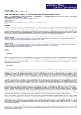
This frame of a high resolution Fundus Fluorescein Angiogram (FFA) shows clearly
the capillary non-fusion and dilated arterioles in a central retinal vein occlusion.
SNEC has both the diagnostic and therapeutic capabilities
to manage these patients well. These patients would require
specialised electrophysiological testing, which includes the
electroretinogram (ERG) and visual evoked potentials (VEP), to
clinch the diagnosis. They would also need Goldmann visual
fields to monitor their condition over the long term. These
diagnostic tests are all available at SNEC in a special laboratory.
These patients need to be followed up long term to exclude
associated conditions such as cataracts and glaucoma. We are
also performing ongoing research, in collaboration with the
Singapore Eye Research Institute, looking at genetic testing for
these patients.
This is a case of a bilateral macular retinal dystrophy. The autofluorescein fundal
photographs demonstrate that the patients maculae are in jeopardy with only an
island of viable nerve cells bilaterally.
06
MEDIC AL UPDATE
The Management of Head and Neck Cancers
By Dr Tan Hiang Khoon, Consultant, Surgical Oncology, National Cancer Centre Singapore
Head and neck cancers including both nasopharyngeal carcinoma
(NPC) and Head and Neck Squamous Cell Carcinoma (HNSCC) is
one of the most common cancers in Singapore with about 500
cases per year, of which nearly eighty percent are treated at the
National Cancer Centre Singapore (NCCS). Although the term
head and neck cancer comprises cancers of different etiology
and from different subsites, they do share a common anatomical
region, which is characterised by a plenitude of crucial structures
with vital physiological function (e.g. swallowing, breathing,
facial expression) packaged into a very small confined space
that is aesthetically important. This unique relation between
space, function and aesthetics accounts for the gravity of
symptomatology caused by the tumour, as well as, the possible
deleterious effects of the prescribed treatment. As such, the
management of Head and Neck Cancers demands careful
consideration of the extent of tumour involvement, the best
treatment option and its oncological outcome, possible functional
impairment and aesthetic effect. This complex task is best
undertaken by a team of experts comprising surgical oncologists,
medical oncologists, radiation oncologists, radiologists, plastic
surgeons, maxillofacial prosthodontists, dentists, physical
therapists, speech therapists, nurses, dietitians and social workers.
This multi-disciplinary approach underpins the management
philosophy of every head and neck cancer managed in NCCS.
What is New?
Radiotherapy
Improvement in computer technology and innovation has
contributed to many of the recent advances in the field of
radiotherapy. For instance, the availability of faster computers has
made intensity-modulated radiotherapy (IMRT) possible. With
multiple fields IMRT, radiation oncologists can now manipulate
radiation beams that can contour around the tumour providing
precise targeted therapy with minimal collateral damage to
important organs and tissues such as spinal cord and orbit.
At the NCCS, nasopharyngeal cancer (NPC) patients are treated
using IMRT as a standard radiotherapy treatment. In addition,
patients in the advanced stage and who are fit also have concurrent
chemotherapy to further improve disease control as has been
demonstrated in a randomised trial that we have recently
conducted and published in Journal of Clinical Oncology (fig. 1) [1].
We have also established ourselves as one of the main treatment
centres in the world contributing to good trial data on NPC.
Recently, image-guided radiotherapy (IGRT) has added an
additional dimension to the already impressive 3-D nature of
IMRT, that of the dimension of ‘time’. Before treatment, the exact
Figure 1
location of the tumour is ascertained by imaging (by X-rays or
with CT). Instead of only a single snap shot in time, with IGRT,
the patient is imaged continuously at all points in a complete
respiratory cycle so that the full extent of all body and organ
movements are captured for planning in the computer. Using
sophisticated techniques like respiratory gating and fiduciary
markers, these machines will shoot only when the tumour moves
into the parameter coordinates within the treatment field. This
allows consistent delivery of radiation to the targeted area taking
into consideration minute movement of tissue during respiratory
cycle. This enables the radiation oncologist to minimise toxicity,
and increase precision without sacrificing tumour control.
Looking into the future, NCCS is looking into building a proton
therapy facility. The use of particle therapy (in this case proton)
has the advantage that it has a very narrow Bragg peak, which
minimises damage to surrounding normal tissue and thus for the
reasons mentioned above, is particularly useful in the treatment
of head and neck cancer.
Molecular Targeted Therapy
The increased understanding of the molecular biology of Head
and Neck Cancer has led to efforts to develop compounds that
target these molecular pathways. The epidermal growth factor
receptor (EGFR) and its ligands {epidermal growth factor (EGF)
and tumour growth factor (TGF-α) are fundamental for cell
proliferation, motility, adhesion, invasion and angiogenesis.
07
MEDIC AL UPDATE
Interestingly, these receptors are over-expressed up to 90% of
HNSCC. One of the EGFR antibodies, cetuximab, has shown great
promise in the treatment of recurrent or metastatic HNSCC in
combination with cisplatin and 5-FU [2]. Equally, it has been shown
to increase overall survival of advanced HNSCC when used in
combination with radiotherapy compared to radiotherapy alone
Figure 2
(Fig 2) [3]. In other words, there is level I evidence that cetuximab
has the ability to potentiate the effect of both chemotherapy
and radiotherapy. Importantly, this was achieved without
significantly increasing the treatment toxicity. It will be therefore
be most interesting to see if EGFR antibody can enhance the
efficacy of concomitant chemoradiation. To specifically address
this question, NCCS has initiated an international multi-center
Phase III, double-blind, placebo-controlled trial to compare
post-operative adjuvant concurrent chemoradiotherapy with
or without nimotuzumab (a new generation EGFR receptor
antibody) for stage III/IV head and neck squamous cell cancer.
This NMRC sponsored trial has 22 participating centers from 12
different countries to accrue 710 patients and will interrogate the
role of EGFR antibody in the setting of post-operative adjuvant
therapy. We envisage that the trial will open towards the end of
2009. The successful execution of a trial of this magnitude will
further cement NCCS role as an important trial centre for head
and neck cancer globally.
Imaging – The Evolving Role of PET
PET imaging exploits the glucose metabolic pathway, through
the use of the most commonly used PET radio-tracer [F-18]
08
fluoro-deoxyglucose (FDG). Various molecular derangements
in malignant cells, including increased glycolytic rates and
upregulated glucose transporters, result in increased cellular
uptake of FDG. A FDG-PET scan can detect and localise such
abnormal concentrations of FDG.
In NCCS, we perform many oncologic PET/CT imaging which
combines PET with CT within a single scanner. PET/CT has been
used to 1) attain diagnosis, 2) evaluate staging, 3) assess
response to chemotherapy/radiotherapy in head and neck
cancer and 4) detect disease recurrence. In terms of attaining
diagnosis, PET/CT is particularly useful in the clinical setting
of cervical lymph node metastases with ‘unknown’ primary.
This functional imaging technique can detect small volume
or submucosal lesions that may be missed in pan-endoscopy
and structural imaging technique such as CT and MRI. To
accurately determine the tumour staging is important not only
to prognosticate but also helps decide on the type of treatment
prescribed. As a whole body imaging technique, PET/CT is
invaluable in the detection of distant metastases. It is also very
useful to help determine the nature of cervical lymph nodes that
are of borderline significance by size criteria in structural imaging.
In the setting of post-chemo/radiotherapy, PET/CT is normally
performed 8-12 weeks after the completion of treatment and
has been shown to have very high negative predictive value. In
other words, if PET/CT post-treatment showed no SUV uptake
in the primary tumour site or cervical lymph nodes, the patient
is likely to have complete response even if clinical examination
and structural imaging may suggest remnant unresolved mass.
Similarly, PET/CT can be useful in detecting recurrences posttreatment particularly in patients with post-radiation fibrosis that
render clinical examination difficult or where the interpretation of
structural imaging are difficult due to altered anatomy.
Role of Surgery
Surgery remains the treatment of choice for cancer of the oral
cavity. It is also the first line treatment in advanced tumours
of the larynx or hypopharynx where organ preservation is no
longer an option. In oropharyngeal cancer that has invaded the
mandible, surgery is again the best treatment option for attaining
local control. Over the last two decades, the progress made in
microvascular techniques and the evolution of free flaps, have
‘liberated’ the hands of surgical oncologists who can now attempt
more extensive resections with wide surgical margins that were
previously difficult to close.
Laryngeal Conservation Surgery:
The advent of transoral endoscopic laryngeal surgery has
opened up debates in the management of early laryngeal
MEDIC AL UPDATE
salvage surgery after failed chemoradiation in nonlaryngeal carcinomas could provide long term survival
(40% at 5 year) with acceptable morbidity [6]. A clear and
pragmatic criterion for selecting patients who are most
likely to benefit from salvage surgery is the key to reduce
unnecessary morbidity and mortality. This should be the
theme for future study.
Robotic Surgery:
Figure 3
carcinomas, an entity that was conventionally treated with
radiotherapy. Recent reports from large series of early laryngeal
carcinomas that underwent endoscopic laser resection showed
local control in the region of 90% [4]. This is comparable with
outcome of treatment by radiotherapy from historical data.
Furthermore, in the event of local recurrence after initial
resection, there is an option for re-excision either by endoscopic
laser surgery or open partial laryngectomy. Alternatively, it is still
possible to attempt radiotherapy to achieve tumour eradication
without sacrificing the larynx. In contrast, in patients previously
treated by radiotherapy upfront, salvage organ preservation
surgery could be difficult. This is partly because post-radiation
recurrences in HNSCCs, tend to be multi-focal and sometimes
sub-mucosal and thus mandate a wider margin of excision,
which would make organ preservation difficult [5]. However,
radiotherapy confers better voice quality post-treatment whilst
the voice quality can be unpredictable after organ preservation
surgery. As such, both radiotherapy and surgery clearly have
their place in the organ preservation protocol of early ca larynx.
Figure 3 outlined the treatment paradigm schematically.
Salvage Surgery:
As concomitant chemoradiation becomes the standard of care
in the non-surgical management of advanced Head and Neck
Cancer, the role of salvage surgery after failed chemoradiation
will be increasingly important. Total laryngectomy for laryngeal
carcinoma that recurs after chemoradiation is probably the
most commonly performed salvage surgery and, in most
studies, has shown good loco-regional control. Salvage surgery
for other head and neck cancers (particularly Ca hypopharynx)
are less common and the benefit of extensive surgery (e.g.
total pharyngolarygectomy) in a heavily treated field, with
tissues at the edge of tolerable toxicity, remains controversial.
However, recent report suggested that in highly selected cases,
Another recent development is the application of
robotic surgery in head and neck cancer. The precision
and the dexterity of the robotic arms make transoral
excision of hard-to-reach sites (e.g. nasopharynx, larynx,
hypopharynx, and oropharynx) technically feasible.
However, this surgical modality remains experimental in most
centres and detail study of cost vs. efficacy is required before it
can become more widely accepted.
Tumour Biology – The New Frontier
It is hard to fully grasp the impact that advances in molecular
biology will have on the management of head and neck
cancer. The discovery of new molecular targets provides new
possibilities of treatment. As mentioned above, EGFR targeted
therapy is one such example. Other molecular targets such as
vascular endothelial growth factor (VEGF) and ErbB2 are also
undergoing further evaluation for the treatment of head and
neck cancer.
A clear and pragmatic
criterion for selecting
patients who are
most likely to benefit
from salvage surgery
is the key to reduce
unnecessary morbidity
and mortality. This
should be the theme
for future study.
09
MEDIC AL UPDATE
An exciting prospect that
molecular biology can offer
would be the stratification of
patients that would permit
prognostication of disease and
tailoring of treatment. Kian Ang
et al. has demonstrated tumours
with EGFR expression above
the median level, in a cohort of
patients enrolled in a randomised
trial, had significantly worse
overall survival and disease free
survival (Fig 4) [7].
Figure 4
The causal effect between Human papilloma virus and
oropharyngeal squamous cell carcinomas has only recently
been established [8]. This is an important finding for the
following reasons: 1) HPV positive oropharyngeal SCCs
tend to be associated with younger patients; with a rising
incidence in developed countries and now comprise about
40% of all oropharyngeal SCCs in United States, 2) they are
more radiosensitive and carry a better prognoses compared
to HPV negative oropharyngeal SCCs and 3) potentially, like
HPV associated cervical cancers, they may be prevented by
vaccination against the cancer causing strains of HPV.
These are a few examples of how tumour biology can impact
on the current paradigm of cancer management. The onus is
now on the clinicians to harness the potential benefit that this
explosion of new knowledge can provide. This will be the greatest
challenge facing head and neck oncologists, or for that matter,
any oncologists in the coming decades.
References
1) Wee J, Tan EH, Tai BC, Wong HB, Leong SS, Tan T, Chua ET, Yang
E, Lee KM, Fong KW et al: Randomized trial of radiotherapy
versus concurrent chemoradiotherapy followed by adjuvant
chemotherapy in patients with American Joint Committee
on Cancer/International Union against cancer stage III and
IV nasopharyngeal cancer of the endemic variety. J Clin Oncol
2005, 23(27):6730-6738.
2) Vermorken JB, Mesia R, Rivera F, Remenar E, Kawecki A, Rottey
S, Erfan J, Zabolotnyy D, Kienzer HR, Cupissol D et al: Platinumbased chemotherapy plus cetuximab in head and neck cancer.
The New England journal of medicine 2008, 359(11):1116-1127.
3) Bonner JA, Harari PM, Giralt J, Azarnia N, Shin DM, Cohen RB,
Jones CU, Sur R, Raben D, Jassem J et al: Radiotherapy plus
cetuximab for squamous-cell carcinoma of the head and neck.
The New England journal of medicine 2006, 354(6):567-578.
4) Ansarin M, Santoro L, Cattaneo A, Massaro MA, Calabrese L,
Giugliano G, Maffini F, Ostuni A, Chiesa F: Laser surgery for early
glottic cancer: impact of margin status on local control and
organ preservation. Archives of otolaryngology--head & neck
surgery 2009, 135(4):385-390.
10
5) Zbaren P, Nuyens M, Curschmann J, Stauffer E: Histologic
characteristics and tumor spread of recurrent glottic carcinoma:
analysis on whole-organ sections and comparison with tumor
spread of primary glottic carcinomas. Head & neck 2007,
29(1):26-32.
6) Tan HK, Giger R, Auperin A, Bourhis Jean, Janot F, Temam S:
Salvage surgery after concomittant chemoradiation in head and
neck squamous cell carcinomas - stratification for postsalvage
survival. Head & neck 2009 (in press)
7) Ang KK, Berkey BA, Tu X, Zhang HZ, Katz R, Hammond EH, Fu KK,
Milas L: Impact of epidermal growth factor receptor expression
on survival and pattern of relapse in patients with advanced head
and neck carcinoma. Cancer research 2002, 62(24):7350-7356.
8) D’Souza G, Kreimer AR, Viscidi R, Pawlita M, Fakhry C, Koch
WM, Westra WH, Gillison ML: Case-control study of human
papillomavirus and oropharyngeal cancer. The New England
journal of medicine 2007, 356(19):1944-1956.
SER VICE PACK AGES & UPDATES
KKH Inpatient Screening Highlights Poor Osteoporosis
Awareness
A proactive screening of inpatients over 70 years old by KK Women’s and Children’s
Hospital brought to light significant findings about the health and wellness of elderly
women in Singapore. Conducted since July 2008, the study highlighted in particular the
poor knowledge and awareness of osteoporosis and falls risk.
The screening found that out of 244 women over 70, just 4% were aware or diagnosed
with osteoporosis. Yet a geriatric nursing assessment at the hospital found more than
half to be at high risk of the condition.
A falls-risk assessment of the same group of women revealed that nearly 70% were at
high or moderate risk of falls, which increases the risk of osteoporotic fractures, and
related complications. An estimated 800 to 900 hip fractures occur every year due to
osteoporosis in Singapore. According to statistics by the International Osteoporosis
Foundation, about 1 in 5 out of these died within a year of sustaining an osteoporotic
hip fracture and 1 in 3 became wheelchair-bound or bedridden.
Berg Balance Scale: A test used to measure the balance of this
group of elderly women by assessing their performance of
functional tasks such as ‘sit-to-stand’ and picking up an object
from the floor. It is a 14-item scale, scored out of 56 points.
An estimated 800 to 900
hip fractures occur every
year due to osteoporosis
in Singapore.
At KKH, patients found to be at high risk for osteoporosis are advised to go for a bone
mineral density (BMD) measurement. A vast majority of the patients who took this test
were found to need medication, while other patients who were borderline cases were
advised to increase calcium and vitamin D intake and exercise regularly.
The study also highlighted that 47% of women patients above 70 years have 1 or 2
co-morbid conditions, and up to 20% of them have more than 5. Hypertension, High
Cholesterol and Diabetes Mellitus constituted the most prevalent co-morbidities.
11
SER VICE PACK AGES & UPDATES
Emphasising the need to address and prevent osteoporosis, Dr Ang Seng Bin, Senior
Resident Physician, Ambulatory Geriatric Service said: “The correlation between
osteoporosis and fracture is as strong if not stronger than the correlation between
hypertension and stroke or hyperlipidemia and heart attack.”
• Others refers to Thyroid Disease, Rheumato & Immuno Disease, Respiratory
Disease, and Osteoartritis
The KKH Inpatient Screening
Programme
The inpatient screening programme
at KKH assesses patients for various
age-related conditions, including
blood pressure, cholesterol, diabetes,
dementia, osteoporosis and falls
risk. The 45-minute assessment also
draws attention to specific health and
care issues a patient faces, enabling
appropriate follow-up for diagnosis,
treatment, discharge-planning or
care-planning. With effect from
February 2009, the screening
has been extended to include all
inpatients above 65 years.
12
SER VICE PACK AGES & UPDATES
New Brace Offers Better Treatment Option For Sciolosis
KKH First Accredited Treatment Centre in Asia to Offer SpineCor
A new corrective brace for the abnormal curvature of the spine,
idiopathic scoliosis, offers patients greater flexibility and comfort
leading to better outcomes.
The SpineCor brace comprises a series of straps and elastic bands
that can be custom-fitted for each patient and worn discreetly
under clothing. Patients wearing the SpineCor can also participate
in sports including gymnastics and ballet. The protocol dictates
that the brace is worn for a minimum of 18 months.
Prior to SpineCor, patients had to wear a rigid, custom-made
Thoracolumbosacral Orthosis (TLSO), which was worn under
clothing, for 22 hours a day, until the child reaches skeletal
maturity.
The comfort level of SpineCor and that fact that it is more discreet
under clothing makes it easer for the patients to follow the protocol
for treatment thus preventing their condition from worsening.
“The poor compliance with the TLSO forced us to look for an
alternative,” said Dr Kevin Lim, Consultant Orthopaedic Surgeon
at KKH. “Many of our patients are visibly upset at the prospect
of having to wear the TLSO. This is not surprising as the brace is
warm and uncomfortable”.
Patients who opt for the SpineCor brace attend a weekly clinic for
patients to reinforce the corrective movements. Patients are also
taught how to keep the flexibility and strength of the spine and
trunk, which is essential for maintaining a good posture after the
brace is removed. Since its introduction in mid-January 2009, 8
patients have chosen the SpineCor brace over the TLSO.
The poor compliance with the TLSO forced
us to look for an alternative... Many of our
patients are visibly upset at the prospect of
having to wear the TLSO. This is not surprising
as the brace is warm and uncomfortable.
KKH is the first accredited treatment centre in Asia to
introduce SpineCor. KKH’s Rehabilitation Department
is also the first in Asia to be accredited as a SpineCor
Physiotherapy Centre.
KKH Sleep Disorders Centre 1st in Asia to be Accredited by
Regional Bodies
As a recognition of the quality of its
programme, the Sleep Disorders Centre of
KK Women’s and Children’s Hospital (KKH)
has become the first sleep service in Asia to
achieve accreditation by the Thoracic Society
of Australia and New Zealand (TSANZ) and
the Australasian Sleep Association (ASA).
The accreditation not only endorses the
international standards and practices
followed by KKH’s Sleep Disorders Service, but
also sets the benchmark for the management
of paediatric sleep disorders in Singapore.
The accreditation of the centre follows
a rigorous audit of the organisation and
administration, staffing and direction,
policies and procedures, staff development
and education, facilities and equipment, and
quality assurance programmes of the Sleep
Disorders Service.
The KKH Sleep Disorders Centre
KKH’s Sleep Disorders Centre sees newborns to 16-year-olds. The 3-room facility
runs over 500 studies a year and is managed by sleep technologists, specifically
trained in paediatrics.
The Service cares for over 1000 outpatients annually for disorders including
obstructive sleep apnea hypopnea syndrome, central and alveolar
hypoventilation syndromes, parasomnias, behavioural sleep disorders, periodic
limb movement disorders, narcolepsy, insomnia, and other non-respiratory
related sleep disorders.
It offers a wide range of tests commonly required and includes overnight
attended video Polysomnography (PSG), Video EEG PSG, Mean sleep latency
testing (MSLT), CPAP/BiPAP/Oxygen Titration studies and overnight Oximetry.
The TcCO2, EEG studies integrated with current PSG software are also available.
In line with its multidisciplinary approach, the Service involves specialists
from multiple disciplines. Specialists from Neurology, Psychology and
Otolaryngology support the Respiratory Medicine Service.
13
Continuing M edical E ducation
General Practitioners Seminar On Laser Vision Correction
General Practitioners Seminar On Laser Vision Correction
Audience/Level
Family Physicians
Date
Saturday, 15 August 2009
Venue
SNEC Auditorium, Level 4, Tower Block
Registration
Ms Cassandra Ang / Ivy Law
Mail to: The Organising Secretariat, GENERAL PRACTITIONERS SEMINAR ON LASER VISION CORRECTION
Singapore National Eye Centre, 11 Third Hospital Avenue, Singapore 168751
Tel: (65) 6322 8315
Fax: (65) 6220 7807
Email: [email protected]
Closing Date
1 August 2009
Course Outline
To update family practitioners on the latest techniques and developments in laser vision correction for myopia,
astigmatism, presbyopia and hyperopia. Aspheric LASIK, presbyopic LASIK, Intralase LASIK, AcuFocus corneal
inlay and other new technologies will be discussed.
Course Faculty
Prof Donald Tan
Medical Director, SNEC
Head and Senior Consultant, Corneal and External Eye Disease Service, SNEC
Head, Department of Ophthalmology, Yong Loo Lin School of Medicine, NUS
Dr Cordelia Chan
Senior Consultant, Corneal and External Eye Disease Service, SNEC
Dr Chan Tat Keong
Senior Consultant, Cataract and Comprehensive Ophthalmology Service, SNEC
Dr Lim Li
Senior Consultant, Corneal and External Eye Disease Service, SNEC
Deputy Director, Singapore Eye Bank
Dr Ti Seng Ei,
Senior Consultant, Corneal and External Eye Disease Service, SNEC
Dr Peter Tseng
Head and Senior Consultant, Cataract and Comprehensive Ophthalmology Service, SNEC
Dr Wee Tze Lin
Senior Consultant (Part-time), Refractive Surgery Service, SNEC
Assoc Prof Leonard Ang
Visiting Consultant, Corneal and External Eye Disease Service, SNEC
Assoc Professor, Department of Ophthalmology, Yong Loo Lin School of Medicine, NUS
Dr Chua Wei Han
Consultant, Refractive Surgery Service, SNEC
Dr Jodhbir S Mehta
Consultant, Corneal and External Eye Disease Service, SNEC
Continued ...
14
Continuing M edical E ducation
SINGHEALTH
CONTINUING
MEDICAL
EDUCATION
PROGRAMME
Contact Lens Update For Family Physicians
... Continued
Dr Raymond Loh
Consultant, SNEC-CGH Eye Service, SNEC
Dr Chan Choi Mun
Associate Consultant, Vitreo-Retinal Service, SNEC
Dr Chng Nai Wee
Associate Consultant, Cataract and Comprehensive Ophthalmology Service, SNEC
Dr Mohamad Rosman Othman
Associate Consultant, Refractive Surgery Service, SNEC
Dr Chan Wing Kwong
Visiting Senior Consultant, Refractive Surgery Service, SNEC
Contact Lens Update For Family Physicians
Audience/Level
Family Physicians
Date
Saturday, 12 September 2009
Venue
SNEC Auditorium, Level 4, Tower Block
Registration
Ms Cassandra Ang / Ivy Law
Mail to: The Organising Secretariat, CONTACT LENS UPDATE FOR FAMILY PHYSICIANS
Singapore National Eye Centre, 11 Third Hospital Avenue, Singapore 168751
Tel: (65) 6322 8315
Fax: (65) 6220 7807
Email: [email protected]
Closing Date
29 August 2009
Course Director
Dr Lim Li
Senior Consultant, Corneal and External Eye Disease Service, SNEC
Deputy Director, Singapore Eye Bank
Course Outline
This is a 2 hours course that will provide participants with updates and practical information on contact lenses.
Topics such as types of contact lenses, contact lens complications and managements will be covered.
Course Faculty
Dr Lim Li
Senior Consultant, Corneal and External Eye Disease Service, SNEC
Deputy Director, Singapore Eye Bank
Dr Cordelia Chan
Senior Consultant, Corneal and External Eye Disease Service, SNEC
Dr Ti Seng Ei,
Senior Consultant, Corneal and External Eye Disease Service, SNEC
Assoc Prof Leonard Ang
Consultant, Corneal and External Eye Disease Service, SNEC
Assoc Professor, Department of Ophthalmology, Yong Loo Lin
School of Medicine, NUS
15
CONTINUING MEDIC AL EDUC ATION
SINGHEALTH
CONTINUING
MEDICAL
EDUCATION
PROGRAMME
1st SGH Obesity & Metabolic Unit Symposium
Time
Programme
Time
Programme
13:00 – 13:50
Registration & Lunch
15:15 – 15:30
Question & Answer
13:55 – 14:00
Welcome Address
15:30 – 15:45
Tea Break
14:00 – 14:25
Medical Treatment of the Obese Diabetic
15:45 – 16:10
Tackling Diabetic Dyslipidemia 2009
14:25 – 14:50
Exercises for Metabolic Disease
16:10 – 16:35
Bariatric Surgery: A Cure for the Obese
Diabetic?
14:50 – 15:15
Food for Thought: Metabolic Syndrome
16:35 – 17:00
Tour of LIFE Centre
Date
Time Venue CME Points : 11 July 2009, Saturday
: 1 pm - 5 pm (inclusive of lunch)
: SGH Postgraduate Medical Institute
: To be confirmed
Registration is by invitation only. Email your name, MCR no.,
clinic name and contact no. to [email protected]
or call 6326 6267. To download flyer or registration form,
please visit www.pgmi.com.sg.
GPEP HOTLINE : 6557 2233
HOTLINE NUMBERS
SOC FAST TRACK APPOINTMENT CONTACT NUMBERS
SGH Singapore General Hospital
KKH KK Women’s and Children’s Hospital CGH Changi General Hospital
NCCS National Cancer Centre Singapore
NDCS National Dental Centre Singapore
6321 4402
6294 4050
6788 3003
6436 8288
6324 8798
NHC
NNI
NNI
SNEC
National Heart Centre Singapore
National Neuroscience Institute @ SGH National Neuroscience Institute @ TTSH Singapore National Eye Centre
6436 7848
6321 4402
9637 9718
6322 9399
DIRECT WARD REFERRAL CONTACT NUMBERS
SGH Singapore General Hospital KKH KK Women’s and Children’s Hospital 16
6321 4822
6394 1183
CGH Changi General Hospital 6850 1648
Download