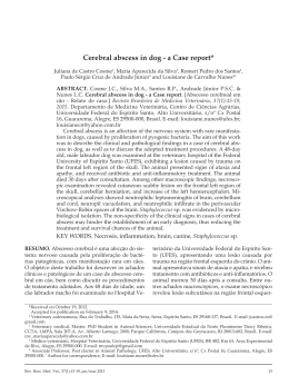
ISSN 1807-5274
Rev. Clín. Pesq. Odontol., Curitiba, v. 6, n. 1, p. 107-112, jan./abr. 2010
Licenciado sob uma Licença Creative Commons
HOPELESS TO HOPEFUL: a clinical study on management
of periodontal abscess with grade II furcation involvement –
endodontic and periodontal interdisciplinary approach:
case report
TÍTULO
Da condenação à esperança: estudo clínico sobre o manejo do abscesso
periodontal com envolvimento de furca grau II – abordagem
interdisciplinar endodôntica e periodontal: relato de caso
KT Chandrashekar[a], Chhavi Saxena[b]
[a]
Professor and Head, Department of Periodontics, Dashan Dental College and Hospital, Udaipur, Rajasthan - India, e-mail:
[email protected]
[b]
Graduate Student, Department of Periodontics, Dashan Dental College and Hospital, Udaipur, Rajasthan - India, e-mail:
[email protected]
Abstract
OBJECTIVES: To present a case report of management of grade II furcation involvement associated
with a periodontal abscess of mandibular right first molar utilizing an alloplastic bone graft. DISCUSSION:
The case was diagnosed as periodontal abscess with grade-II furcation involvement and was primary
periodontal lesion with secondary endodontic involvement. Treatment included scaling, root planing &
curettage and root canal treatment of the involved tooth. Periodontal flap surgery was done with bone
graft placement in the furcation area. RESULTS: Patient was evaluated after onear. Radiographic
examination showed healing of furcation defect as well as resolution of periapical inflammation. Clinical
evaluation revealed resolution of furcation involvement as well as reduction of tooth mobility.
Keywords: Periodontal abscess. Endo-periodontal lesion. Furcation defects. Bone grafts.
Resumo
OBJETIVOS: Apresentar um caso de comprometimento grau II de furca, lesão primariamente
periodontal com comprometimento endodôntico secundário, em primeiro molar inferior. O
tratamento constituiu de alisamento radicular, curetagem e tratamento endodôntico do dente
comprometido. Cirurgia de retalho periodontal foi efetuada, com colocação de enxerto ósseo
Rev Clín Pesq Odontol. 2010 jan/abr;6(1):107-112
Chandrashekar KT, Saxena C.
108
na área da furca. RESULTADOS: o paciente foi avaliado após um ano, sendo que a imagem
radiográfica demonstrou cicatrização do defeito da furca, bem como resolução da inflamação
periapical. A avaliação clínica demonstrou resolução da lesão da furca, bem como redução
da mobilidade dentária.
Palavras-chave: Abscesso periodontal. Lesões endo-periodontais. Defeitos de furca. Enxertos ósseos.
INTRODUTION
Periodontal abscess is the third most
frequent dental emergency (1). It is defined as a
localized purulent infection within the tissue
adjacent to the periodontal pocket that may lead
to the destruction of periodontal ligaments and
alveolar bone (2). In periodontitis, a periodontal
abscess represents a period of active bone
destruction, although such events also occur
without abscess formation (1). Often it is difficult
to differentiate the cause of an abscess, as it may
be of pulpal or periodontal origin. These conditions
have to be promptly managed, failure of which
could lead to tooth loss.
Abscesses are frequently found in furcations
and management of periodontal abscess has been a
challenge for many years. For example, in 17th century,
Louis XIV of France was treated for his periodontal
abscesses with masses of mixed bread and milk, in
order to soften the swelling and to allow drainage of
the abscess (1). The treatment of acute periodontal
abscess usually includes two stages: the management
of acute lesion; and the appropriate treatment of the
original or residual lesion, once the acute situation
has been controlled (3).
Pulpal and periodontal problems are
responsible for more than 50% of tooth mortality
(4). The effect of periodontal disease on dental pulp
was first described by Turner & Drew in 1919. The
relationship between periodontal and pulpal disease
was first described by Simring and Goldberg in
1964 (5). Since then the term ‘endo-perio lesion’
has been used to describe lesions due to inflammatory
products found in varying degrees in both the
periodontium and pulpal tissues. The major
pathways for communication and therefore for
extension of disease from a periodontal pocket to
the pulp are through patent dentinal tubules, lateral
canals and the apical foramen (4). According to the
classification given by Simon, Glick and Franklin S.
Weine (1972) (6) these lesions can be classified as:
1) Primary endodontic lesions;
2) Primary periodontal lesions;
3) Primary endodontic lesion with
secondary periodontal involvement;
4) Primary periodontal lesion with
secondary endodontic involvement;
5) True combined lesions.
Regenerative treatments of furcation may
be difficult because of complicated anatomy,
inacessesibility of patient to the area, small size of
furca foramen and accumulation of microbial
plaque (2). Different regenerative treatments like
bone graft (auto graft, allograft, xenograft and
alloplastic materials) and guided tissue regeneration
methods (resorbable and non resorbable
membranes) have been used in periodontal defects.
The purpose of this article is to describe
a case of periodontal abscess with grade- II furcation
involvement, a primary periodontal lesion with
secondary endodontic involvement and its
management which involved an interdisciplinary
approach of endodontic and periodontics.
CASE REPORT
A 31-year-old male patient reported to the
Outpatient Department of Periodontics, Darshan
Dental College, Udaipur, with chief complaint of
swelling of gums and discomfort in right lower back
region. The swelling was present since the last four
days. Oral hygiene status of patient was fair. Swelling
was seen in relation to tooth 46 (Figure 1). Grade II
mobility was evident in the 46, with pain and difficulty
in mastication. Tooth was tender on percussion and
vitality test was negative. Periodontal examination
revealed a clinical probing pocket depth of 4 to 7 mm
(mean 5.5 mm), with class II furcation involvement as
measured with a Naber’s probe (Figure 2). Intra oral
periapical radiograph revealed periapical radiolucencies
in distal root and furcation area of 46 (Figure 3).
Rev Clín Pesq Odontol. 2010 jan/abr;6(1):107-112
Hopeless to hopeful
Figure 1 - Periodontal abscess in relation to 46
Phase I therapy, periodontal regenerative procedure
using alloplastic osteoconductive bone graft material
was instituted. Biograft HTTM was selected as the
material of choice because of its unique combination
of hydroxyapatite and β-triclacium phosphate
combination. Under local anesthesia, a full thickness
mucoperiosteal flaps were elevated (Figure 5). On
surgical debridement, a Grade II furcation involvement
was evident which was filled with the Biograft- HTTM
bone graft material (Figure 6). Flaps were sutured
with 4-0 black silk sutures and periodontal dressing
was applied and post operative instructions were
given. Post operative healing was good with minimal
discomfort. Post operative medications included
analgesic Diclofenac sodium 50 mg twice a day for
five days. Post operative healing was satisfactory.
Follow up was done for one week, one month, three
months, six months and 12 months.
Figure 2 - Furcation involvement in relation to 46
Figure 4 - Image after root canal treatment
Figure 3 - Radiograph showing radiolucency in furcation
and distal root periapical area
Emergency treatment included drainage of
the abscess with prescription of antibiotic regimen
(OrnidazoleTM 500mg & OfloxacinTM 200 mg
combination) and analgesic DiclofenacTM sodium 50
mg twice a day for five days. Patient was revaluated
after five days, as the swelling and inflammation
subsided; root canal treatment was initiated at the
second appointment (Figure 4). After evaluation of
109
Figure 5 - Surgical exposure of 46 after RCT
Rev Clín Pesq Odontol. 2010 jan/abr;6(1):107-112
Chandrashekar KT, Saxena C.
110
Figure 8 - Image of 46 after 1 year showing bone
healing in the furcation and periapical area
of distal root
DISCUSSION
Figure 6 - Placement of Biograft HTTM in the furcation
defect area
Recall appointments consisted of
reinforcement of oral hygiene instructions, scaling
if required and periapical radiographs of the
involved tooth. Patient revealed no bleeding on
probing, no suppuration or episode of abscess and
no mobility of tooth (Figure 7). The radiological
evaluation revealed increased bone density in the
periapical and furcation area after one year (Figure
8). Indicating successful resolution of infection
and bone fill in the residual defect of furcation.
Figura 7 AQUI
Figure 7 - Clinical furcation probing after 1 year
Diagnosis of a periodontal abscess is
primarily based on the symptoms presented by the
patient and the signs found during oral examination,
careful medical and dental history and radiographic
examination (6). Symptoms range from light
discomfort to severe pain, tenderness of gingivae,
swelling, tooth mobility, tooth elevation, sensitivity
of the tooth to palpation. Suppuration either
spontaneous or after pressure on abscess, combined
with rapid tissue destruction and deep pocket
formation can be seen (7). Radiographic examination
may reveal a normal appearance or some degree of
bone loss, ranging from a widening of periodontal
space to a dramatic radiographic bone loss.
The entry of bacteriae into the soft tissue
pocket wall could be the first event to initiate the
periodontal abscess. Inflammatory cells are then
attracted by chemotactic factors released by the
bacteria, and the concomitant inflammatory
reactions leads to destruction of the connective
tissues, the encapsulation of the bacterial infection
and the production of pus. The microflora related
with periodontal abscess is complex, dominated
by Gram negative, strict anaerobe rods. P.
gingivalis, P. intermedia and F. nucleatum are the
prevalent bacterial species (8, 9).
Management of periodontal abscesses
recommended protocol (1) is: drainage through
the pocket, or stab incision to the most fluctuant
area. After one week the definitive treatment
Rev Clín Pesq Odontol. 2010 jan/abr;6(1):107-112
Hopeless to hopeful
could be carried out. In the treatment of a
periodontal abscess associated with furcation
involvement, therapy for the furcation should
commence quickly and definitively following
reappraisal of the periodontal damage after the
relief of pain and swelling, that is, emergency
management of the infection (10). Clinical tests
are imperative for obtaining correct diagnosis and
differentiating between endodontic and periodontal
disease. Treatment, decision making and prognosis
depend primarily on the diagnosis of the specific
endodontic and/or periodontal disease. The main
factors to be considered are pulp vitality and type
and extent of the periodontal defec (6).
The tooth with a necrotic pulp may pose
as a risk factor in initiation of periodontal disease.
A periodontal–endodontic combination problems
are much more frequent in posterior teeth,
particularly in molars then anterior teeth because
of greater number of auxillary and furcation canals
present in molars. Vertucci and Williams reported
that 46% of mandibular first molars had auxillary
canals in furcation region (9).
The management of class II furcation
involvement presents a unique clinical problem.
Reasons for compromised results in furcation
areas include the lack of proper access for
instrumentation as well as for proper maintenance
care due to the complex furcation anatomy and
consequently a persistence of pathogenic
microflora. The early work concentrated more on
resective procedures intended to eliminate the
pocket by furcationplasty and root resection.
The most favorable outcome of any furcation
therapy would be the regeneration of the lost
attachment apparatus, which would result in the
closure of the furcation. The various regenerative
approaches utilized in the management of class II
furcation involvements include root surface
biomodification, coronally positioned flaps, the
use of various bone replacement grafts, and
Guided Tissue Regeneration procedures (2).
According to Weine (9), if a patient with
open or closed furcation area displays the classic
symptom of pulpitis, despite the absence of decay
or extensive restorations, an endodontic procedure
must be considered until another logical alternative
is discovered. As the tooth involved was non-vital
endodontic treatment was carried out so that bacteria
are eliminated from the root canal system by
mechanical instrumentation and irrigation with
111
normal saline and sodium hypochlorite substantially
decreased the amount of bacteria in pulpal spaces.
In the present case the pulp was non-vital
and radiological evaluation revealed periapical
radiolucencies in relation to distal root and in furcation
area. There was no evidence of carious lesion on the
involved tooth, so the lesion was diagnosed as primarily
periodontal lesion with secondary endodontic
involvement. Treatment of the residual lesion caused
by periodontal abscess was treated by using bone
graft which was a combination of hydroxyapatite and
beta – tricalcium phosphate.
Similar evidence of successful treatment
of class II furcations using porous hydroxyapatite
has been documented earlier also which was
substantiated by improvements in clinical linear
measurements and reentry evidence of incorporation
of the implant in the surrounding bone (11).
CONCLUSION
Endodontic-periodontal lesions present a
diagnostic and treatment dilemma. Pathologic
changes in endodontic and periodontal tissues affect
each other. Pulp necrosis is a risk factor that damages
adjacent periodontal structures. Primary periodontal
diseases with secondary endodontic involvement
require both endodontic and periodontal therapies.
Their prognosis depends primarily upon the severity
of the periodontal disease and the response to
periodontal treatment. The suggested treatment of
periodontal abscess with Grade-II furcation by root
canal treatment followed by periodontal flap
procedure with bone graft placement in the furcation
of the involved tooth could result in complete
healing of tooth which was considered hopeless.
CONFLICT OF INTEREST
STATEMENT
The authors declared no conflict of
interest in the present manuscript.
INFORMED CONSENT STATEMENT
The patients signed an informed
consent, kept in the records, in the archives of
the Dashan Dental College.
Rev Clín Pesq Odontol. 2010 jan/abr;6(1):107-112
112
Chandrashekar KT, Saxena C.
REFERENCES
1. Herrera D, Roldan S, Sanz M. The periodontal
abscess: a review. J Clin Periodontol. 2000;27(6):
377-86.
2. Newman T, Klokkevold, Carranza FA. Clinical
Periodontology. 10th ed. Philadelphia: Saunders;
2006.
3. Ahl DR, Hilgeman JL, Snyder JD. Periodontal
emergencies. Dent Clin North Am. 1986;30
(3):459-72.
4. Chen SY, Wang HL, Glickman GN. The influence
of endodontic treatment upon periodontal
wound healing. J Clin Periodontol. 1997;24
(7):449-56.
5. Simring M, Goldberg M. The pulpal pocket
approach: retrograde periodontitis. J Periodontol.
1964;35(1):22-48.
6. Rotstein I, Simon JH. Diagnosis, prognosis and
decision-making in the treatment of combined
periodontal endodontic lesions. Periodontol.
2000;34(3):165-203.
7. Consensus report: abscesses of the periodontium
Ann. Periodontol. 1999;4(1):83.
8. Meng HX. Periodontic-Endodontic lesion. Ann
Periodontol. 1999;4(1):84-9.
9. Weine FS. Endodontic therapy. 4th ed. St. Louis:
The CV Mosby Co; 1989.
10. Mhairi R W. The pathogenesis and treatment of
endo-perio lesions. CPD Dentistry. 2001;2(3):91-5.
11. Kenney EB, Lekovic V, Elbaz JJ, Kovacvic K,
Carranza Jr FA, Takei HH. The use of a porous
hydroxylapatite implant in periodontal defects.
Treatment of Class II furcation lesions in lower
molars. J Periodontol 1988;59(2):67-72.
Received: 08/15/2009
Recebido: 15/08/2009
Accepted: 10/25/2009
Aceito: 25/10/2009
Rev Clín Pesq Odontol. 2010 jan/abr;6(1):107-112
Baixar