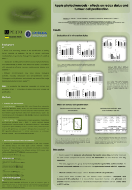
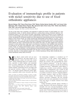
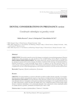
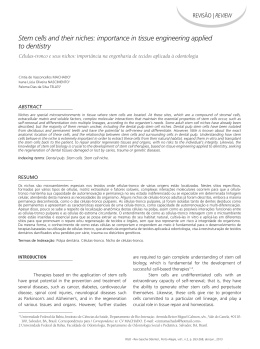
Pesqui Odontol Bras
2002;16(3):209-215
Implantodontia
Surface topography of hydroxyapatite affects
ROS17/2.8 cells response
A topografia de superfície da hidroxiapatita afeta
a resposta de células ROS17/2.8
Adalberto Luiz Rosa*
Márcio Mateus Beloti*
Richard van Noort**
Paul Vincent Hatton**
Anne Jane Devlin**
ABSTRACT: Hydroxyapatite (HA) has been used in orthopedic, dental, and maxillofacial surgery as a bone substitute.
The aim of this investigation was to study the effect of surface topography produced by the presence of microporosity
on cell response, evaluating: cell attachment, cell morphology, cell proliferation, total protein content, and alkaline
phosphatase (ALP) activity. HA discs with different percentages of microporosity (< 5%, 15%, and 30%) were confected
by means of the combination of uniaxial powder pressing and different sintering conditions. ROS17/2.8 cells were cultured on HA discs. For the evaluation of attachment, cells were cultured for two hours. Cell morphology was evaluated
after seven days. After seven and fourteen days, cell proliferation, total protein content, and ALP activity were measured. Data were compared by means of ANOVA and Duncan’s multiple range test, when appropriate. Cell attachment
(p = 0.11) and total protein content (p = 0.31) were not affected by surface topography. Proliferation after 7 and 14 days
(p = 0.0007 and p = 0.003, respectively), and ALP activity (p = 0.0007) were both significantly decreased by the most irregular surface (HA30). These results suggest that initial cell events were not affected by surface topography, while
surfaces with more regular topography, as those present in HA with 15% or less of microporosity, favored intermediary
and final events such as cell proliferation and ALP activity.
UNITERMS: Hydroxyapatite; Porosity; Surface topography; ROS17/2.8 cells.
RESUMO: A hidroxiapatita (HA) tem sido utilizada como revestimento de implantes e para substituição de tecido ósseo.
O objetivo deste estudo foi avaliar o efeito da topografia de superfície da HA, resultante da presença de microporosidade, sobre a adesão, a morfologia e proliferação celulares, a medida de proteína total e a atividade de fosfatase alcalina.
Discos de HA com diferentes porcentagens de microporosidade (< 5%, 15% e 30%) foram fabricados por uma combinação das técnicas de pressão uniaxial e sinterização. Células ROS17/2.8 foram cultivadas sobre os discos de HA. Para a
adesão, as células foram cultivadas por duas horas. A morfologia foi avaliada após sete dias. A proliferação, medida de
proteína total e atividade de ALP foram avaliadas após sete e quatorze dias. Os dados foram comparados por ANOVA e
teste de Duncan quando apropriado. A adesão (p = 0,11) e a medida de proteína total (p = 0,31) não foram afetadas pela
topografia de superfície. A proliferação após sete e quatorze dias (p = 0,0007 e p = 0,003, respectivamente), e a atividade de ALP (p = 0,0007) foram significantemente menores na superfície irregular (HA30). Esses resultados sugerem que
eventos iniciais não são afetados pela topografia, enquanto superfícies com topografias mais regulares (microporosidade de 15% ou menos) favoreceram eventos intermediários e finais, como proliferação e atividade de ALP.
UNITERMOS: Hidroxiapatita; Porosidade; Topografia de superfície; Células ROS17/2.8.
INTRODUCTION
Hydroxyapatite (HA) has received considerable
attention as a material for implants and bone augmentation procedures, since it bonds chemically directly to bone after implantation6. Synthetic HA for
bone substitution can be produced by a combination
of a powder pressing technique and sintering. Powder pressing techniques allow the obtainment of
“green bodies”, which are then sintered at high temperatures to produce the fusion of powder particles.
Changes in the powder compaction and sintering
conditions can result in a variety of chemical and porosity characteristics of HA16,20. Porosity can be classified, according to the diameter of pores, in macroporosity (> 10 mm) and microporosity (< 10 mm). It
has been suggested that these structural modifications of HA influence cellular activity16,20.
* Department of Surgery, Maxillofacial Traumatology and Periodontology, School of Dentistry of Ribeirão Preto, University of São Paulo.
** Centre for Biomaterials and Tissue Engineering, University of Sheffield, England.
209
Rosa AL, Beloti MM, van Noort R, Hatton PV, Devlin AJ. Surface topography of hydroxyapatite affects ROS17/2.8 cells response. Pesqui Odontol Bras 2002;16(3):209-215.
Dziedzic et al.8 (1996) reported that HA samples
with different microporosities result in different
bone-HA interfaces after implantation in rats. Preliminary results from in vitro studies using a primary culture of rat bone marrow cells and a rat
osteosarcoma cell line have demonstrated that an
increase in micropore percentage negatively influences HA biocompatibility18,19.
Variations of micropore percentage result in different surface topographies3,6 and authors have
agreed that topography must be considered in
terms of amplitude and organization13. Anselme et
al.1 (1997) observed a lower proliferation of human
osteoblasts cultured on less organized Ti surfaces.
Considering that the surface characteristics of
HA necessary for optimal osseointegration are still
not fully understood, the aim of this study was to
carry out an in vitro evaluation of the effect of varying the surface topography of HA on the response
of osteoblast-like cells. For this, HA samples with
less than 5%, 15%, and 30% of microporosity were
evaluated using osteoblast-like cells derived from
rat osteosarcoma (ROS17/2.8). These cells were
used because they express numerous osteoblastic
characteristics, including high levels of alkaline
phosphatase (ALP) activity, synthesis of type I collagen, and osteocalcin10. The following parameters
were evaluated: cell attachment, cell morphology,
cell proliferation, total protein content, and ALP
activity.
MATERIAL AND METHODS
Preparation of HA samples
HA discs (with the diameter of 12 mm and
height of 2 mm) with different degrees of microporosity (< 10 mm), as shown in Table 1, were
confected by means of a combination of uniaxial
powder pressing and different sintering conditions. This procedure allows the confection of HA
with identical chemical composition and crystallinity but with different porosities20,21. The discs were
made of a high purity and crystalline HA powder
TABLE 1 - Axial load, sintering condition, and microporosity percentage of the HA samples.
Load (ton)
Sintering (ºC/hour)
Microporosity
percentage
HA5
6
1,250/3
<5
HA15
2
1,225/3
15
HA30
2
1,150/3
30
Sample
210
(Plasma Biotal, Tideswell, UK). The HA discs utilized in this study were divided in three groups:
HA5, with less than 5% of microporosity, HA15,
with 15% of microporosity, and HA30, with 30% of
microporosity, as calculated from the actual density compared with the theoretical density of HA.
The HA surface was evaluated by scanning
electron microscopy (SEM - JSM-5410, Jeol, MA,
USA). Surface roughness (Ra) was measured using
a surface profilometer (Prazis Rug-03, São Paulo,
Brazil), and an average was calculated for each
disc, taking into account three measurements.
The average Ra of each microporosity group was
determined based on measurements of three discs
(n = 3). All discs were cleaned in an ultrasonic bath
and autoclaved before utilization in the cell culture
experiments.
Cell culture
ROS17/2.8 cells were cultured in HAM’S F12
(Gibco - Life Technologies, Grand Island, NY, USA)
supplemented with 8% fetal bovine serum,
50 mg/ml penicillin-streptomycin (Gibco), glutamine 5.4 ´ 10–7 M (Gibco), and 0.3 mg/ml amphotericin B (Gibco). Cells were cultured at a concentration of 0.825 ´ 104 cells/well on HA discs in 24-well
culture plates (Falcon, Franklin Lakes, NJ, USA).
During the culture period, cells were incubated at
37ºC in a humidified atmosphere of 5% CO2 and
95% air, and the medium was changed every
forty-eight hours. In each plate, empty wells were
only used as a control of culture conditions.
Cell attachment
For the evaluation of attachment, cells were
cultured for two hours on HA discs (n = 5, for each
microporosity). The culture medium was removed,
and the wells were washed three times with PBS at
37ºC to eliminate unattached cells. The adherent
cells were then enzymatically released (1 mM
EDTA + 0.25% trypsin - Gibco) from the HA discs,
and counted using a hemacytometer. Cell attachment was expressed as a percentage of the initial
number of cells.
Cell morphology
For the evaluation of morphology, cells were
cultured for seven days on HA discs and processed
for SEM. First, cells were fixed with 1.5% glutaraldehyde (Sigma, St. Louis, MO, USA) buffered in
0.1 M sodium cacodylate (Sigma), and stained in
1% osmium tetroxide (Sigma). Then, cells were dehydrated through a graded series of alcohol, fol-
Rosa AL, Beloti MM, van Noort R, Hatton PV, Devlin AJ. Surface topography of hydroxyapatite affects ROS17/2.8 cells response. Pesqui Odontol Bras 2002;16(3):209-215.
lowed by critical point drying at 32ºC and 1,300
psi for one hour. After that, samples were sputter-coated with gold (10 nm) before examination
under a SEM.
Cell proliferation
For the evaluation of proliferation, cells were
cultured for seven and fourteen days on HA discs
(n = 5 for each day and each microporosity). The
cells were released and counted as described in
cell attachment.
Total protein content
Total protein content after seven and fourteen
days in culture on HA discs (n = 5 for each
microporosity) was calculated according to the
modified Lowry et al.14 (1951) method. The culture
medium was removed, the wells were washed three
times with PBS at 37ºC and were filled with 2 ml of
0.1% sodium lauryl sulfate (Sigma). After thirty
minutes, 1 ml of the solution from each well was
mixed with 1 ml of Lowry solution (Sigma) and left
for twenty minutes at room temperature. After this
period, it was added to 0.5 ml of the solution of
phenol reagent of Folin and Ciocalteau (Sigma). It
stood for thirty minutes at room temperature to allow color development, and absorbance was then
spectrophotometrically measured (CE3021 - Cecil,
Cambridge, UK) at 680 nm. The total protein content was calculated from a standard curve of bovine albumin and expressed as µg/ml. These data
were normalized by the number of cells counted after seven and fourteen days, respectively.
ALP activity
ALP activity was assayed as the release of
r-nitrophenol from r-nitrophenyl, using a commercial kit (Sigma), and the specific activity was calculated. From the same solutions used for calculating total protein content, 0.1 ml aliquots were
assayed for measuring ALP activity. Absorbance
was spectrophotometrically measured at 410 nm,
and ALP activity was calculated from a standard
curve obtained from different solutions of r-nitrophenol and sodium hydroxide 0.02 N. Results were
calculated as µmol r-nitrophenol/hour, and data
were expressed as ALP activity normalized by the
total protein content and by the number of cells
counted after seven and fourteen days, respectively.
Statistical analysis
Data were submitted to analysis of variance
(ANOVA) and Duncan’s multiple range test, when
appropriate. Differences at p ≤ 0.05 were considered statistically significant.
RESULTS
HA samples
Ra measurements presented the following results: HA5 = 0.38 µm, HA15 = 0.48 µm and
HA30 = 0.49 µm. ANOVA revealed that Ra was not
affected by the manufacturing conditions of HA
(F = 2.79; gl = 2; p = 0.14). It was possible to observe, from SEM micrographs, that the amount of
microporosity produced surfaces with different topographies (Figure 1). Thus, HA5 and HA15 presented more regular surfaces while HA30 presented a very irregular surface topography
(Figure 1).
Cell attachment
Cell attachment was not affected by HA surface
topography (ANOVA: gl = 2; F = 2.65; p = 0.11).
Data are presented in Table 2.
Cell morphology
HA5 e HA15 presented more cells than HA30.
Cells observed on HA5 and HA15 presented a polygonal morphology with dorsal ruffles and a large
number of filaments connecting distant cells (Figures 2A and 2B). On HA30 discs, cells did not
cover all surfaces. In addition, the formation of a
monolayer was observed, with cells predominantly
presenting a more flattened morphology, showing
almost no dorsal ruffles and filaments (Figure 2C).
Cell proliferation
The number of cells was affected by surface topography after both seven and fourteen days
(ANOVA: gl = 2; F = 14.31; p = 0.0007 e gl = 2;
F = 9.61; p = 0.0032, respectively) in the following
order: HA5 = HA15 > HA30. Data are presented in
Figure 3.
Total protein content
Total protein content after seven and fourteen
days was not affected by both surface topography
(ANOVA: gl = 2; F = 1.21; p = 0.31) and period of
culture (ANOVA: gl = 1; F = 1.58; p = 0.22). Data
are presented in Table 3.
211
Rosa AL, Beloti MM, van Noort R, Hatton PV, Devlin AJ. Surface topography of hydroxyapatite affects ROS17/2.8 cells response. Pesqui Odontol Bras 2002;16(3):209-215.
A
TABLE 2 - Attachment of ROS17/2.8 cells, expressed as
a percentage of the initial number of cells, on hydroxyapatite (HA) discs after two hours. Data are presented as
mean ± standard deviation (n = 5).
Cell attachment
HA5
HA15
HA30
% of the initial
number of cells
6.65 ± 4.15
7.4 ± 3.99
3.7 ± 1.38
HA5 = less than 5% of microporosity; HA15 = 15% of
microporosity; HA30 = 30% of microporosity.
HA5 = HA15 > HA30. However, it was not affected
by the period of culture (ANOVA: gl = 1; F = 0.21;
p = 0.65). Data are presented in Table 4.
B
C
FIGURE 1 - Scanning electron microscopy (SEM) of the
hydroxyapatite (HA) surfaces. (A) HA5 (5% of microporosity); (B) HA15 (15% of microporosity) and (C) HA30
(30% of microporosity).
ALP activity
ALP activity after seven and fourteen days was
affected by surface topography (ANOVA: gl = 2;
F = 9.69; p = 0.0007) in the following order:
212
DISCUSSION
Early in vitro studies on the interaction between
osteoblasts and biomaterials were essentially concerned with the effect of diverse range materials on
cell response, without paying attention to the influence of surface characteristics23. However, it is
now understood that the superficial properties of
biomaterials play a critical role in the establishment of cell-biomaterial interfaces and that manufacturing conditions affect the superficial properties of HA1,20.
The present results revealed that different combinations of powder pressing and sintering produced HA samples with different microporosities
and, consequently, different superficial appearances. Despite these differences, Ra, a factor
known to influence cell response, was not significantly affected by the manufacturing process of
the HA samples. Hence, any differences in cell response observed in this study would be a consequence of differences in the microporosity of HA
samples.
Osteogenesis, induced by osteoblastic cells, is
characterized by a sequence of events, involving
cell attachment and cell proliferation, followed by
the expression of osteoblastic phenotype6. This
study evaluated the response of ROS 17/2.8 osteoblast-like cells, originated from rat osteosarcoma,
cultured on HA discs with different surface topographies. The present results revealed that all HA
samples, regardless of surface topography, are
biocompatible, considering that they allowed cell
attachment, proliferation, and expression of markers of osteoblastic differentiation.
The effect of surface characteristics on cells
may be the result of the surface characteristics
themselves or the result of the reaction that occurs
as the surface is conditioned by the medium and
Rosa AL, Beloti MM, van Noort R, Hatton PV, Devlin AJ. Surface topography of hydroxyapatite affects ROS17/2.8 cells response. Pesqui Odontol Bras 2002;16(3):209-215.
80
Cell number x 10 4
A
HA5
60
HA15
HA30
40
20
0
0
7
14
Period (days)
B
FIGURE 3 - Proliferation of ROS 17/2.8 cultured on
hydroxyapatite (HA) discs. Data are presented as average values, and bars indicate standard deviation (n = 5).
HA5 = less than 5% of microporosity; HA15 = 15% of
microporosity; HA30 = 30% of microporosity.
TABLE 3 - Total protein content (µg/104 cells) produced
by ROS17/2.8 cells cultured on hydroxyapatite (HA)
discs. Data are presented as mean ± standard deviation
(n = 5).
Period (days)
HA5
HA15
HA30
7
4.37 ± 0.65
7.06 ± 0.39
7.08 ± 7.08
14
4.44 ± 0.59
3.55 ± 0.12
6.13 ± 0.51
HA5 = less than 5% of microporosity; HA15 = 15% of
microporosity; HA30 = 30% of microporosity.
C
TABLE 4 - Alkaline phosphatase (ALP) activity (µmol
p-nitrophenol/hour) normalized by the total protein
content and the number of ROS17/2.8 cells cultured on
hydroxyapatite (HA) discs. Data are presented as
mean ± standard deviation (n = 5).
Period (days)
HA5
HA15
HA30
7
2.84 ± 0.39
2.32 ± 1.01
0.58 ± 0.65
14
1.99 ± 0.92
2.44 ± 0.43
1.65 ± 0.13
HA5 = less than 5% of microporosity; HA15 = 15% of
microporosity; HA30 = 30% of microporosity.
FIGURE 2 - Morphology of ROS 17/2.8 cells after seven
days in culture on hydroxyapatite (HA) discs, evaluated
under scanning electron microscopy (SEM). (A) HA with
less than 5% of microporosity (HA5); (B) HA with 15% of
microporosity (HA15), and (C) HA with 30% of microporosity (HA30).
serum15. This initial interaction produces a layer of
macromolecules that modify cell response. Fibronectin, a cell adhesion protein present in serum,
has been shown to mediate cell attachment and
spread on artificial substrates by interacting with
glycosaminoglycans and with the cytoskeleton7.
This study did not show any effect of surface topography on cell attachment. While it may occur, 2
hours – which was the period used in this evaluation – is a very short time to observe the development of a difference in protein adhesion that could
affect cell attachment. Besides the period of incubation, it is also possible that cell attachment in
213
Rosa AL, Beloti MM, van Noort R, Hatton PV, Devlin AJ. Surface topography of hydroxyapatite affects ROS17/2.8 cells response. Pesqui Odontol Bras 2002;16(3):209-215.
tissue culture is not seriously influenced by any
characteristic of the material, since it has been
shown that materials with different degrees of
biocompatibility produce little or no differences in
cell attachment17.
In the present study, the amount of protein was
normalized by the number of cells. Hence, these
results evaluated the cell secretory activities that
were not affected by surface topography. Similarly,
Castellani et al.4 (1999) did not find significant differences in the production of protein by rat bone
marrow cells cultured on titanium with different
surface topographies. Thus, it is possible that the
total protein production, regardless of the chemical composition of the biomaterial, is not affected
by surface topography.
Cell proliferation and ALP activity were affected
by surface topography. HA samples with more regular surfaces such as HA5 e HA15 allowed greater
cell proliferation and ALP activity than HA samples
with irregular surface (HA30). These results suggest that more regular surfaces would facilitate cell
growth and the expression of osteoblast phenotype
markers. Cell morphology confirmed these findings, since cells cultured on more regular surfaces
presented a polygonal morphology with dorsal ruffles and a large number of filaments. This morphology has been described as osteoblast feature22.
On the contrary, cells cultured on irregular surfaces presented a flattened morphology showing
almost no dorsal ruffles and filopodia. Other studies pointed out low cell proliferation and ALP activity on less organized surfaces1,6, showing that the
regularity and uniformity of the HA surface are important factors that affect cell proliferation and
differentiation2,12. Therefore, differences in surface
topography result in different degrees of HA biocompatibility, which confirms our previous results
that revealed that HA30 presents, comparatively,
low biocompatibility18,19.
An explanation for these findings is that the
physicochemical properties of the ceramic surface
can affect the reorganization of proteins on ceramic specimens and change the profile of adsorbed proteins8. The type of substrate determines
which integrins and extracellular matrix proteins
are expressed by osteoblasts, providing information about how implant materials may affect
osteoblastic differentiation23. The differences in
protein adsorption can result in very different initial cell responses23, and it is known that some proteins, such as fibronectin, have an important role
in osteoblastic differentiation11. It is possible that
organized surfaces favor the selective adsorption of
proteins responsible for osteoblastic differentiation in serum. As a result, biomaterials that present more regular surfaces are more biocompatible.
CONCLUSIONS
• Varied manufacturing conditions produced HA
samples with different microporosities. Surface
roughness was not affected by the presence of
micropores. However, surfaces with smaller
percentages of microporosity presented more
regular surface topography.
• All HA samples, regardless of surface topography, are biocompatible. However, samples
with more regular surface topography were
more biocompatible, which was evidenced by
the greater cell proliferation and ALP activity
observed on their surfaces, when compared
with samples with irregular surface topography.
ACKNOWLEDGMENTS
This work was supported by the State of São
Paulo Research Foundation (FAPESP), Grant no.
98/06851-0.
REFERENCES
1. Anselme K, Bigerrelle M, Noel B, Dufresne E, Judas D, Iost A,
Hardouin P. Qualitative and quantitative study of human
osteoblast adhesion on materials with various surface
roughnesses. J Bimed Mater Res 1997;49(2):155-66.
2. Bowers K, Keller JC, Randolph B, Wick D, Michaels C.
Optimization of surface micromorphology for enhanced
osteoblast responses in vitro. Int J Oral Maxillofac Impl
1992;7(3):302-10.
3. Boyan DB, Hummet TW, Dean DD, Schwartz Z. Role of material surfaces in regulating bone and cartilage cell response. Biomaterials 1996;17(2):137-46.
214
4. Castellani R, De Ruijter JE, Renggli H, Jansen JA. Response of rat bone marrow cells to differently roughened titanium discs. Clin Oral Impl Res 1999;10(5):369-78.
5. Curtis ASG, Wilkinson CDW. Reactions of cells to topography. J Biomater Sci Polymer Edn 1998;12(9):1313-29.
6. Deligianni DD, Katsala ND, Koutsoukos PG, Missirlis YF.
Effect of surface roughness of hydroxyapatite on human
bone marrow cell adhesion, proliferation, differentiation
and detachment strength. Biomaterials 2001;22(1):87-96.
7. Doillon CJ, Silver FH, Berg RA. Fibroblast growth on a porous collagen sponge containing hyaluronic acid and fibronectin. Biomaterials 1987;8(3):195-200.
Rosa AL, Beloti MM, van Noort R, Hatton PV, Devlin AJ. Surface topography of hydroxyapatite affects ROS17/2.8 cells response. Pesqui Odontol Bras 2002;16(3):209-215.
8. Dziedzic DM, Sawa IH, Wilkinson DS, Davies JE. Osteoconduction on, and bonding to, calcium phosphate ceramic
implants. Mater Res Soc Symp Proc 1996;414:147-56.
9. Frayssinet P, Rouquet N, Fages J, Durand M, Vidalain PO,
Bonel G. The influence of sintering temperature on the
proliferation of fibroblastic cells in contact with HA-bioceramics. J Biomed Mater Res 1997;35(3):337-47.
10. Garcia AJ, Ducheyne P, Boettinger D. Effect of surface reaction stage on fibronectin-mediated adhesion of osteoblast-like cells to bioactive glass. J Biomed Mater Res
1998;40(1):48-56.
11. Globus RK, Doty SB, Lull JC, Holmuhamedov E, Humphries MJ, Damsky CH. Fibronectin is a survival factor
for differentiated osteoblasts. J Cell Sci 1998;111(pt10):
1385-93.
12. Keller JC, Collins JG, Niederauer GG, McGee TD. In vitro
attachment of osteoblast-like cells to osteoceramic materials. Dent Mater 1997;13(1):62-8.
13. Lopez J, Hanseli G, Le Bosse JC, Mathia T. Caractérisation
factale de la rugosité tridimensionnelle de surface. J
Phys III France 1994;4:2201-19.
14. Lowry OH, Rosebrough NJ, Farr AL, Randall RJ. Protein
measurement with the Folin phenol reagent. J Biol
Chem 1951;193:265-75.
15. Martin JY, Schawartz Z, Hummert TW, Schraub DM,
Simpson J, Lankford JJr, et al. Effect of titanium surface roughness on proliferation, differentiation, and pro-
16.
17.
18.
19.
20.
21.
22.
23.
tein synthesis of human osteoblast-like cells (MG63). J
Biomed Mater Res 1995;29(3):389-401.
Ong JL, Hoppe CA, Cardenas HL, Cavin R, Carnes DL, Sogal A, et al. Osteoblast precursor cell activity on HA surfaces of different treatments. J Biomed Mater Res
1998;39(2):176-183.
Puleo DA, Hollerman LA, Domerus RH, Bizios R. Osteoblast responses to orthopedic implant materials in vitro.
J Biomed Mater Res 1991;25(6):11-23.
Rosa AL, Hatton PV, Van Noort R. The influence of porosity
on the in vitro biocompatibility of hydroxyapatite. J Dent
Res 1997;76(5):1081.
Rosa AL, Devlin JA, Hatton PV, Van Noort R. In vitro biocompatibility of microporous hydroxyapatite. J Dent
Res 1999;78(5):409.
Rosa AL, Shareef MY, Van Noort R. Efeito das condições de
preparação e sinterização sobre a porosidade da hidroxiapatita. Braz Oral Res 2000;14(3):273-7.
Shareef MY, Messer PF, Van Noort R. Fabrication, characterization and fracture study of a machinable hydroxyapatite ceramic. Biomaterials 1993;14(1):69-75.
Vrouwenvelder WCH, Groot CG. Histological and biochemical evaluation of osteoblasts cultured on bioactive
glass, hydroxyapatite, titanium alloy, and stainless
steel. J Biomed Mater Res 1993;27(4):465-75.
Zeng H, Chittur KK, Lacefield WR. Analysis of bovine serum albumin adsorption on calcium phosphate and titanium surfaces. Biomaterials 1999;20(4):377-84.
Recebido para publicação em 10/12/01
Enviado para reformulação em 06/06/02
Aceito para publicação em 25/06/02
SOCIEDADE BRASILEIRA
DE PESQUISA ODONTOLÓGICA
Divisão Brasileira da IADR
Visite a página da
SBPqO na Internet:
http://www.sbpqo.org.br
SBPqO - Av. Professor Lineu Prestes, 2.227
CEP 05508-900 - Cidade Universitária - São Paulo - SP
Tel./Fax: (0**11) 3091-7855 - E-mail: [email protected]
215
Download