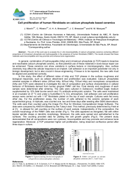
Effect of calcium hydroxide as intracanal medication on the apical
sealing ability of mineral trioxide aggregate (MTA): an in vitro
apexification model
Efeito da medicação intra-canal de hidróxido de cálcio na capacidade seladora do mineral trióxido
agregado (MTA): um modelo de apicificação in vitro
Tânia Afonso1, Maria Lusitana Sá de Andrade Moura Pega1, André Luiz da Costa Michelotto2,
Ana Margarida Coelho Abrantes3, Bárbara Oliveiros3, Eunice Virgínia Palmeirão Carrilho1,
Filomena Rabaça Roque Botelho3, Manuel Marques Ferreira1
1
Department of Dentistry, Faculty of Medicine, University of Coimbra, Coimbra, Portugal; 2School of Dentistry, Tuiuti University of Paraná,
Curitiba-PR, Brazil; 3Institute of Biomedical Research in Light and Image, Faculty of Medicine, University of Coimbra, Coimbra, Portugal.
Abstract
Objective – To evaluate the effect of calcium hydroxide premedication on the apical seal of White MTA, placed as an apical barrier in permanent teeth with simulated immature apices. Furthermore, we intended to compare potential changes, under the influence of calcium hydroxide, in the apical seal of MTA over time. Methods – Thirty-four single-rooted extracted teeth were prepared in order to simulate a
divergent open apex. Two experimental groups of 10 teeth were created: group 1 (G1) and group 2 (G2) with and without calcium hydroxide
intracanal medication previous to the placement of MTA apical plug. Two control groups, positive and negative, each with 7 teeth were
created. On the 7th and on the 28th day after placement of the MTA apical plug, the apices of the teeth were submersed in a solution of
sodium pertechnetate (99mTcO4Na) for 3 hours. The radioactivity was measured using a gamma camera. Results – Results revealed statistically
significant differences between the 2 control groups and the 2 experimental groups with respect to the microleakage. Within the experimental
groups no statistically significant differences were found; nor between the two observed periods. Conclusion – Based on the results obtained
in this study, it was concluded that intracanal medication with calcium hydroxide did not affect the sealing ability of WMTA, placed as an
apical plug, neither on the 7th, nor on the 28th day.
Descriptors: Dental cements; Tooth apex; Calcium hydroxide; Endodontics; Root canal filling materials
Resumo
Objetivo – Avaliar o efeito do hidróxido de cálcio como medicação intracanal, no selamento apical do MTA branco, colocado como uma
barreira apical em dentes permanentes com ápices imaturos simulados. Métodos – Trinta e quatro dentes unirradiculares extraídos foram preparados para simular um ápice aberto divergente. Foram separados em dois grupos experimentais (n=10): G1 e G2 com e sem medicação intracanal de hidróxido de cálcio antes da colocação do tampão apical com o MTA e dois grupos controle, positivos e negativos (n=7 em cada).
Em 7 e 28 dias após a colocação do tampão apical de MTA, os ápices dos espécimes foram submersos em uma solução de pertecnetato de
sódio (99mTcO4Na) por 3 horas. A radioatividade foi medida usando uma câmara gama. Resultados – Os resultados revelaram diferenças estatisticamente significativas entre os grupos controle e os grupos experimentais com relação a microinfiltração. Não houve diferença entre os
grupos experimentais nos dois períodos. Conclusão – Com base nos resultados obtidos neste estudo, concluiu-se que a medicação intracanal
com hidróxido de cálcio não influenciou na capacidade seladora do MTA.
Descritores: Cimentos dentários; Ápice dentário; Hidróxido de cálcio; Endodontia; Materiais restauradores do canal radicular
Introduction
Given its high success rate the treatment of choice for
these cases has been considered the placement of a mineral trioxide aggregate (MTA) apical plug4-8. Before placing the MTA apical barrier, the manufacturer recommends the use of calcium hydroxide [Ca(OH)2] for one
week as intracanal dressing and its subsequent removal9.
The use of calcium hydroxide has been considered an
important step in the reduction of the intracanal microbial
flora10. Because of the detrimental effects of an acidic
pH, caused by the inflammation of periapical tissues, on
various physical properties of MTA, it could be advisable
to delay the placement of the MTA plug to a second session, using Ca(OH)2 as inter-appointment intracanal medication in order to achieve additional disinfection and
neutralization of an acidic environment2,11.
Before placing the apical plug, the removal of Ca(OH)2
has been recommended to allow proper adhesion between
the filling material and the root dentin12-13. However, it has
The occurrence of pulp necrosis in an immature permanent tooth caused by trauma or by pulpal disease can
be considered an endodontic challenge due to several
inherent characteristics of these teeth1-2.
The immature permanent tooth has an incomplete root
formation, characterized by unreached final root length
and thus an unfavourable crown/root ratio; thin root dentinal wall; wide canal space; apical divergence, absence
of an apical constriction and an open apex2.
The traditional techniques of mechanical and chemical
disinfection of root canals in teeth with an incomplete
root formation are limited. Instrumentation increases the
risk of weakening the thin dentinal walls. The divergent
apical architecture and the lack of an apical stop make
complete debridement and control of the filling nearly
impossible3.
J Health Sci Inst. 2012;30(4):318-22
318
G1 (n=10) a 4.0 mm apical plug of MTA was placed
into the canals previously medicated with calcium hydroxide for a period of 7 days and the root segments
were double coated with nail varnish except for the apical 1.0 mm. In G2 (n=10) a 4.0 mm apical plug of MTA
was placed into the canals without prior calcium hydroxide use and the root segments were double coated with
nail varnish except for the apical 1.0 mm.
After drying the canals of the samples of the group (G1),
calcium hydroxide paste (Calcicur® – Voco GMBH, Cuxhaven, Germany) was applied with a size 30 K-file. After
filling the canal with the medication, a dry cotton pellet
was placed at its entrance and the canal space was sealed
with a temporary restorative material (Cavit® – 3M ESPE,
Seefeld, Germany) put in the access cavity. After one week
the dressing was removed through irrigation with NaOCl
1.0% and a #30 K-file and the canals were drying with
paper points, orthograde mineral trioxide aggregate (MTA)
– Pro Root MTA® (White MTA) (Dentsply Maillefer, Ballaigues, Switzerland) apical plugs with a thickness of 4.0
mm were placed the root canals of all groups except positive group, whose root segments were left unfilled. The
mixture of MTA was performed according to the manufacturer’s instructions with a powder/distilled water ratio
of 3/1. The carrier MAP System (Micro Apical Placement
System) (Produits Dentaires SA, Vevey, Switzerland) was
used for placement of MTA into the canals subsequently
condensed with Schilder pluggers (Dentsply Maillefer,
Ballaigues, Switzerland), with stop placed at 11.0 mm.
MTA placement was performed under an operating microscope (Leica CLS® 150MR) by the same operator. Afterward a cotton pellet embedded in 0.9% NaCl was put
at the entrance of the canal and temporary restorative
material Cavit® (3M ESPE, Seefeld, Germany) was used to
seal the access cavity and samples were stored in 100%
relative humidity for 7 days to allow MTA to fully set.
On the 7th and 28th day after placement of the MTA
two coats of nail polish were applied to the external surface of each root except for the apical 1.0 mm, unlike
the negative control group, where the root ends were
also sealed with nail polish. The apical region (3.0 mm)
of the samples were submersed into a solution of sodium
pertechnetate (99mTcO4Na), placed in radioimmunoassay
tubes, during 3 hours. Afterward, the removal of the radioisotope solution present on the outer surface of the
root segments followed, by keeping them under running
water and removing layers of varnish with a scalpel. Prior
to new immersion on 28th day, the samples were again
coated with nail varnish and its removal preceded the
placement of the root segments before the gamma camera. The evaluation of the apical microleakage was determined indirectly by capturing the radioactivity emitted
by the samples through the gamma camera (GE 400 AC,
Milwaukee, USA) controlled by an acquisition computer
(GenieAcq). For each tooth a static image was acquired
for three minutes for a 512x512 matrix size. Regions of
interest (ROIs) in each image were drawn over each tooth,
to obtain the total counts and the counts per minute
(cpm). The procedure was repeated at 7 and 28 days. The
mean counts obtained in each image were used to determine the degree of infiltration.
been shown that complete removal of calcium hydroxide
from the root canal walls is very difficult to achieve if not
impossible, at least with the techniques so far available1,13-16.
The question that naturally arises is whether the remaining
calcium hydroxide may affect the sealing ability of the MTA
apical plug and consequently the results of the treatment.
The aim of this experimental in vitro study was: to evaluate
the influence of the calcium hydroxide paste, applied as
dressing over a period of 7 days, on the apical sealing of the
MTA apical barrier placed in permanent teeth with simulated
immature apices and to compare potential changes of the
apical sealing of the MTA plug over time, under the influence
of this dressing. For this purpose it was used an experimental
model of a tooth with open apex, and the infiltration was
assessed on the 7th and the 28th day after placing the MTA
apical barrier, using 99mTcO4Na as a tracer.
Methods
Thirty-four single-root extracted human teeth were
used. The inclusion criteria were teeth with mature apices, single canal, with no caries lesions, visible fractures
nor fissures, without any restoration, with no signs of internal or external resorption, without calcification and
without pronounced apical curvature. Preoperative radiographs served to confirm these criteria. The cleaning
of the external surface, including the removal of soft tissues and calculus were carried out with the aid of Gracey
curettes. The crowns of all teeth were removed with a
cylindrical diamond drill, mounted on turbine with refrigeration, in order to obtain 17.0 mm in length. Then
2.0 mm of the roots tip were removed using also a cylindrical diamond drill, mounted on turbine with refrigeration, to obtain a final length of 15.0 mm. The teeth
had been kept in a floral foam moistened with 0,9% sodium chloride (NaCl) (saline solution Braun®) to create
an environment similar to the existent in vivo, with the
intent to simulate the soft periapical tissues. The coronal
access was performed by a round bur and the patency
of the root canals was established with a #15 K-file. A
divergent open apex was created by retrograde preparation with ProFile Orifice Shaper® OS#1 (05/20), OS#2
(06/30) (Dentsply Maillefer, Ballaigues, Switzerland) inserted 8.0 mm and operated on a X-SMARTTM (Dentsply
Maillefer, Ballaigues, Switzerland) electric motor with
constant speed (300rpm) and torque (1N/cm) control.
The root canals were biomechanically prepared by instrumentation and irrigation with sodium hypochlorite
(NaOCl) at 1.0%. Orthograde preparation was carried
out with ProFile® instruments (04/20-35) to D15 (Dentsply Maillefer, Ballaigues, Switzerland). Following the canal preparation thirty-four root segments were randomly
divided into four groups: two experimental groups respectively with 10 teeth each and two control groups
with 7 teeth each. In the positive control group (PG)
(n=7), the samples were left unfilled and were double
coated with nail varnish except for the apical 1.0 mm.
In the negative control group (NG) (n=7) a 4.0 mm apical
barrier of MTA was placed into the canals without any
previous medication and all root segments were coated
with two coats of nail varnish, including the apices. In
Afonso T, Pega MLSAM, Michelotto ALC, Abrantes AMC, Oliveiros B,
Carrilho EVP et al.
319
J Health Sci Inst. 2012;30(4):318-22
Statistical analyses were performed at a significance
level of 5%, using the software SPSS, version 19. The statistical tests applied were nonparametric. To compare the
groups each time a Mann-Whitney test with multiple
comparisons adjusted was used, and to compare the data
obtained in different periods for each group a Wilcoxon
test was applied.
The means of the results obtained in both experimental
groups (G1 with prior use of calcium hydroxide as medication and G2 – without prior medication) on 7th and
28th day are presents on Graph 1. Despite the lack of
statistically significant differences, as mentioned above,
G1 shows less leakage on both days. Nevertheless, G1
presents greater infiltration on the 28th day than on the
7th day, whereas in G2 the results are inverse.
Results
Discussion
Statistical analysis showed statistically significant differences between the control groups and the experimental
groups. Graph 1 presents a general comparison of the
radioactivity measured on the 7th and 28th day.
The present experimental study was designed to evaluate the effect of Ca(OH)2 on the apical seal with MTA,
placed as an apical plug in immature simulated teeth.
The significantly higher leakage associated with the positive control group compared to the other groups on 7th
and 28th day shows that, in the absence of the MTA apical
plug, the solution containing the 99mTc was able to penetrate, and that MTA functions well as filling material, significantly reducing infiltration. In turn, the negative control
group showed significantly lower values of infiltration,
demonstrating that the two varnish layers were effective
in sealing the root surfaces, thus preventing side microleakage. There were no statistically significant differences
related to the apical leakage between the group previously
treated with Ca(OH)2 and the group that was not previously medicated. This lack of statistically significant difference was noted both on 7th and 28th day after placing
the White MTA (WMTA) apical plug. These results are in
agreement with those obtained in the in vitro study of
Hachmeister et al.1 (2002) using a microbiological model
for detection of apical leakage, who did not find any
effect of pretreatment with Ca(OH)2 in the sealing performance of Gray MTA (GMTA) placed as an apical plug.
Despite the lack of statistical significance in the present
study, there was found a lower microleakage in the specimens of G1 compared to the G2 at both periods, on
the 7th day and 28th days. A possible reason for this finding
could be that calcium hydroxide may form a layer between MTA and dentin, thus blocking the dentinal tubules.
Another aim of the present study was to evaluate the sealing ability of MTA apical plug over time. Mileti et al.17
(2002) emphasize the need to determine infiltration at
different periods as the sealing ability of a given material
can change over time. In the present study, there were
not found any statistically significant differences between
the two selected periods. On the 28th day, a slight increase
of infiltration in G1 and a slight decrease of infiltration in
G2 were observed, although on both days the previously
medicated (G1) presents slightly lower values of infiltration
than the group not previously medicated (G2) (Graph 1).
Given these results, it is possible raise the question if over
a certain time the sealing ability of MTA could improve,
possibly due to the fact that the final setting is only completed at 21 days18. However, improvement was not observed in the group previously treated with calcium hydroxide, which may be due to the fact that over time
dissolution of Ca(OH)2 may occur, resulting in gaps, leading to higher infiltration values. Thus, the remaining calcium hydroxide may reduce the infiltration only temporarily. Perhaps the selection of the second period should
Graph 1. Comparison of the occurred apical microleakage between
groups on 7th day and 28th day (CP3M – Counts per 3 minutes) (PG = positive control group; NG = negative control
group; G1 = experimental group, prior use of calcium hydroxide as medication; G2 = experimental group without prior
medication)
The positive control group presented significantly higher
radioactivity (p<0.01), compared with other groups. On
the other hand, the negative control group presented a
significantly lower radioactivity (p<0.005), compared
with other groups.
The statistically significant differences between control
groups and experimental groups were observed at both
phases, on the 7th and 28th day. Significant differences
were not found between the two experimental groups,
neither on the 7th day (p=0.8) nor on the 28th day (p=1)
after placement of the MTA apical plug. In all groups statistically significant differences were not found between
the two periods (7th day and 28th day) (Table 1).
Table 1. Means and standard deviations of each group on the
7th and 28th day and p-values related to the differences
found between both periods
group
n
7th day
standard
means
PG
NG
G1
7
7
10
0,94
0,03
0,21
0,46
0,01
0,10
1,05
0,04
0,24
0,37
0,02
0,16
0,9165
0,1797
0,8139
G2
10
0,29
0,13
0,27
0,22
0,4802
deviation
J Health Sci Inst. 2012;30(4):318-22
28th day
standard
mean
p-value
deviation
320
Effect of calcium hydroxide on the apical sealing ability of MTA
dontitis, replacing the usual procedure of apexification
by regeneration procedures as the main therapeutic option29-30. However, despite the emergent potential of pulp
regeneration, apexification with MTA will still be indicated
in cases where the regenerative endodontic procedures
do not result in maturogenesis2,29.
be after the 28 days to provide more time for the complete
dissolution of calcium hydroxide and assess the influence
of its use on long term MTA apical sealing. However, because of the lack of statistical significance, the observed
differences may also be due to chance or unidentified
variables. Several studies were performed to evaluate the
influence of residual calcium hydroxide for various filling
materials, with different results. Based on these studies,
hypotheses have emerged about the possible means of
influence of Ca(OH)2 on obturation cements and MTA.
Tsimas and Tsivilis19 (2001) report that a chemical reaction
can occur between the residual Ca(OH)2 and different
filling materials, which may affect their sealing ability.
Residual calcium hydroxide can also prevent the entry of
filling materials in the dentinal tubules, leading to a lower
capacity of sealing and consequent greater infiltration. At
the same time, just as the calcium hydroxide can affect
the entry in the dentinal tubules of the filling materials it
can also affect the penetration of tracers, especially dyes,
and may thus result in lower leakage values as found in
some studies14,20-23. However, this initial improvement of
apical seal may be temporary, since the calcium hydroxide
can form calcium carbonate by a chemical reaction,
which is an absorbable substance, thus resulting in possible cracks in these areas and increased microleakage in
the long term12,14,16. Further, it is hypothesized that
Ca(OH)2 can be incorporated in the filling materials, thereby decreasing its permeability21. Additionally, calcium
hydroxide may produce a residual expansion of filling
materials and thus increase its sealing ability20. It is not
known what effect residual Ca(OH)2 may have on the
formation of the hydroxyapatite-type-layer on the surface
of MTA, thought to promote an initial chemical and posterior mechanical bond between dentin and MTA24. The
methodologies used to determine infiltration differ widely
between the existing studies, thus limiting the interpretation of the results. Several methods have been used to assess the apical sealing ability of different materials and
techniques, by determining apical microleakage. Many
infiltration studies use subjective methods, thereby increasing the margin of error. Concerning dye-leakage studies using methylene blue dye, false negative results may
have occurred, because this substance may lose its colour
in contact with calcium hydroxide25-26. The use of the radionuclide 99mTc, as a tracer, and the gamma camera for
detecting the radiation emitted by the teeth allows obtaining quantitative data and comparing different periods,
using the same sample without need to carry out any
cutting of teeth. The radioisotope 99mTc is an emitter of
pure gamma radiation, with low energy (140 KeV) and
presents as an advantage its short half-life of 6 hours27.
After its production, 99mTc presents stability over a period
of 4-8 hours28. The methodology of determining the leakage via detection of radioactivity by gamma camera may
be advantageous, allowing a quantitative analysis and
comparison between different periods. As for future perspectives, it is noteworthy that several authors, based on
several published case reports, where maturogenesis occurred after application of regenerative procedures, advocate a paradigm shift regarding the clinical management
of necrotic immature permanent teeth with apical perioAfonso T, Pega MLSAM, Michelotto ALC, Abrantes AMC, Oliveiros B,
Carrilho EVP et al.
Conclusion
According to the results obtained in the present study,
dressing with calcium hydroxide did not affect the apical
sealing ability of the MTA plug on the 7th and 28th day
and there were no statistically significant differences of
apical leakage between the different selected periods,
the 7th and 28th day after placing the apical barrier with
MTA. In vivo studies are recommended to obtain data
with clinical relevance.
References
1. Hachmeister DR, Schindler WG, Walker WA, Thomas DD. The
sealing ability and retention characteristics of mineral trioxide aggregate in a model of apexification. J Endod. 2002;28(5):386-90.
2. Waterhouse PJ, Whitworth JM, Camp JH, Fuks AB. Pediatric endodontics: endodontic treatment for the primary and young permanent dentition. In: Hargreaves KM, Cohen S. Cohen´s pathways of
the pulp. 10th ed. Saint Louis: Mosby Elsevier; 2011.
3. Bidar M, Disfani R, Gharagozloo, Khoynezhad S, Rouhani A.
Medication with calcium hydroxide improved marginal adaptation
of mineral trioxide aggregate apical barrier. J Endod. 2010;36(10):
1679-82.
4. Witherspoon DE, Ham K. One-visit apexification: technique for
inducing root-end barrier formation in apical closures. Pract Proced
Aesthet Dent. 2001(6);13:455-60.
5. Simon S, Rilliard F, Berdal A, Machtou P. The use of mineral trioxide aggregate in one-visit apexification treatment: a prospective
study. Int Endod J. 2007;40(3):186-97.
6. Sarris S, Tahmassebi JF, Duggal MS, Cross IA. A clinical evaluation of mineral trioxide aggregate for root-end closure of non-vital
immature permanent incisors in children: a pilot study. Dent Traumatol. 2008;24(1):79–85.
7. Holden DT, Schwartz SA, Kirkpatrick TC, Schindler WG. Clinical
outcomes of artificial root-end barriers with mineral trioxide aggregate
in teeth with immature apices. J Endod. 2008;34(7):812-7.
8. Mente J, Hage N, Pfefferle T, Koch MJ, Dreyhaupt J, Staehle HJ
et al. Mineral trioxide aggregate apical plugs in teeth with open apical foramina: a retrospective analysis of treatment outcome. J
Endod. 2009;35(10):1354-8.
9. Parirokh M, Torabinejad M. Mineral trioxide aggregate: A comprehensive literature review. Part III: Clinical applications, drawbacks, and mechanism of action. J Endod. 2010;36(3):400-13.
10. Law A, Messer H. An evidence-based analysis of the antibacterial effectiveness of intracanal medicaments. J Endod. 2004; 30(10):
689-94.
11. Parirokh M, Torabinejad M. Mineral trioxide aggregate: a comprehensive literature review. Part I: chemical, physical, and antibacterial properties. J Endod. 2010;36(1):16-27.
12. Kim SK, Kim YO. Influence of calcium hydroxide intracanal medication on apical seal. Int Endod J. 2002;35(7):623-8.
13. Balvedi RPA, Versiani MA, Manna FF, Biffi JCG. A comparison
of two techniques for the removal of calcium hydroxide from root
canals. Int Endod J. 2010;43(9):763-8.
321
J Health Sci Inst. 2012;30(4):318-22
23. Kubo CH, Valera MC, Gomes APM, Mancini MNG, Camargo
CHR. The effect of endodontic materials on the optical density of
dyes used in marginal leakage studies. Braz Oral Res. 2008;22(1):
25-30.
14. Porkaew P, Retief DH, Barfield RD, Lacefield WR, Soong SY. Effects of calcium hydroxide paste as an intracanal medicament on
apical seal. J Endod. 1990;16(8):369-74.
15. Lambrianidis T, Margelos J, Beltes P. Removal efficiency of calcium hydroxide dressing from the root canal. J Endod. 1999;25(2):
85-8.
24. Sarkar NK, Caicedo R, Ritwik P, Moiseyeva R, Kawashima I.
Physicochemical basis of the biologic properties of mineral trioxide
aggregate. J Endod. 2005;31(2):97-100.
16. Silva JM, Andrade Junior CV, Zaia AA, Pessoa OF. Microscopic
cleanliness evaluation of the apical root canal after using calcium
hydroxide mixed with chlorhexidine, propylene glycol, or antibiotic
paste. Oral Surg Oral Med Oral Pathol Oral Radiol Endod. 2011;
111(2):260-4.
25. Kontakiotis EG, Wu M-K, Wesselink PR. Effect of calcium hydroxide dressing on seal of permanent root filling. Endod Dent Traumatol. 1997;13(6):281-4.
26. Wu M-K, Kontakiotis EG, Wesselink PR. Decoloration of 1%
methylene blue solution in contact with dental filling materials.
J Dent. 1998;26(7):585-9.
17. Miletic I, Ribarić SP, Karlović Z, Jukić S, Bošnjak A, Anić I. Apical leakage of five root canal sealers after one year of storage.
J Endod. 2002;28(6):431-2.
27. Canalda-Sahli C, Brau-Aguade E, Sentis-Vilalta J, Aguade-Bruix
S. The apical seal of root canal sealing cements using a radionuclide
detection technique. Int Endod J. 1992;25(5):250-6.
18. Torabinejad M, Hong CU, McDonald F, Pitt Ford TR. Physical
and chemical properties of a new root-end filling material. J Endod.
1995;21(7):349–53.
28. Mazzi U, Schibli R, Pietzch H-J, Kunstler J-U. 99mTechnetium
Chemistry. In: Zolle I., editor. Technetium-99m pharmaceutics: preparation and quality control in nuclear medicine. Berlim; New York:
Springer; c2007.
19. Tsimas S, Tsivilis S. Science and technology of cement. Athens:
Department of Mechanical Engineering, National Polytechnics University; 2001.
20. Kumar TS. Influence of calcium hydroxide as an intracanal medicament on apical leakage following obturation using three different sealers [dissertation]. [Ramanagara]: Rajv Gandhi University
of Health Sciences; 2006.
29. Murray PE, Garcia-Godoy F, Hargreaves KM. Regenerative endodontics: a review of current status and a call for action. J Endod.
2007;33(4):377-90.
30. Huang GTJ. A paradigm shift in endodontic management of immature teeth: conservation of stem cells for regeneration. J Dent.
2008;36(6):379-86.
21. Agrawal N, Kanodia S, Parmar G. Effect of calcium hydroxide
as an intracanal dressing on apical seal – an in vitro study. Endodontology. 2010;22(1):59-66.
22. Tanomaru Filho M, Figueiredo FA, Tanomaru JMG. Effect of different dye solutions on the evaluation of the sealing ability of mineral trioxide aggregate. Braz Oral Res. 2005;19(2):119-22.
Corresponding author:
Manuel Marques Ferreira
Departamento de Odontologia
Faculdade de Medicina
Universidade de Coimbra
Av. Bissaya Barreto – Bloco de Celas
3000-075 Coimbra, Portugal
E-mail: [email protected]
Received September 3, 2012
Accepted October 4, 2012
J Health Sci Inst. 2012;30(4):318-22
322
Effect of calcium hydroxide on the apical sealing ability of MTA
Download