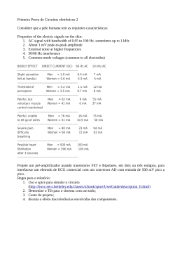
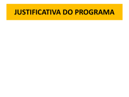
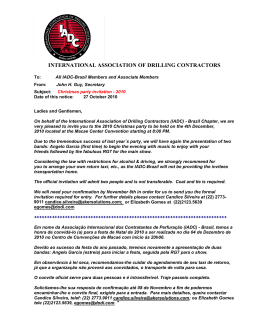
PROGRAMA DE PÓS-GRADUAÇÃO STRICTO SENSU
DOUTORADO EM ODONTOLOGIA
FABIANO PAIVA VIEIRA
PROPRIEDADES BIOLÓGICAS E FÍSICO-QUÍMICAS DE
BIOMATERIAIS EXPERIMENTAIS PARA PROTEÇÃO DO
COMPLEXO DENTINO-PULPAR
Londrina
2015
FABIANO PAIVA VIEIRA
PROPRIEDADES BIOLÓGICAS E FÍSICO-QUÍMICAS DE
BIOMATERIAIS EXPERIMENTAIS PARA PROTEÇÃO DO
COMPLEXO DENTINO-PULPAR
Tese apresentada ao Programa de PósGraduação em Odontologia da Universidade
Norte do Paraná - UNOPAR, como requisito
parcial à obtenção do título de Doutor em
Odontologia.
Área de Concentração: Dentística
Orientador: Prof. Dr. Sergio da Silva Cava
Co-orientador: Prof. Dr. Alcides Gonini Júnior
Co-orientador: Prof. Dr. Cesar Henrique Zanchi
Londrina
2015
AUTORIZO A REPRODUÇÃO TOTAL OU PARCIAL DESTE TRABALHO, POR
QUALQUER MEIO CONVENCIONAL OU ELETRÔNICO, PARA FINS DE
ESTUDO E PESQUISA, DESDE QUE CITADA A FONTE.
Dados Internacionais de catalogação-na-publicação
Universidade Norte do Paraná
Biblioteca Central
Setor de Tratamento da Informação
V721p
Vieira, Fabiano Paiva
Propriedades biológicas e físico-químicas de biomateriais experimentais para
proteção do complexo dentinho-pulpar / Fabiano Paiva Vieira Londrina: [s.n],
2015.
113f.
Tese (Doutorado). Odontologia. Dentística. Universidade Norte do Paraná.
Orientador: Prof. Dr. Sérgio da Silva Cava
Co-orientador: Prof. Dr. Alcides Gonini Júnior
Co-orientador: Prof. Dr. Cesar Henrique Zanchi
1- Odontologia - tese - doutorado - UNOPAR 2-Propriedades físicas e
químicas 3- Propriedades biológicas 4- Cimentos odontológicos 5- Capeamento
da polpa dentária I- Cava, Sérgio da Silva, oriente. II- Gonini Junior, Alcides,
orient. III- Zanchi, Cesar Henrique, orient. IV- Universidade Norte do Paraná.
CDU 616.314-089.27/.28
FABIANO PAIVA VIEIRA
PROPRIEDADES BIOLÓGICAS E FÍSICO-QUÍMICAS DE
BIOMATERIAIS EXPERIMENTAIS PARA PROTEÇÃO DO
COMPLEXO DENTINO-PULPAR
Tese apresentada ao Programa de PósGraduação em Odontologia da Universidade
Norte do Paraná - UNOPAR, como requisito
parcial à obtenção do título de Doutor em
Odontologia.
Área de Concentração: Dentística
BANCA EXAMINADORA
____________________________________
Prof. Dr. Sergio da Silva Cava
Universidade Federal de Pelotas
____________________________________
Prof. Dr. Evandro Piva
Universidade Federal de Pelotas
____________________________________
Prof. Dr. Rafael Ratto de Moraes
Universidade Federal de Pelotas
____________________________________
Profª. Drª. Giana da Silveira Lima
Universidade Federal de Pelotas
____________________________________
Profª. Drª. Cristiane Wienke Raubach Ratmann
Universidade Federal de Pelotas
Pelotas, _____de ___________de 2015.
Dedico este trabalho a Deus, meus familiares e
amigos.
AGRADECIMENTOS
Agradeço a Deus, meus pais, Waterloo Vieira Fonseca e Elizabeth
Paiva Vieira, minha companheira Raiane de Barros, seus pais José Cesar Buschetti e
Elizete de Barros Buschetti.
Agradeço aos amigos da panela Helen e Albery, Renato e Giseli.
Agradeço aos amigos do Instituto Federal do Paraná pelo apoio,
Marcelo Poleti e Thaís, Tânia Simões e André, Berenice Tatibana, Juliana Vizoto,
Paulo Rossato e Carlos Bertoncelo, Marcelo Estevam, Amir Limana, Silvana Sona,
Geraldo Teixeira, Luiz, Dina, Mônica e todos colegas de trabalho.
Agradeço aos amigos da Universidade Federal de Pelotas pelo apoio,
Sergio Cava, Cesar Tino, Evandro Piva, Neftalí Carreño, Rafael Moraes, Carolina,
Guilherme, Faili, Hellen, Wellington, Cristina, Fernanda e Tati.
Agradeço aos amigos da UNOPAR pelo apoio, Alcides Gonini Jr.,
Gleydson, Alessandro e demais colegas.
Agradeço aos funcionários do CEME Sul – FURG.
VIEIRA, Fabiano Paiva. Propriedades biológicas e físico-químicas de
biomateriais experimentais para proteção do complexo dentino-pulpar. 41. [Tese
de Doutorado]. Programa de Pós-Graduação em Odontologia – Universidade Norte
do Paraná, Londrina, 2015.
RESUMO
O objetivo deste estudo foi avaliar propriedades físicas-químicas e biológicas de
cimentos experimentais para capeamento pulpar. Foram desenvolvidos cimentos
resinosos de presa dual com diferentes tipos de partículas de carga inorgânica,
agregado trióxido mineral (MTA), titanato de cálcio e aluminatos de cálcio (CA)
distintos. Estas partículas foram caracterizadas por espectroscopia de infravermelho,
espectroscopia por dispersão de energia de raios X, difração de raios X e microscópio
eletrônico de varredura. A resistência à tração diametral, potencial hidrogeniônico (pH)
e citotoxicidade dos cimentos experimentais foram avaliadas e comparadas com as
do MTA. Para a avaliação da cinética de conversão foram realizadas análises em
espectroscopia no infravermelho em tempo real (RT-FTIR). Os resultados mostraram
o potencial do material experimental em comparação com as principais propriedades
físicas e biológicas do MTA, os mais críticos para a triagem inicial de novos materiais.
As pastas CA e CLQ (clinker-Fillapex Angelus®) à base de resina apresentaram
propriedades semelhantes ou superiores às do MTA.
Palavras-chave: Propriedades físicas e químicas. Propriedades biológicas. Cimentos
odontológicos. Capeamento da Polpa Dentária.
VIEIRA, Fabiano Paiva. Biological and physicochemical properties of
experimental biomaterials for the pulp-dentin complex protection. 41. [Tese de
Doutorado]. Programa de Pós-Graduação em Odontologia – Universidade Norte do
Paraná, Londrina, 2015.
ABSTRACT
Objective: The aim of this study was to evaluate physical-chemical and biological
properties of experimental pulp capping cements. Methods: Dual-cured resin cements
with different types of inorganic filler particles, mineral trioxide aggregate (MTA),
calcium titanate and distinct calcium aluminate (CA) were developed. These inorganic
filler particles were characterized by infrared spectroscopy, spectroscopy and energy
dispersive X-ray, X-ray diffraction and scanning electron microscope. The diametral
tensile strength, hydrogen potential (pH) and cytotoxicity of experimental cements
were evaluated and compared with the MTA. Real-time degree of conversion was
performed in a Fourier transform infrared spectrometer. The results showed the
experimental material potencial in comparison with the MTA key physical and
biological properties, the critical ones to initial screening of new materials. The resin
based CA and CLQ (clinker-Fillapex Angelus®) pastes had similar or superior
properties to those of MTA.
Key-words: Physical and chemical properties. Biological properties. Dental cements.
Dental Pulp Capping.
SUMÁRIO
1 INTRODUÇÃO ....................................................................................................... 9
2 REVISÃO DE LITERATURA - CONTEXTUALIZAÇÃO ........................................ 11
3 PROPOSIÇÃO ....................................................................................................... 14
4 ARTIGO ............................................................................................................... 15
5 CONCLUSÃO ........................................................................................................ 35
REFERÊNCIAS ......................................................................................................... 36
9
1 INTRODUÇÃO
O tecido pulpar é envolto em tecido duro e rodeado por células
formadoras deste [1], os odontoblastos, que são células organizadas como uma
camada de células em paliçada ao longo da interface entre a polpa dentária e dentina.
Eles são responsáveis pela formação da dentina fisiológica primária e da secundária,
também participam da manutenção desse tecido duro ao longo da vida útil do dente,
sintetizando dentina reacionária em resposta a estímulos adversos ou condições
patológicas [2,3].
Algumas condições como a cárie, o trauma ou o procedimento de
preparo dentário podem expor a polpa dentária. Uma das alternativas para esta
condição é o capeamento pulpar, em que um medicamento é colocado diretamente
sobre a polpa exposta (capeamento pulpar direto), ou um material forrador de
cavidade é colocado sobre a cárie residual (capeamento pulpar indireto) em uma
tentativa de manter a vitalidade pulpar e evitar um tratamento mais extenso exigido
pela terapia endodôntica [4]. O potencial para a cura por formação de uma ponte
dentinária é bom, desde que a polpa não esteja inflamada [5].
A era da terapia de polpa vital tem sido bastante reforçada com a
introdução de vários materiais de capeamento pulpar [6]. Apesar destas alternativas,
apenas o hidróxido de cálcio tem uma longa história de indução à formação de pontes
de dentina para promover a recuperação pulpar bem sucedida [5]. Porém, a literatura
sugere que o agregado trióxido mineral (MTA) é o material de escolha para
capeamento pulpar em dentes permanentes em comparação com os materiais usados
atualmente [7-11].
Resinas compostas estão surgindo como materiais alternativos para
capeamento pulpar, mas a cura é mais lenta, e relativamente pouca pesquisa clínica
está disponível para análise [12,5]. Alguns monómeros liberados por estas resinas são
citotóxicos e induzem efeitos genotóxicos [13]. A fim de melhorar a biocompatibilidade
destes materiais resinosos, foi proposta a utilização de monômeros de elevado peso
molecular [14,15]. Além disso, um material resinoso de capeamento pulpar pode ter a
vantagem adicional de ligação química com a resina utilizada para a restauração,
assim minimizar a ocorrência de falhas no material de capeamento e na interface
destes materiais [16], pois alguns autores sugerem que a aplicação do ácido na
superfície do agregado trióxido mineral afeta a sua micromorfologia e a força de
10
ligação deste ao composito [17]. A formulação resinosa também permitiria uma
aplicação imediata [18].
Alguns estudos têm demonstrado que um cimento fotopolimerizável à
base de agregado trióxido mineral apresenta resultados semelhantes ao MTA®
(Angelus, Londrina, PR, BR) [18,19]. Outra pesquisa com produtos comerciais,
TheraCal® (Bisco Inc., Schaumburg, IL, USA), também indicam propriedades
semelhantes ou até melhores que o ProRoot MTA® (Dentsply Tulsa Dental Specialties,
Tulsa, OK, USA) [20].
Assim, este estudo propõe a utilização de aluminatos de cálcio [21]
como partículas de carga, que podem ser considerados como material potencialmente
alternativo ao agregado trióxido mineral [22,23] e materiais resinosos com monómeros
de elevado peso molecular [15], a fim de criar materiais com potencial de utilização na
proteção do complexo dentino-pulpar. Portanto, as propriedades físico-químicas e
biológicas destes serão testadas.
11
2 REVISÃO DE LITERATURA – CONTEXTUALIZAÇÃO
A polpa dentária é um tecido conjuntivo especializado que, como a
maioria dos tecidos humanos, apresenta uma capacidade de regeneração limitada.
[24]. A cárie, o trauma ou o procedimento de preparo dentário podem expor esta polpa
dentária. A morbidade associada ao tratamento das exposições pulpares pode exigir
um tratamento endodôntico, mas um procedimento alternativo para este é o
capeamento pulpar, em que um medicamento é colocado diretamente sobre a polpa
exposta (capeamento pulpar direto), ou um material forrador de cavidade é colocado
sobre cárie residual (capeamento pulpar indireto) em uma tentativa de manter a
vitalidade pulpar e evitar um tratamento mais extenso exigido pela terapia
endodôntica. As chances de sobrevivência dos dentes são excelentes, se o dente é
assintomático e bem selado, mesmo que uma cárie residual permaneça [4]. O
potencial para a cura por formação de uma ponte dentinária é bom, desde que a polpa
não esteja inflamada [5].
Embora haja muitos produtos, uma recente revisão sistemática
concluiu que não existem provas suficientes quanto ao material de capeamento pulpar
mais adequado [25]. Alguns materiais de capeamento pulpar utilizados para a
proteção do complexo dentino-polpar são o hidróxido de cálcio, cimento de óxido de
zinco e eugenol (ZOE), cimento de fosfato de cálcio, ionômero de vidro / ionômero de
vidro modificado por resina, agregado trióxido mineral (MTA), Biodentine ® (Septodont,
St. Maurdes Fossés, France), novo cimento endodôntico (NEC, Shahid Beheshti
University, Tehran, Iran), agente resinoso de capeamento pulpar direto com hidróxido
de cálcio (Ca-MTYA) dentre outros materiais e métodos [6]. Apesar destas
alternativas, apenas o hidróxido de cálcio tem uma longa história de indução à
formação de pontes de dentina para promover a recuperação pulpar bem sucedida
[5].
Na área da terapia da polpa vital, o agregado trióxido mineral parece
ser equivalente e possivelmente superior ao clássico CaOH em termos de
capeamento pulpar direto [11]. Os dados disponíveis mostram que a mistura de
agregado trióxido mineral com água resulta na formação de hidróxido de cálcio e um
ambiente de alto pH. A literatura mostra que o agregado trióxido mineral tem um efeito
antibacteriano e antifúngico [26], que é um material biocompatível e não é mutagênico
[27].
12
A literatura sugere que o agregado trióxido mineral é o material de
escolha para capeamento pulpar em dentes permanentes em comparação com os
materiais usados atualmente [9,10]. As informações atuais sugerem que o agregado
trióxido mineral é um material bioativo e possui a habilidade de criar um ambiente ideal
para a recuperação pulpar. A partir do momento que o agregado trióxido mineral é
colocado em contato direto com os tecidos humanos, sugere-se que o material induz
a formação de hidróxido de cálcio que libera íons de cálcio para a fixação e
proliferação celular, cria um ambiente antibacteriano pelo seu pH alcalino, modula a
produção de citocinas, estimula a diferenciação e migração de células produtoras de
tecido mineralizados e forma hidroxiapatita ou apatita carbonatada na superfície do
cimento e fornece um selamento biológico [28,9]. O tempo de presa do agregado
trióxido mineral é uma das desvantagens deste material. Além disso, a literatura
sugere que a presença de diferentes soluções afeta as propriedades físicas do
agregado trióxido mineral [26].
Resinas compostas estão surgindo como materiais alternativos para
capeamento pulpar, porém pouca pesquisa clínica está disponível para análise [12,5].
Estas resinas odontológicas são biomateriais comumente usados para restaurar
esteticamente a estrutura e função dos dentes prejudicados pela cárie, a erosão ou
fratura. Monômeros residuais liberados das restaurações de resina, como resultado
de processos de polimerização incompleta, podem interagir com os tecidos orais.
Alguns monómeros são citotóxicos, induzem efeitos genotóxicos, influem no ciclo
celular e a resposta das células do sistema imune inato, inibem as funções dos
odontoblastos ou retardam os processos de diferenciação e de mineralização
odontogênicos em células derivadas de polpa, incluindo células tronco [13].
A fim de melhorar a biocompatibilidade de materiais resinosos,
monómeros de elevado peso molecular, como o dimetacrilato etoxilado bisphenol A
glicol (Bis-EMA 30), foram sugeridos pela literatura [14,15]. Assim, em um estudo
sobre novos cimentos para capeamento pulpar, a comparação das propriedades
físico-químicas e biológicas entre o agregado trióxido mineral, um cimento de
ionômero de vidro (CIV) e outros novos cimentos experimentais baseados na
combinação sinérgica de materiais existentes (híbrido, pasta e resinoso) foi explorada
e o cimento resinoso experimental apresentou desempenho semelhante ou superior
aos materiais comerciais e experimentais avaliados [15].
13
Além disso, um material resinoso de capeamento pulpar pode ter a
vantagem adicional de ligação química com a resina utilizada para a restauração,
assim minimizar a ocorrência de falhas no material de capeamento e na interface
destes materiais [16].
Vários materiais são sugeridos para capeamento pulpar, mas ainda
não há um material com as propriedades necessárias para o desempenho ideal. A
combinação das propriedades de escolha de diferentes materiais pode permitir o
desenvolvimento de novos cimentos com propriedades aprimoradas e melhorar os
resultados das atuais estratégias terapêuticas da polpa [24].
A utilização de diferentes partículas de carga, aluminatos de cálcio
(CA) e titanatos de cálcio (CaTiO3), e materiais resinosos com monómeros de elevado
peso molecular [15], poderá criar materiais biocompatíveis, compósitos, com potencial
para alcalinizar o meio e aumentar os níveis extracelulares de íons Ca 2+ [22,29,30],
propriedades que favorecem a formação de pontes de tecido mineralizado e sua
utilização no capeamento pulpar direto.
14
3 PROPOSIÇÃO
O propósito deste estudo é avaliar determinadas propriedades físicoquímicas e biológicas de cimentos experimentais para proteção do complexo dentinopulpar. Assim, será testada a seguinte hipótese:
A utilização de aluminatos de cálcio (CA) e titanatos de cálcio
(CaTiO3) como partículas de carga associadas a materiais resinosos com monômeros
de elevado peso molecular pode resultar em materiais, compósitos, com propriedades
físico-químicas e biológicas equivalentes ao MTA.
15
ARTIGO
BIOLOGICAL AND PHYSICOCHEMICAL PROPERTIES OF EXPERIMENTAL
BIOMATERIALS FOR THE PULP-DENTIN COMPLEX PROTECTION
(A ser submetido ao periódico Dental Materials)
16
Essential title page information
Title. Properties of experimental pulp capping cements.
Fabiano Paiva Vieira a1, Sergio da Silva Cavab, Cesar Henrique Zanchic, Alcides Gonini
Júniord, Evandro Pivac, Héllen de Lacerda Oliveirae, Wellington Luiz de Oliveira da
Rosae, Adriana Fernandes da Silvac.
a
Federal Institute of Paraná, Campus Londrina, PR, Brazil.
b
Department of Materials Engineering, School of Engineering, Federal University of
Pelotas, RS, Brazil.
c
Department of Operative Dentistry, School of Dentistry, Federal University of Pelotas,
RS, Brazil.
d
Head professor, Department of Dentistry, University of Northern Paraná - Londrina,
Paraná, Brazil.
e
school of Dentistry, Federal University of Pelotas, RS, Brazil.
¹Corresponding author. Fabiano Paiva Vieira. Federal Institute of Paraná, Londrina
Campus, PR, Brazil, Rua João XXIII, 600, Praça Horace Wells, Jardim Dom Bosco,
CEP.: 86.060-370. Fone: 55 43 33519644, e-mail: [email protected].
17
ABSTRACT
Objective: The aim of this study was to evaluate physical-chemical and biological
properties of experimental cements. Methods: Dual-cured resin cements with different
types of inorganic filler particles, mineral trioxide aggregate (MTA), calcium titanate
and distinct calcium aluminate (CA) were developed. These inorganic filler particles
were characterized by infrared spectroscopy, spectroscopy and energy dispersive Xray, X-ray diffraction and scanning electron microscope. The diametral tensile strength,
hydrogen potential (pH) and cytotoxicity of experimental cements were evaluated and
compared with the MTA. Real-time degree of conversion was performed in a Fourier
transform infrared spectrometer. Results: The results showed the experimental
material potencial in comparison with the MTA key physical and biological properties,
the critical ones to initial screening of new materials. The resin based CA and CLQ
(clinker-Fillapex®, Angelus, Londrina, PR, BR) pastes had similar or superior properties
to those of MTA. Significance: the proposed materials have as advantage to be able
to bind chemically to the restorative composite resin to form a stronger interface.
Another advantage would be the technical simplification of the pulp-dentin complex
protection, requiring only two steps and less time.
Key-words: Physical and chemical properties. Biological properties. Dental cements.
Pulp-Dentin Complex Protection.
18
1. Introduction
The dental pulp is a highly vascularized and innervated connective
tissue responsible for maintaining the tooth vitality and able to respond to injuries [31]
and is encased in hard tissue and surrounded by hard tissue-forming cells [1].
Odontoblasts are post-mitotic cells organized as a layer of palisade cells along the
interface between the dental pulp and dentin. They are responsible for the formation
of the physiological primary and secondary dentins. They also participate to the
maintenance of this hard tissue throughout the life of the tooth by synthesizing
reactionary dentin in response to pathological conditions [2].
The consequences of pulp exposure from caries, trauma or tooth
preparation misadventure can be severe. The morbidity associated with treating pulp
exposures is consequential, often requiring either extraction or endodontic therapy, but
an alternative procedure to these options is pulp capping, in which a medicament is
placed directly over the exposed pulp (direct pulp cap), or a cavity liner or sealer is
placed above residual caries (indirect pulp cap) in an attempt to maintain pulp vitality
and avoid the more extensive treatment dictated by extraction or endodontic therapy.
The chances for tooth survival are excellent if the tooth is asymptomatic and well
sealed, even if residual caries remains [4]. The potential for healing by formation of a
dentinal bridge is good, in case the pulp is not inflamed [5].
The era of vital-pulp therapy has been greatly enhanced with the
introduction of various pulp capping materials [6]. The highest level of current best
evidence has revealed that calcium-enriched mixture cement is a suitable endodontic
biomaterial for vital pulp therapy treatments of primary molars as well as
mature/immature permanent teeth with reversible/irreversible pulpitis [32]. It appears
that mineral trioxide aggregate is the best choice material for pulp capping in
permanent teeth compared with currently used materials [9,10,11].
Another option for pulp capping, resin-based composites, may be
promising, however more and long-term researches are necessary [12,5]. Studies on
the molecular toxicology of substances released by resin-based dental restorative
materials clearly support that the majority of these molecules are able to cause
cytotoxic and genotoxic effects at concentrations relevant to those released into the
oral cavity. These effects include irreversible disturbance of basic cellular functions,
such as cell proliferation, enzyme activities, cell morphology, membrane integrity, cell
19
metabolism and cell viability [33]. To improve the biocompatibility of resinous materials,
new monomers with a high molecular weight have been proposed, reducing
opportunities for monomer diffusion through dentin and toxicity [14,15]. Furthermore,
a resinous capping material may have the additional advantage of chemical bonding
with the composite resin used for restoration, minimizing the occurrence of failures at
the capping material/restorative material interface [15,16], situation caused by acidic
treatment of the mineral trioxide aggregate surface [17]. Resinous formulation can
allow light-cure, immediate setting and better working properties [18].
An experimental light-cure mineral trioxide aggregate has been
developed to have similar properties to mineral trioxide aggregate, but with better
working properties [18,19]. Other research reported that TheraCal® (Bisco Inc.,
Schaumburg, IL, USA), another light-curable MTA-like material for pulp capping,
displayed higher calcium-releasing ability and lower solubility than either ProRoot
MTA® or Dycal® (Dentsply Tulsa Dental Specialties, Tulsa, OK, USA). These properties
offer major advantages in direct pulp-capping treatments [20,34].
Therefore, this study proposed the use of calcium aluminate (CA) and
calcium titanate (CaTiO3) and resinous materials with high molecular weight
monomers to create biocompatible materials [15,22,29,30] with the potential to alkaline
environment and to release calcium ions (Ca2+), properties that favor the formation of
mineralized tissue bridges [9] and their use in the pulp-dentin complex protection.
Thus, the biological and physico-chemical properties of these biocompatible materials
will be tested.
2. Materials and methods
2.1 Formulation of experimental materials
Calcium aluminate (CA) and calcium titanate (CaTiO3) are used as the
inorganic filler particles to create the experimental groups, they have the potential to
increase the biocompatibility of the material developed, to rise the environmental pH
and extracellular levels of Ca2+ ions [22,29,30], properties that favor the formation of
mineralized tissue bridges and the potential outcome of the proposed material.
20
2.1.1 Synthesis and characterization of inorganic particles
Currently, some methods are employed for the production of the
interest CA and calcium titanate crystalline phase, this study used the Pechini method
to obtain calcium titanate and combustion for the synthesis of CA, because they have
the advantages of being simple synthesis techniques, performed at low temperatures
and with a good control of the powders’ composition [35,36].
Calcium nitrate tetrahydrated (Ca(NO3)2• 4H2O) and aluminum nitrate
nonahydrated (Al(NO3)3•9H2O) and urea (CO(NH2)2) were used to produce CA. Those
two reagents and the fuel were obtained from Sigma-Aldrich (St. Louis, USA) and used
without any further treatment. The amount of each component required for the
chemical reaction to obtain tricalcium aluminate was calculated based on the total of
valencies of oxidizing and reducing reagents and fuel [35].
These reagents already weighed on the hot plate was taken onto a
heating plate at 90°C and subsequently to the preheated muffle furnace at 400°C [37].
The material obtained in this reaction was heat treated at 800°C or 1200°C during four
hours to promote formation of CA crystalline phases [21,35]. A 45 µm size opening
particle analysis sieve was used to reduce the filler particle agglomerates obtained at
the end of the described process.
Titanium isopropoxide [Ti(OC3H7)4] (Vetec, RJ, Brazil), absolute ethyl
alcohol (Vetec, RJ, Brazil), anhydrous citric acid (C6H8O7) (Synth, SP, Brazil), calcium
nitrate tetrahydrate (Ca(NO3)2•4H2O) (Sigma-Aldrich, St. Louis , USA) and ethylene
glycol (C2H6O2) (Vetec, RJ, Brazil) were used for the production of calcium titanate.
Stoichiometric calculations set the required amount of each element in the chemical
reaction to obtain calcium titanate, the citric acid/ethylene glycol mass ratio was fixed
at 60:40.
Thus, the amount of each reagent was weighed and added in the same
sequence of reagents, as described above, in a beaker with constant agitation on a
heating plate slowly heated at 100ºC to promote citrate polymerization by the
polyesterification reaction and to evaporate the solvent, adjusting the viscosity [36,38].
The obteined material, a polymeric resin, was placed in conventional furnace at 300°C
for 2 hours, with a heating rate of 1°C/min, promoting the pulverization of the polymeric
resin and formation of the precursor powder.
21
Finally, these material were heat treated at 700°C for seven hours,
with a heating rate of 20°C/min, in microwave oven to obtain the filler particles [36]. A
45 µm size opening particle analysis sieve was used to reduce the filler particle
agglomerates obtained at the end of the described process.
The characterization of the filler particles was performed by X-ray
diffraction analysis (XRD), Fourier transform infra-red (FT-IR) spectroscopy, energy
dispersive X-ray (EDX) and scanning electron microscopy [21]. The crystalline phases
analysis by X-ray diffraction (XRD) was carried out using the diffractometer Rigaku
D/Max2500 PC (Rigaku Corporation, Tokyo, Japan) and Cu K_ radiation at 30 mA and
30 kV, detector rotation between 10° and 80°, with a sampling pitch of 0.02° and scan
speed of 2°/min.
Then, the materials were analyzed using a Fourier Transform infrared
spectroscopy (FT-IR, Shimadzu Prestige21 Spectrometer, Shimadzu, Japão), with the
Happ-Genzel apodization, at a range of 4000 and 600 cm-1, spectral resolution of 4
cm−1 and 10 scans per spectrum. Background noise was removed prior to analysis by
background scans.
Elemental constitution of each phase identified was carried out by
energy dispersive X-ray (EDX) analysis with a EDX fluorescence spectrometer
(Shimadzu, Japão). The filler particles were viewed under a scanning electron
microscope (SEM; Model 5400, JEOL, Tokyo, Japan) and particles microstructure,
typical particle agglomerates and grain morphology were assessed in back scatter
electron mode at 1000X magnification.
2.1.2 Experimental groups
Six groups were proposed, the first one is the standard for comparison,
MTA® (Angelus, Londrina, PR, BR), and the others were formulated using clinkerFillapex® (Angelus, Londrina, PR, BR), calcium aluminates (CA) and calcium titanate
(CaTiO3) obtained according to the description above without any other treatment.
Experimental cements and their respective compositions are presented in Table 1.
22
Table 1 -. Tested materials, their composition and proportion
Material
Composition
Powder: Portland cement, tricalcium silicate, dicalcium
silicate, tricalcium aluminate, tetracalcium ironaluminate,
MTA Angelus®
bismuth oxide.
Liquid: distilled water
Proportion/
Curing mode
Powder / Liquid 3:1 Chemical
Paste CLQ
(CDC-Bio)
Paste 1: 60% clinker-Fillapex Angelus®, 20% Bis-EMA 10,
20% PEG 400
Initiator: 1% DHEPT, 0.8% EDAB, 0.4% CQ
Inhibitor: 0.05% butylated hydroxytoluene.
Paste 2: 60% Fluoride Ytterbium 20% Bis-EMA 10, 20%
Bis-EMA 30
Initiator: 1.5% Benzoyl Peroxide
Inhibitor: 0.05% butylated hydroxytoluene
Paste 1 / Paste 2-1:
1
Dual: Chemical and
photoactivation
Paste CA 800
(CDC-Bio)
Paste 1: 60% CA (800ºC), 20% Bis-EMA 10, 20% PEG
400
Initiator: 1% DHEPT, 0.8% EDAB, 0.4% CQ
Inhibitor: 0.05% butylated hydroxytoluene.
Paste 2: 60% Fluoride Ytterbium 20% Bis-EMA 10, 20%
Bis-EMA 30
Initiator: 1.5% Benzoyl Peroxide
Inhibitor: 0.05% butylated hydroxytoluene
Paste 1 / Paste 2-1:
1
Dual: Chemical and
photoactivation
Paste 1: 60% CA (1200ºC), 20% Bis-EMA 10, 20% PEG
400
Initiator: 1% DHEPT, 0.8% EDAB, 0.4% CQ
Paste CA 1200 Inhibitor: 0.05% butylated hydroxytoluene.
Paste 2: 60% Fluoride Ytterbium 20% Bis-EMA 10, 20%
(CDC-Bio)
Bis-EMA 30
Initiator: 1.5% Benzoyl Peroxide
Inhibitor: 0.05% butylated hydroxytoluene
Paste 1 / Paste 2-1:
1
Dual: Chemical and
photoactivation
Paste CA
(CDC-Bio)
Paste 1: 60% CA (1200ºC), 20% Bis-EMA 10, 20% PEG
400
Initiator: 1% DHEPT, 0.8% EDAB, 0.4% CQ
Inhibitor: 0.05% butylated hydroxytoluene.
Paste 2: 60% CA (1200ºC) 20% Bis-EMA 10, 20% BisEMA 30
Initiator: 1.5% Benzoyl Peroxide
Inhibitor: 0.05% butylated hydroxytoluene
Paste 1 / Paste 2-1:
1
Dual: Chemical and
photoactivation
Paste Ti
(CDC-Bio)
Paste 1: 60% Calcium titanate, 20% Bis-EMA 10, 20%
PEG 400
Initiator: 1% DHEPT, 0.8% EDAB, 0.4% CQ
Inhibitor: 0.05% butylated hydroxytoluene.
Paste 2: 60% Fluoride Ytterbium 20% Bis-EMA 10, 20%
Bis-EMA 30
Initiator: 1.5% Benzoyl Peroxide
Inhibitor: 0.05% butylated hydroxytoluene
Paste 1/Paste 2-1:
1
Dual: Chemical and
photoactivation
MTA: mineral trioxide aggregate. Bis-EMA: dieterdimethacrylate. PEG 400: poly-ethyleneglycol (400)
dimethacrylate. DHEPT: dihidroxietil-p-toluidine. EDAB: ethyl-4-dimethylamino benzoate. CQ –
camphorquinone. CA calcium aluminate.
23
2.2 Kinetics of Polymerization by RT-FTIR Spectroscopy
The degree of conversion from the experimental materials were
evaluated using real-time Fourier transform infrared spectroscopy-attenuated total
reflectance (FTIR-ATR) (ZnSe crystal, P IKE Technologies, Madison, WI). A support
standardized the distance between the fiber tip of the light curing unit (LED, Radii ®
Curing Light, SDI, Bayswater, Victoria, Australia) and the sample at 5 mm. The
IRSolution software (Shimadzu, Columbia, MD) setup was the Happ-Genzel
apodization, at a range of 1750 and 1550 cm -1, resolution of 4 cm-1 and mirror speed
of 2.8 mm/s and was used in monitoring mode during photoactivation to scan every 1
second. A standarded amount of the sample (0,1g) was manipulated for 60 s and
directly dispensed on the ZnSe crystal, then a initial scanning was performed, before,
the photoactivation and sample scanning for 60 s. The degree of conversion was
calculated considering the intensity of carbon–carbon double-bond stretching vibration
(peak height) at 1635 cm-1 and using, as an internal standard, the symmetric ring
stretching at 1610 cm-1 from the polymerized and unpolymerized samples as
described in previous literature [39]. The plotted data were analised for curve fitting
and a logistic non-linear regression was performed. These data fitting was used to
calculate the polymerization rate (RP (s-1)) as the degree of conversion at time t
subtracted of degree of conversion at time t-1. The coefficient of determination was
greater than 0.97 for CA, CA 800 e CLQ curves, but smaller to the CA1200 (0,919)
and Ti (0,918) curves, and the fitting failed for this last one [40].
2.3 Diametral Tensile Strength (DTS) Test
The diametral tensile strength was performed in a universal testing
machine (EMIC 2000, Equipamentos e Sistemas de Ensaio LTDA., São José dos
Pinhais, PR, Brasil), applying 100kgf load at a speed of 1.0 mm / min. Standard disks
(n=10, Ø=6,0 ± 0,1mm ; h=3,0 ± 0,1mm) were prepared for each experimental group,
their borders were gently polished with 600-grit abrasive paper (Norton Abrasivos
Brasil, São Paulo, SP, Brazil) and they were stored at 37ºC and 100% humidity for 24h
and a digital caliper (Mitutoyo 500-144B, Suzano SP, Brazil) was used to measure the
disks before the test. The resistance value of diametral tensile strength (σt) was
expressed in Mpa.
24
2.4 Post-Setting pH Changes
The evaluations of hydrogenic potential (pH) were performed at 3, 24,
48, 72 hours, 7 and 14 days, using a digital pH meter (608 Analion PM Plus, Ribeirão
Preto, SP, Brazil), calibrated with reference solutions. Standard disks (n=15; Ø=4,0 ±
0,1mm; h=1,0 ± 0,1mm) were prepared for each experimental group. All disks were
individually stored in Eppendorf tubes containing 1 ml distilled water and incubated at
37 ° C during all test period.
2.5 Cytotoxicity
An immortalized cell line, 3T3/NIH mouse fibroblasts, in culture
medium (Dulbecco's Modified Eagle’s Medium with 4,5g/L Glucose and L-Glutamine –
DMEM, Lonza, Walkersville,MD, USA) supplemented with 10% fetal bovine serum and
1% antibiotics (10,000 IU/mL of penicillin G and 10,000 mg/mL of streptomycin; Gibco
Laboratories Inc., Grand Island, NY, USA) was used in the cytotoxicity assay. The cells
were seeded in culture dishes and maintained in an incubator (37ºC, 5% of CO2) until
getting subconfluent.
Thus, a 96-well plate received 2 x 10⁴ cells in 200μL of culture medium
and was incubated with controlled temperature and pressure, in a humid environment
at 37 ° C, 95% air and 5% CO2 for 24 hours. After this period, there was adhesion of
cells at the bottom of the culture plate, forming a cell monolayer which was deposited
on the eluates. This was obtained simultaneously by the immersion of the standard
disks (n=6, Ø=5,5 ± 0,1mm ; h=1,0 ± 0,1mm) of each material individually in Eppendorf
micro-tubes containing 1 ml of DMEM culture medium, using the same parameters for
incubation at 37 ° C, 5% CO2 for 24 hours. These eluates replaced the medium of the
test wells and the plate incubated again for the same period under the same conditions
(37, 5% CO2 and 24h).
After 24 hours of eluate action on cells, this medium from each well
was replaced by 20μL of 3- (4,5-dimethylthiazol-2-yl) -2,5-diphenyl tetrazolium bromide
(MTT) solution (2mg / ml DMEM) and the plate incubated again for 4 hours to allow its
metabolism, to assess cell viability by the MTT assay, which is based on the ability of
viable cells to reduce it metabolically, by mitochondrial succinic dehydrogenase
enzyme activity, to a blue-purple color formazan crystal that accumulates in the
25
cytoplasm. After the incubation period, the medium was replaced with 200μL of
dimethylsulfoxide (DMSO) to resuspend the formazan.
In addition to these wells corresponding to each material tested, was
used a positive control (wells with cells without addition of eluates), a negative control
(wells without cells, with DMEM only) and compounds used in development of
experimental cements alone. This plate was analyzed by spectrophotometry Universal
ELISA reader (ELX 800; BIO-TEK Instruments, Winooski, VT, USA), in a wavelength
of 570nm where the results were evaluated considering the absorbance values as
viability cell indicator.
2.6 Statistical Analysis
Statistical analysis were performed using GraphPad Prism version
5.00 for Windows (GraphPad Software, San Diego, California USA) according to the
characteristics of the data and tests, the level of significance of 5% were applied for all
tests. Kolmogorov-Smirnov Normality Test was applied to evaluate data’s Gaussian
distribution. Then One-way analysis of variance and Tukey’s test were aplicaded on
parametric data and Kruskal-Wallis test e Dunn's Multiple Comparison Test on nonparametric data.
3. Results
Test results are presented in a descriptive way with graphs and tables.
3.1 Characterization of inorganic particles
Particles microstructure, typical particle agglomerates and grain
morphology were assessed by the scanning electron micrographs (SEM) in back
scatter electron mode at 1000X magnification (Fig. 2). Particle agglomerates and grain
size varied, showing irregular morphology.
The powders were analysed by X-ray diffraction (XRD) to identify the
present crystalline phases. All diffraction peaks were identified as belonging to the
described phase in agreement with the related literature. Figure 3 illustrates the XRD
patterns of the samples.
26
a
b
c
d
e
Figure 2 - Scanning electron micrographs (SEM) in back scatter electron mode at
1000X magnification of the stoichiometric as-prepared powders, showing particles
microstructure, typical particle agglomerates and grain morphology of (a) CA calcined
at 800°C, (b) CA calcined at 1200°C, (c) clinker-Fillapex Angelus®, (d) MTA Angelus®,
(e) calcium titanate.
The crystalline phases analysis of CA calcined at 800°C indicated the
presence of small crystallites confirmed by the broad bands diffuse and the minor
peaks in the X-ray diffraction pattern (Fig. 3a), however it evidenced the formation of
crystalline phases, mayenite (C12A7 or Ca12Al14O33) and tricalcium aluminate (C3A or
27
Ca3Al2O6), as well in the CA calcined at 1200°C, although more evidently with higher
peaks in the pattern of X-ray diffraction indicating larger grain and particles size (Fig.
3a) [21,41].
The crystalline phase analysis of MTA Angelus® and clinker-Fillapex
Angelus® showed tricalcium silicate (C3S, Ca3SiO5) indicated by the main peaks at
32.48º and 34.30º and dicalcium silicate (C2S, Ca2SiO4) indicated by the main peaks
at 32.12º and 32.50º 2θ (Fig. 3b). MTA powder showed peaks at 27.36º, 33.24º and
46.30º 2θ representing bismuth oxide (BO, Bi2O3), and peaks at 47.62º 2θ indicating
tricalcium aluminate (C3A) (Fig. 3b) [42].
*
#
a
# Ca12Al7
* C3A
#
#
** *
* *
##
#
º
b
º×
#
×
#
*
*
*
#
c
#
#
#
#
#
* CA
# C3S
d
# CaTiO3
º Ti2O3
× Ca4Ti3O10
#
×
º
#
#
#
#
#
*
× Ti-O
#
#
º
#
#
×
#
#
× #
# ×
#
º BO
# C3S
× C2S
* C3A
#
#
*
*
Figure 3 – Characterization of the crystalline phases by X-ray diffraction (XRD) and
Fourier Transform infrared spectroscopy (FT-IR), (a) CA calcined at 800°C / 1200°C
XRD, (b) clinker-Fillapex Angelus® and MTA Angelus® XRD, (c) calcium titanate XRD
and (d) all samples FT-IR.
The crystalline phase analysis of the Ti powder showed calcium
titanate indicated by the main peaks at 32.9º, 47º and 59,2º (CaTiO3, perovskite). The
powder showed peak at 37.36º and 53.74º 2θ representing calcium oxide (CaO) and
28
titanium oxide (Ti2O3), respectively (Fig. 3c) [29,38].
Fourier Transform infrared spectroscopy (FT-IR) analysis of the
powders is presented in Figure 3d. The characterization of the crystalline phases of
CA calcined at 800°C and 1200°C showed a similar pattern, as well as MTA Angelus®
and clinker-Fillapex Angelus®, as expected. Aluminate phases were identified by the
absorption peaks in the region 900-750 cm-1, which are attributed to the stretching
vibration of the tetrahedral interconnected grid of AlO4 [21]. Absorption peaks around
875 cm-1 indicated tricalcium silicate (C3S) [34]. Strong absorption peaks below 700
cm−1 were observed, which are attributed to the stretching vibration of the Ti–O bond
[29], representing titanate phases (Fig. 3d).
The elemental analysis of the powders was carried out by energy
dispersive X-ray (EDX), then their constitution and the elements' proportion is
presented in Table 2.
Table 2 -. Results of the elemental analysis of each filler particles
Material
MTA
CA 800
CA 1200
CLQ
TI
Ca(%)
70,3
77,5
76,5
91,1
76,1
Al(%)
Si(%)
6,1
Ti(%)
Bi(%)
21,4
Fe(%)
1,5
22,5
23,5
7,9
23,5
Elemental analysis by energy dispersive X-ray (EDX) of MTA Angelus® and the filler particles, CA
calcined at 800°C / 1200°C, clinker-Fillapex Angelus® and calcium titanate. The elements proportion
Ca (Calcium), Al (Aluminium), Si (Silicon), Ti (Titanate), Bi (Bismuth) and Fe (Iron) are expressed in
percentage.
This table shows the predominance of calcium element in the powders,
which is expected to be released by the application of the material.
3.2 Kinetics of Polymerization by RT-FTIR
The degree of conversion and rate of polymerization profiles of the
experimental cements are presented in Figure 4. These data showed a higher
performance of the Paste CLQ and CA 800. The others, Paste CA 1200, Ti and CA,
presented their performance in ascending order, respectively, in the degree of
conversion (Fig. 4).
29
Figure 4 - Degree of conversion and rate of polymerization profiles of the experimental
cements.
3.3 Diametral Tensile Strength (DTS) Test
The results of the diametral tensile strength (DTS) test are presented
in Figure 5.
There was no statistical difference between the performance of the
Pastes that had the best results, Paste CA 800, CA 1200 and CLQ, nor between the
8
6
4
2
TA
M
LQ
C
Ti
12
0
A
C
A
C
0
80
0
A
0
C
Diametral Tensile strength (MPa)
performance of the others, Paste CA, Ti and MTA (Table 3).
Material
Figure 5 - Results of the diametral tensile strength (DTS) test
30
Table 3 -. Results of the diametral tensile strength (DTS) test
Material
CA
CA800
CA1200
TI
CLQ
MTA
Diametral
Tensile
strength
(MPa)
4,827
(0,281)ab
6,397
(0,992)c
6,106
(0,933)c
4,448
(0,968)a
5,684
(1,284)bc
3,801
(0,588)a
One-way analysis of variance and Tukey’s test. Different lowercase letters in rows indicate statistically significant
difference (p<0.05).
3.4 Post-Setting pH Changes
The results of the pH measurements for the different materials at the
time intervals of 3 h, 24 h, 48 h, 72 h, 7 and 14 days are presented in Figure 6. During
this period, there was an initial elevation trend in the pH values, but it proved small and
still followed by subsequent slight variation or stabilization tendency of the pH values
of each material (Fig. 6).
14
CA
CA 800
CA 1200
Ti
CLQ
MTA
13
pH
12
11
10
9
8
7
0
7
14
Time interval (Days)
Figure 6 - Results of the pH measurements for the different materials at the time
intervals of 3 h, 24 h, 48 h, 72 h, 7 and 14 days
Despite the MTA present the best performance in this test, the
evaluation of the range of values of each material in the study period showed a
statistically significant increase in pH values only for Pastes CA and CLQ, although it
is a small numeric variation (Table 4). The comparison of pH values of the materials in
31
each time period evaluated showed that at the beginning, in the evaluation of 3h, only
the Paste Ti had no statistical difference with the MTA. However, in the last study
period, 14 days, Pastes Ti, CLQ and CA also showed no statistically significant
difference from the MTA, showing a tendency to the balance of performance between
some materials (CA, Ti, CLQ and MTA) in the long time. The Paste CA 800 had the
worst performance in this test (Table 4).
Table 4 -. Mean and standard deviations (SD) of pH for the different materials at the
time intervals of 3 h, 24 h, 48 h, 72 h, 7 and 14 days.
Time
CA
CA 800
CA1200
TI
CLQ
MTA
interval
3h
10,58ab
9,91a
9,88a
11,09bc
10,16a
11,33c
A(0,08)
A(0,12)
A(0,10)
A(0,11)
A(0,11)
A(0,08)
24h
48h
72h
7d
14 d
10,94ab
10,22a
10,48a
11,52b
A(0,33)
A(0,16)
10,94ab
A(0,21)
11,00ab
AB(0,21)
AB(0,07)
A(0,15)
11,07ab
AB(0,30)
10,01c
A(0,37)
10,43ac
A(0,21)
11,50bde
AB(0,08)
11,10ad
B(0,13)
11,79e
A(0,14)
11,08ab
10,07c
A(0,37)
10,42acd
A(0,22)
11,50b
AB(0,34)
AB(0,24)
11,12bd
B(0,13)
A(0,16)
10,21ac
A(0,49)
B(0,13)
11,11bd
B(0,22)
A(0,10)
10,53ab
A(0,74)
AB(0,66)
10,97abc
B(0,65)
A(0,55)
10,94ab
9,80c
AB(0,37)
A(0,60)
11,37ac
B(0,51)
A(0,95)
10,01b
11,76d
11,26ac
11,70b
11,54d
12,02c
Kruskal-Wallis test e Dunn's Multiple Comparison Test. Different uppercase letters in columns and lowercase letters
in rows indicate statistically significant difference (p<0.05).
3.5 Cytotoxicity
Results of the cell viability in the different groups after 24h and 48h in
contact with cement eluates are presented in Figure 7.
The analysis of the values in both time periods showed that there were
statistical difference between the tested materials, CA, Ti, MTA and the control,
presenting the worst results (Table 5).
32
Cell viability 24h
Cell viability 48h
3
1
1
2
1
1
A
3A
C
3A
C
C
24
h
3A
80
0
A
C
C
2
0
24
12
h
00
24
h
TI
24
C
LQ h
24
M
TA h
24
h
B
LD
24
C h
C
24
h
0
3
48
h
80
3A 0
48
12
h
00
48
h
TI
48
C
LQ h
48
M
TA h
48
h
B
LD
48
C h
C
48
h
2
Absorbance
2
3
C
Absorbance
3
Material
Material
Figure 7 - Results of the cell viability after 24 h and 48 h in contact with cement eluates
Table 5 -. Mean and standard deviation (SD) for cell viability in the different groups
after 24 h and 48 h in contact with cement eluates.
Time
interval
CA
CA800
CA1200
TI
CLQ
MTA
BLD
CC
24h
1,734
(0,264)a
2,030
(0,295)ab
2,008
(0,261)ab
1,621
(0,271)a
2,007
(0,298)ab
1,102
(0,147)c
2,321
(0,108)b
2,401
(0,065)b
48h
0,944
(0,338)a
1,594
(0,130)ac
1,450
(0,092)ac
1,128
(0,374)ab
1,607
(0,081)ac
1,011
(0,139)ad
1,749
(0,131)bc
2,210
(0,319)c
One-way analysis of variance and Tukey’s test for 24h / Kruskal-Wallis test e Dunn's Multiple Comparison Test
aplicaded for 48h, different letters indicate statistically significant difference (p<0.05).
4. Discussion
Interpretations of the data, their eventual implications and limitations
were related to literatura.
The characterization of the inorganic particles showed a mixture of
phases as a result of the synthesis process. The preparation of CA powder via solution
combustion synthesis using only urea as fuel produced a mixture of CA phases, C3A
and C12A7 [41, 43] and the aditional annealing promoted the degree of crystallinity and
grain growth and the formation of CA crystalline phases [35,21].
In the same way, the chemical method employed for the synthesis of
CaTiO3 perovskite generated amorphous carbon powders from residual organic
compounds, pulverized citric acid and ethylene glycol. The microwave oven system
33
used to annealing promotes the rapid phase formation also related with the TiO2
formation, which is able to absorb partially the microwave radiation [38].
The crystalline phase analysis of MTA Angelus® and clinker-Fillapex
Angelus® also showed a mixture of phases, aspect noticed even in the micrographs
of the MTA powder. Therefore, this condition was balanced between the samples,
although it could have influenced some differences in the test results.
Kinetics of polymerization analisys by RT-FTIR showed the higher
performance of more fluid and translucent pastes, CLQ and CA 800, with the higher
values of degree of conversion and rate of polymerization, respectively. This can be
explained by the factors related to the filler particle. The first factor is the powder
density difference. To obtain the same weight of different powders, the volume required
for each will depend on their respective densities, so the use of a low-density powder
will require greater volume, creating a less fluid and translucent pastes. The other
factor is the homogenic distribution of particle size. These factors influence the paste
flow capacity, as well as its translucency. The performance in the polymerization
kinetics increases in direct proportion to the translucency and paste flow capacity.
Thus, the most opaque and less fluid pastes showed the worst performance, Paste
CA, CA 1200 and Ti. The ternary photoinitiation system (DHEPT, EDAB and CQ) used
contributed to this result.
Furthermore, despite the use of calcium aluminate in different pastes,
the results varied widely because of the factors previously described, which is related
with the calcination temperature, that promotes difference in particle size and phase
distribution, as cited before (Fig. 4).
Despite the low values of the Pastes CA and Ti in the polymerization
kinetics, they had a similar performance than the MTA in diametral tensile test,
whereas the Pastes CA 1200, CA 800 and CLQ had a superior performance, in
accordance with others findings in the literature [15]. These results showed the
potencial of the dual cure system by chemical and photoactivation, using a ternary
photoinitiation system (DHEPT, EDAB and CQ).
Moreover, the superior mechanical properties of the aluminate is due
to the presence of CA phases, mainly C12A7, which hydrates rapidly, improving the
cement setting time properties. Particularly, the Paste CA 800 has small crystallites
confirmed by the broad bands diffuse and the minor peaks in the X-ray diffraction
pattern, and thus more reactivity, which improves the setting time. Thus, this paste
34
presents a better mechanical properties than CA 1200, as the effect of augmented
annealing temperature, increasing the particle size and grain [21].
The diametral tensile strength was used to test the physical properties
of these materials due to their possible future clinical application, considering it
probably would suffer significant pressure during restorative procedures [15]. Then, in
this aspect, the MTA may be replaced by the proposed materials, because these
showed equivalent or better results. Moreover, they still have a potential advantage to
be able to bind chemically to the restorative composite resin to form a stronger
interface [16]. Another advantage would be the technical simplification of the pulp
capping, requiring only two steps and less run time, it is not necessary to wait the
setting time. However, the chemical and biological properties are essential to this
potential aplication, so the results related to them will be discussed below.
The results of the pH measurements of all tested materials showed a
good potential to alkalize the environment and a tendency of stabilization of the values
in relation to the initial ones during the study period. Although the values are slightly
higher than other reported, these results were corroborated, especially the described
tendency of stabilization of values for a certain period when using resin material [15,
34]. Despite the best numeric performance of the MTA, the equivalent results of Pastes
CA and Ti, suggest that both materials are promising for growth factor release from
dentin, which has been implicated in signaling events for pulp repair and may favor
maintenance of the potential antimicrobial effects for a period of time [15], aspect not
considered in this work.
Likewise the chemical properties, the cell viability data in cytotoxicity
test showed an equivalent performance of Pastes CA, Ti and MTA, which suggests a
potencial similar biocompatibility of the materials. This property was increased by the
resin component with its lower diffusion characteristics, which had the best result in
this test. Furthermore, the good performance of Pastes CA 800, CA 1200 and CLQ,
and the literature corroborates these findings, which may in part be a consequence of
its high dimensional stability and stable pH post-setting [15].
The opposite performance of Pastes CA, Ti and MTA in cytotoxicity
test and in post-setting pH changes suggests an adverse effect of the large increase
of pH on the biocompatibility of the material [15,23]. Higher cell viability values in the
presence of MTA in 24h evaluation can be found in the literature [44], however as it
increases the time of evaluation, the cell viability found values decreased [45]. Thus,
35
more research are needed to clarify this issue.
Nevertheless, the results showed the experimental material potencial
in comparison with the MTA key physical and biological properties, the critical ones to
initial screening of new materials. The resin based CA and CLQ (clinker-Fillapex
Angelus®) pastes had similar or superior properties to those of MTA, corroborating
literature findings [15, 22, 23, 34].
5. Conclusion
Calcium aluminate (CA) and calcium titanate (CaTiO3) used as filler
particles in resin with high molecular weight monomers have the potencial to create a
biomaterial for pulp capping with similar physicochemical and biological properties to
those of MTA.
Significance: the proposed materials have as advantage to be able to
bind chemically to the restorative composite resin to form a stronger interface. Another
advantage would be the technical simplification of the pulp capping, requiring only two
steps and less time.
Finally, since the physico-chemical and biological properties have
been investigated, it is indicated as future research comparing the radiopacifier
potential of CA filler particles with the bismuth oxide. Once the aluminate s cheaper
and less toxic.
36
REFERÊNCIAS
1.
Zhou M, Kawashima N, Suzuk N, Yamamoto M, Ohnishi K, Katsube KI, et al.
Periostin is a negative regulator of mineralization in the dental pulp tissue.
Odontology. 2014.
2.
Bleicher F. Odontoblast physiology. Exp Cell Res. 2014;325(2):65-71.
3.
Ricucci D, Loghin S, Lin LM, Spångberg LS, Tay FR. Is hard tissue formation
in the dental pulp after the death of the primary odontoblasts a regenerative or a
reparative process? J Dent. 2014;42(9):1156-70.
4.
Hilton TJ. Keys to clinical success with pulp capping: a review of the literature.
Oper Dent. 2009;34(5):615-25.
5.
Mjör IA. Pulp-dentin biology in restorative dentistry. Part 7: The exposed pulp.
Quintessence Int. 2002;33(2):113-35.
6.
Qureshi A, E S, Nandakumar, Pratapkumar, Sambashivarao. Recent
advances in pulp capping materials: an overview. J Clin Diagn Res. 2014;8(1):31621.
7.
Accorinte ML, Loguercio AD, Reis A, Bauer JR, Grande RH, Murata SS, et al.
Evaluation of two mineral trioxide aggregate compounds as pulp-capping agents in
human teeth. Int Endod J. 2009;42(2):122-8.
8.
Mente J, Geletneky B, Ohle M, Koch MJ, Friedrich Ding PG, Wolff D, et al.
Mineral trioxide aggregate or calcium hydroxide direct pulp capping: an analysis of
the clinical treatment outcome. J Endod. 2010;36(5):806-13.
9.
Parirokh M, Torabinejad M. Mineral trioxide aggregate: a comprehensive
literature review--Part III: Clinical applications, drawbacks, and mechanism of action.
J Endod. 2010;36(3):400-13.
10.
Hilton TJ, Ferracane JL, Mancl L, (NWP) NP-bRCiE-bD. Comparison of CaOH
with MTA for direct pulp capping: a PBRN randomized clinical trial. J Dent Res.
2013;92(7 Suppl):16S-22S.
11.
Jefferies S. Bioactive and biomimetic restorative materials: a comprehensive
review. Part II. J Esthet Restor Dent. 2014;26(1):27-39.
12.
Schuurs AH, Gruythuysen RJ, Wesselink PR. Pulp capping with adhesive
resin-based composite vs. calcium hydroxide: a review. Endod Dent Traumatol.
2000;16(6):240-50.
13.
Krifka S, Spagnuolo G, Schmalz G, Schweikl H. A review of adaptive
mechanisms in cell responses towards oxidative stress caused by dental resin
monomers. Biomaterials. 2013;34(19):4555-63.
14.
Zanchi CH, Münchow EA, Ogliari FA, Chersoni S, Prati C, Demarco FF, et al.
Development of experimental HEMA-free three-step adhesive system. J Dent.
2010;38(6):503-8.
15.
Dantas RV, Conde MC, Sarmento HR, Zanchi CH, Tarquinio SB, Ogliari FA, et
al. Novel experimental cements for use on the dentin-pulp complex. Braz Dent J.
2012;23(4):344-50.
16.
Ruiz JL, Mitra S. Using cavity liners with direct posterior composite
restorations. Compend Contin Educ Dent. 2006;27(6):347-51; quiz 52.
17.
Shin JH, Jang JH, Park SH, Kim E. Effect of mineral trioxide aggregate surface
treatments on morphology and bond strength to composite resin. J Endod.
2014;40(8):1210-6.
37
18.
Gomes-Filho JE, de Faria MD, Bernabé PF, Nery MJ, Otoboni-Filho JA,
Dezan-Júnior E, et al. Mineral trioxide aggregate but not light-cure mineral trioxide
aggregate stimulated mineralization. J Endod. 2008;34(1):62-5.
19.
Gomes-Filho JE, de Moraes Costa MT, Cintra LT, Lodi CS, Duarte PC,
Okamoto R, et al. Evaluation of alveolar socket response to Angelus MTA and
experimental light-cure MTA. Oral Surg Oral Med Oral Pathol Oral Radiol Endod.
2010;110(5):e93-7.
20.
Gandolfi MG, Siboni F, Prati C. Chemical-physical properties of TheraCal, a
novel light-curable MTA-like material for pulp capping. Int Endod J. 2012;45(6):571-9.
21.
Veiga FCT. Investigação experimental das transições de fase do aluminato de
cálcio e suas características óticas e estruturais. Pelotas: Universidade Federal de
Pelotas; 2013.
22.
Castro-Raucci LMS. Efeitos de diferentes preparações de cimento de
aluminato de cálcio sobre culturas de células osteogênicas e de células
indiferenciadas da polpa dental. Ribeirão Preto: Universidade de São Paulo; 2013.
23.
Chang KC, Chang CC, Huang YC, Chen MH, Lin FH, Lin CP. Effect of
tricalcium aluminate on the physicochemical properties, bioactivity, and
biocompatibility of partially stabilized cements. PLoS One. 2014;9(9):e106754.
24.
Demarco FF, Conde MC, Cavalcanti BN, Casagrande L, Sakai VT, Nör JE.
Dental pulp tissue engineering. Braz Dent J. 2011;22(1):3-13.
25.
Miyashita H, Worthington HV, Qualtrough A, Plasschaert A. Pulp management
for caries in adults: maintaining pulp vitality. Cochrane Database Syst Rev.
2007(2):CD004484.
26.
Parirokh M, Torabinejad M. Mineral trioxide aggregate: a comprehensive
literature review--Part I: chemical, physical, and antibacterial properties. J Endod.
2010;36(1):16-27.
27.
Torabinejad M, Parirokh M. Mineral trioxide aggregate: a comprehensive
literature review--part II: leakage and biocompatibility investigations. J Endod.
2010;36(2):190-202.
28.
Okiji T, Yoshiba K. Reparative dentinogenesis induced by mineral trioxide
aggregate: a review from the biological and physicochemical points of view. Int J
Dent. 2009;2009:464280.
29.
Dubey AK, Tripathi G, Basu B. Characterization of hydroxyapatite-perovskite
(CaTiO3) composites: phase evaluation and cellular response. J Biomed Mater Res
B Appl Biomater. 2010;95(2):320-9.
30.
Ohtsu N, Sato K, Yanagawa A, Saito K, Imai Y, Kohgo T, et al. CaTiO(3)
coating on titanium for biomaterial application--optimum thickness and tissue
response. J Biomed Mater Res A. 2007;82(2):304-15.
31.
Rosa V, Botero TM, Nör JE. Regenerative endodontics in light of the stem cell
paradigm. Int Dent J. 2011;61 Suppl 1:23-8.
32.
Asgary S, Ahmadyar M. Vital pulp therapy using calcium-enriched mixture: An
evidence-based review. J Conserv Dent. 2013;16(2):92-8.
33.
Bakopoulou A, Papadopoulos T, Garefis P. Molecular toxicology of
substances released from resin-based dental restorative materials. Int J Mol Sci.
2009;10(9):3861-99.
34.
Formosa LM, Mallia B, Camilleri J. The chemical properties of light- and
chemical-curing composites with mineral trioxide aggregate filler. Dent Mater.
2013;29(2):e11-9.
35.
Fumo DA, Morelli MR, Segadães AM. Combustion synthesis of calcium
aluminates. Mater Res Bull. 1996;31(10):1243-55.
38
36.
Tai L-W, Lessing PA. Modified resin–intermediate processing of perovskite
powders: Part I. Optimization of polymeric precursors. Journal of Materials Research.
1992;7(02):502-10.
37.
Kingsley JJ, Suresh K, Patil KC. Combustion synthesis of fine-particle metal
aluminates. J Mater Sci. 1990;25(2):1305-12.
38.
Cavalcante LS, Marques VS, Sczancoski JC, Escote MT, Joya MR, Varela JA,
et al. Synthesis, structural refinement and optical behavior of CaTiO3 powders: A
comparative study of processing in different furnaces. Chemical Engineering Journal.
2008;143:307.
39.
Ogliari FA, de Sordi ML, Ceschi MA, Petzhold CL, Demarco FF, Piva E. 2,3Epithiopropyl methacrylate as functionalized monomer in a dental adhesive. J Dent.
2006;34(7):472-7.
40.
Ogliari FA, Ely C, Petzhold CL, Demarco FF, Piva E. Onium salt improves the
polymerization kinetics in an experimental dental adhesive resin. J Dent.
2007;35(7):583-7.
41.
Ianoş R, Lazău I, Păcurariu C, Barvinschi P. Fuel mixture approach for
solution combustion synthesis of Ca3Al2O6 powders. Cement and Concrete
Research. 2009;39(7):566-72.
42.
Guven Y, Tuna E, Dincol M, Aktoren O. X-ray diffraction analysis of MTA-Plus,
MTA-Angelus and DiaRoot BioAggregate2014 April 1, 2014. 211-5 p.
43.
Andrade TL, Santos GL, Pandolfelli VC, Oliveira IR. Otimização da síntese
das fases de cimento de aluminato de cálcio para fins biomédicos. Cerâmica.
2014;60:88-95.
44.
Silva EJ, Herrera DR, Rosa TP, Duque TM, Jacinto RC, Gomes BP, et al.
Evaluation of cytotoxicity, antimicrobial activity and physicochemical properties of a
calcium aluminate-based endodontic material. J Appl Oral Sci. 2014;22(1):61-7.
45.
Poggio C, Arciola CR, Beltrami R, Monaco A, Dagna A, Lombardini M, et al.
Cytocompatibility and antibacterial properties of capping materials.
ScientificWorldJournal. 2014;2014:181945.
Download