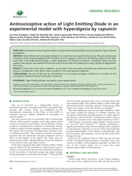
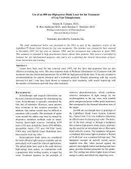
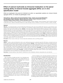
Basic Research—Technology Effects of Diode Laser (810 nm) Irradiation on Root Canal Walls: Thermographic and Morphological Studies Adriana da Costa Ribeiro, MSc,*† Gessé Eduardo Calvo Nogueira, PhD,† João Humberto Antoniazzi, PhD,* Andreas Moritz, PhD,‡ and Denise Maria Zezell, PhD† Abstract This study investigated the thermal effects and the morphological changes after diode laser irradiation (810 nm) of root canals. Samples were irradiated at 2.5 W, 1,989 W/cm2 (group 2) and 1.25 W, 10 Hz, 994 W/cm2 (group 3), with group 1 being the control group of nonirradiated samples. The temperature rise was evaluated using an infrared thermographic camera, and the morphological changes were assayed by scanning electron microscopy (SEM). The SEM images revealed closed dentinal tubules, especially at the apical regions when compared with the control samples. The maximum temperature variations at the apical region were analyzed, and the resulting 95% confidence intervals of the medians (Wilcoxon) ranged from 1.6 to 8.6°C (group 2) and from 1.2 to 3.3°C (group 3). The results suggest that the diode laser can be used for endodontic purposes and show that the method is safe for periodontal tissues at the investigated parameters. (J Endod 2007;33:252–255) Key Words Diode laser, endodontics, scanning electron microscopy, temperature, thermographic camera A mong the laser applications in various fields of dentistry, a range of laser wavelengths has been applied in endodontics to clean and disinfect the canal walls and dentine tubules, to remove the smear layer, and to seal the tubules (1– 6). The use of diode lasers in endodontic treatment has been increasing in recent years, especially because of the antimicrobial abilities of lasers, which have been investigated in vitro (7) and in vivo (8). The thermal effects are among the most important points to be considered in laser root canal irradiation, as the temperature could rise to critical levels, with deleterious effects to the tissues surrounding the tooth. The temperature can rise 10°C above body temperature for less than 1 minute without resulting in damage to periodontal tissue (9, 10). However, if the tissue temperature rises above 60°C, the blood flow is interrupted and bone necrosis is observed (11). To avoid temperature rises during irradiation, the delivery fiber should be constantly moving inside the root canal. The temperature at the external root surface can rise by 6, 12, and 18°C if the fiber remains at a fixed position in the apical region for 1, 2, or 3 seconds, respectively (12). This temperature elevation is a function of intensity output and time of irradiation, as well as the remaining dentin thickness. Recently the results of an in vitro study proposed a time protocol for preventing the increase of the temperature on the root surface. After 5 seconds of irradiation with any diode laser on internal root surfaces, the operator should rest for 5 seconds to prevent the temperature rising above the biological safety limit (13). This study was carried out to investigate thermal effects at the external root surface and the morphological changes on the root canal walls after continuous (CW) and pulsed diode laser root canal irradiation at 810 nm. Materials and Methods From the *Departamento de Endodontia, Faculdade de Odontologia, Universidade de São Paulo, São Paulo, Brazil; † Centro de Lasers e Aplicações, IPEN, Universidade de São Paulo, São Paulo, Brazil; and ‡Dental School, Medical University Vienna, Vienna, Austria. Address requests for reprints to Dr. Denise Maria Zezell, Instituto de Pesquisas Energéticas e Nucleares, Centro de Lasers e Aplicações, Av. Professor Lineu Prestes 2242, Universidade de São Paulo, 05508-900 São Paulo, Brazil. E-mail address: [email protected]. 0099-2399/$0 - see front matter Copyright © 2007 by the American Association of Endodontists. doi:10.1016/j.joen.2006.09.002 Sample Preparation Twenty-four extracted inferior human incisors were included in this study. Calculus and residual tissue were removed from the root surface, and crowns were cut at the cementum-enamel junction using a diamond disk (Buehler, Lake Bluff, USA). The working length was established 1 mm short of the apical foramen. All the canals were enlarged to an apical size of #45 by hand using K-files and cleaned with 0.5% sodium hypochlorite solution after the use of each file. The prepared samples were then randomly assigned among three groups. ● Group 1 (control): Four samples were prepared but not irradiated. The remaining 20 samples were divided into two groups: ● ● Group 2: Laser irradiating samples in continuous mode—2.5 W (peak power); Ø ⫽ 400 m; 1,989 W/cm2. Group 3: Laser irradiating samples in pulsed mode, with a duty cycle of 50%—1.25 W (mean power); 10 Hz; Ø ⫽ 400 m; 994 W/cm2. Laser Irradiation In this study, we used an 808-nm (⫾5 nm) GaAlAs laser (ZAP Softlaser, Pleasant Hill, USA), operating in continuous mode or with a duty cycle of 50% (50 ms on and 50 ms off). The power emitted at the distal end of the fiber (Ø ⫽ 400 m) was measured with a power or energy meter (Fieldmaster, Coherent Inc., Santa Clara, USA) before and after the irradiation of each sample. The fiber was inserted into the root canal to the apex 252 Ribeiro et al. JOE — Volume 33, Number 3, March 2007 Basic Research—Technology Figure 1. Representative image registered by the thermographic system (A). Average of temperature variation at external root surface during root canal laser irradiation of group 2 (B) and group 3 (C) (from upper to below line: maximum temperature; apical; medium; and coronal regions, respectively). and pulled from the apical to the coronal end in helical movements (⬃2 mm/s) (14), five times with breaks of 20 seconds in between each laser dose. All the laser irradiation was performed after the root canal had been dried. SEM Temperature Variation Measurement (⌬T) The ambient conditions were maintained at a room temperature of 21°C and relative humidity of 72%. Temperature changes were registered with a longwave focal plane array (FPA) infrared thermographic camera using a quantum well infrared photodetector (SC 3000, Boston, MA) with a sensitivity of 0.03°C. The camera was calibrated considering the dentine emissivity to be 0.91 within the temperature range of 20 to 100°C with data acquisition of 60 Hz. The safe temperature threshold resulting from irradiation laser in the root canal is considered to be 10°C (11). The samples were fixed using an apparatus that touched the root surface at two opposing points and were placed at a distance of 10 cm from the camera, corresponding to the lens focal length (Fig. 1). The data were processed using commercial software (ThermaCam Research 2001, Boston, MA) to determine the maximum temperature rise at the apical, medium, and cervical regions. The temperature variation (⌬T) of each irradiation cycle was calculated using computer software (Origin 7.0, Northampton, USA). The resulting 95% confidence intervals of the medians were analyzed using the Wilcoxon method. Although the laser interacts directly with dentine during root canal irradiation, the resulting heat from this interaction will propagate and can reach the periodontal structures. The mechanism of heat conversion depends directly on the tissue constituents and the irradiation wavelength. The dentin absorption coefficient is low for the wavelength used in this work (808 ⫾ 5 nm), so scattering is predominant against absorption (15). This fact leads photons to be absorbed far away from the irradiation surface. According to the modified Beer-Lambert law and diffusion theory, the laser intensity exponentially decreases at deeper layers of the tissue; thus, the resulting temperature at the internal root canal walls is higher than at the external root surface (15, 16). Nevertheless, the absorption of the scattered photons at deep layers can result in a temperature rise harmful to the periodontal tissue. For all these reasons, knowledge of the estimated temperature rise for a certain irradiation condition before undertaking any clinical application is extremely necessary. Temperatures reach higher values at the apical region during root canal irradiation when the thickness of the remaining root canal wall is thin (13, 17). In the present study, inferior incisors were used, and the root canals were enlarged to an apical size of #45 to evaluate the temperature variation under the worst conditions. The majority of the studies were carried out using other single-rooted teeth (13, 17–21), but the results obtained should be carefully extrapolated to inferior incisors, as the anatomy and total mass reflect a lower dentine volume. The results obtained in this study could be safety extrapolated for other dental groups, if the operating conditions are respected. Infrared thermography systems have been preferred over the thermocouple method to monitor the temperature rise because the temperature can be analyzed over a large surface area. The real-time video thermal image correlates a false color with temperature change patterns. Added to these advantages are the facilities of image analysis by the software, allowing the identification of maximum temperatures at different points. The accuracy of the thermal imaging system relies on A representative SEM image is shown in Fig. 2; melting and fusing were observed at the apical region, compared with the control group. Discussion Scanning Electron Microscopy The samples were split longitudinally in the labiolingual direction and submitted to an ultrasonic bath for 5 minutes in sterile distilled water. Subsequently, they were dehydrated through a series of graded aqueous ethanol solutions (30, 50, 70, 90, and 96%), for 10 minutes at each concentration, with a final immersion in 100% ethanol solution for 20 minutes. The specimens were sputter-coated and introduced into the vacuum chamber of a scanning electron microscope (SEM; Phillips LX-30, Eindhoven, Holland). The apical micrographs were taken at the half length of the root and 2 mm from the apex. Results Temperature Variation Measurement (⌬T) The maximum temperature at each irradiation time cycle for groups 2 and 3 are illustrated in Fig. 1. The resulting Wilcoxon 95% confidence intervals of the medians at the apical region ranged from 1.6 to 8.6°C for group 2 and from 1.2 to 3.3°C for group 3. The mean temperature rise and maximum temperature variation at the apical region are exhibited in Table 1. JOE — Volume 33, Number 3, March 2007 TABLE 1. Comparison of peak temperature rises (°C) at the apical region Group ⌬T Mean SD* Minimum Maximum Group 2 Group 3 3.8 2.4 29.28 23.71 4.98 1.67 23.1 20.0 42.1 27.7 *SD, standard deviation. Effects of Diode Laser Irradiation on Root Canal Walls 253 Basic Research—Technology Figure 2. Representative SEM image showing melting and fusion at the apical region in group 2 (A) and group 3 (B). At the middle region, the dentinal tubules remained opened in group 2 (D) and group 3 (E). In the control group, the dentinal tubules appeared open at the apical and middle thirds (C and F, respectively). Original magnification 500⫻. the emissivity of the material analyzed; thus, the measurements determined by the software system must be calibrated in accordance with the material that is being analyzed (18). The SEM images revealed melting and fusing at the apical regions. At the middle regions, only partial sealing of the tubules was observed, probably because there was less contact of the fiber with the dentinal walls, as the canal is large at this region compared with its size at the apical region, where the laser energy is more concentrated. These results agree with those of Altundasar et al. (6), who used an Er;Cr:YSGG laser, and can be correlated to the lower microleakage obtained by Karlovic et al. (1) when the root end was irradiated with Er:YAG. The output power displayed on the laser equipment was always higher than the value measured using an external power meter and was always less than 2.5 W when measured after the irradiation. The loss of energy along the fiber is a major problem for the clinician, as the lower than expected output could reduce the probability of a successful treatment. In a more hazardous situation, if the real output is higher than the settings, deleterious effects could result. Thus, the clinician should always measure the output power before treatment and remove debris from the fiber. Any in vitro study does not replicate the clinical environment completely. In vivo, the surrounding tissues, which have much lower thermal conductivity than air, together with blood flow constitute potential heat sinks. Thus, the thermal energy also dissipates more rapidly in vivo than in vitro because of the vital circulation of blood in adjacent structures (22). This study showed that the medians of ⌬T did not exceed 10°C in either of the laser experimental groups. The maximum mean of the temperature variation between the samples irradiated with the laser operating in continuous mode was 8.6°C, whereas in the pulsed group it reached 3.3°C. The peak temperatures were below the critical value of 47 °C, even for group 2 at 2.5 W output power (CW—1,989 W/cm2). The temperature values assumed in group 3 are explained by the lower fluence (pulsed—994 W/cm2), as the laser was operated with a duty cycle of 50%, which reduces the heat transport into tissue. The breaks between the irradiation cycles are very important to avoid a temperature increase above the safety limit. The gap of 20 254 Ribeiro et al. seconds between treatments in this study was greater than the 5 seconds proposed by Gutknecht et al. (13) when the root canal was irradiated by a diode laser at 1 to 1.5 W output power CW. As shown in Fig. 2, the temperature reached the peak in 10 seconds and the rise then slowed, reaching a safety limit after approximately 20 seconds for both groups. This resting time is important to enable the heat dissipation and the cooling of the tissue. The results showed that the diode laser (810 nm) can be used for endodontic applications at the investigated parameters. The temperature will not increase above the safety limit (10°C) for the periodontal tissues; however, after each treatment a 20-second resting time should be considered to prevent an excessive temperature rise in the tissue when the laser is operating in either continuous or pulsed mode. Acknowledgments This study was supported by CNPq (n.478865/2004), Capes/ Procad (n.0349054), Fapesp/CEPID. The authors thank Professor John Girkin from Strathclyde University (UK) for the fruitful discussions and grammar revision and LELO–FOUSP for the laser device. References 1. Karlovic Z, Pezelj-Ribaric S, Miletic I, Jukic S, Grgurevic J, Anic I. Erbium:YAG laser versus ultrasonic in preparation of root-end cavities. J Endod 2005;31:821–3. 2. Carvalho CA, Valera MC, Gouw-Soares S, et al. Effects of Nd:YAG and Er:YAG lasers on the sealing of root canal fillings. J Clin Laser Med Surg 2002;20:215–9. 3. Schoop U, Kluger W, Moritz A, Nedjelik N, Georgopoulos A, Sperr W. Bactericidal effect of different laser systems in the deep layers of dentin. Lasers Surg Med 2004;35:111– 6. 4. Gouw-Soares S, Stabholz A, Lage-Marques JL, et al. Comparative study of dentine permeability after apicectomy and surface treatment with 9.6 micron TEA CO2 and Er:YAG laser irradiation. J Clin Laser Med Surg 2004;22:129 –39. 5. Liu JF. Effects of Nd:YAG laser pulpotomy on human primary molars. J Endod 2006;32:404 –7. 6. Altundasar E, Ozcelik B, Cehreli ZC, Matsumoto K. Ultramorphological and histochemical changes after Er,Cr:YSGG laser irradiation and two different irrigation regimes. J Endod 2006;32:465– 8. 7. Moritz A, Gutknecht N, Goharkhay K, Schoop U, Wernisch J, Sperr W. In vitro irradiation of infected root canals with a diode laser: results of microbiologic, JOE — Volume 33, Number 3, March 2007 Basic Research—Technology 8. 9. 10. 11. 12. 13. 14. infrared spectrometric, and stain penetration examinations. Quintessence Int 1997;28:205–9. Gutknecht N, van Gogswaardt D, Conrads G, Apel C, Schubert C, Lampert F. Diode laser radiation and its bactericidal effect in root canal wall dentin. J Clin Laser Med Surg 2000;18:57– 60. Anic I, Tachibana H, Masumoto K, Qi P. Permeability, morphologic and temperature changes of canal dentine walls induced by Nd:YAG, CO2 and argon lasers. Int Endod J 1996;29:13–22. Ramskold LO, Fong CD, Stromberg T. Thermal effects and antibacterial properties of energy levels required to sterilize stained root canals with an Nd:YAG laser. J Endod 1997;23:96 –100. Eriksson AR, Albrektsson T. Temperature threshold levels for heat-induced bone tissue injury: a vital-microscopic study in the rabbit. J Prosthet Dent 1983;50:101–7. Moritz A, Doertbudak O, Gutknecht N, Goharkhay K, Schoop U, Sperr W. Nd:YAG laser irradiation of infected root canals in combination with microbiological examinations. J Am Dent Assoc 1997;128:1525–30. Gutknecht N, Franzen R, Meister J, Vanweersch L, Mir M. Temperature evolution on human teeth root surface after diode laser assisted endodontic treatment. Lasers Med Sci 2005;20:99 –103. Gutknecht N, Kaiser F, Hassan A, Lampert F. Long-term clinical evaluation of endodontically treated teeth by Nd:YAG lasers. J Clin Laser Med Surg 1996;14:7–11. JOE — Volume 33, Number 3, March 2007 15. Niemz M. Laser tissue interactions: fundamentals and applications. Berlin: Springer; 1996. 16. Fowles GR. Introduction to modern optics, 2nd ed. New York: Holt Rinehartand Winton; 1968. 17. Kimura Y, Yonaga K, Yokoyama K, Kinoshita J, Ogata Y, Matsumoto K. Root surface temperature increase during Er:YAG laser irradiation of root canals. J Endod 2002;28:76 – 8. 18. Mc Cullagh JJ, Setchell DJ, Gulabivala K, et al. A comparison of thermocouple and infrared thermographic analysis of temperature rise on the root surface during the continuous wave of condensation technique. Int Endod J 2000;33:326 –32. 19. Cohen BI, Deutsch AS, Musikant BL, Pagnillo MK. Effect of power settings versus temperature change at the root surface when using multiple fiber sizes with a Holmium YAG laser while enlarging a root canal. J Endod 1998;24:802– 6. 20. Yamazaki R, Goya C, Yu DG, Kimura Y, Matsumoto K. Effects of erbium, chromium: YSGG laser irradiation on root canal walls: a scanning electron microscopic and thermographic study. J Endod 2001;27:9 –12. 21. Deutsch AS, Cohen BI, Musikant BL. Temperature change at the root surface when enlarging a root canal with a holmium:YAG (Ho:YAG) laser, using six different fiberoptic sizes. Gen Dent 2004;52:222–7. 22. Saunders EM. In vivo findings associated with heat generation during thermomechanical compaction of gutta-percha. 1. Temperature levels at the external surface of the root. Int Endod J 1990;23:263–7. Effects of Diode Laser Irradiation on Root Canal Walls 255
Download