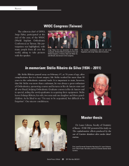
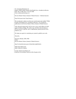
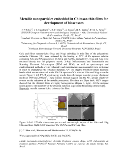
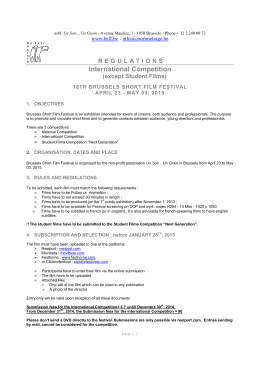
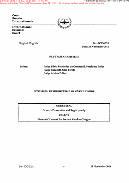
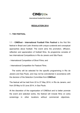
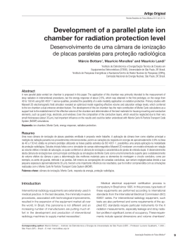
original article
Evaluation of light filter of portable dark chamber and
its influence on radiographic image quality
Marcos Coelho Santiago1
Carolina dos Santos Guimarães2
Márcia Maria Fonseca da Silveira3
Maria Luiza dos Anjos Pontual4
Carlos Estrela5
Cleomar Donizeth Rodrigues6
abstract
chamber (control). The films remained three minutes in the
developer and at every minute the films were approximated
to the display, simulating the clinical condition, followed by
intermediate bath and fixing for six minutes. Photodensitometric assessment was made and the data were analyzed
by Mann-Whitney and Kruskal-Wallis tests. Results: The
first test showed that in relation to the control group there
was no statistically significant differences with the Unemol®
chamber when using IP-21 Insight and E-Speed films and the
VH® chamber when using IP-21 Insight film. The second test
showed a significant statistical difference when comparing
one film to each other. Conclusion: This research suggested that the acrylic display increases the base veiling density,
jeopardizing the quality of the radiographic image.
Introduction: The Ordinance 453 of the Brazilian Ministry
of Health recommends processing intraoral radiographic
films in opaque portable dark chamber without display and
using the temperature x time method. However, the processing using the visual method in dark chambers with acrylic
polymer display is still frequent. The aim of this study was to
evaluate and compare the filtering capacity of four portable
dark chamber (Unemol®, VH®, MPG® and a camera without identification) with the capacity of a conventional dark
chamber (control) and check its influence on image quality in
different radiographic films (Kodak®: DF-58 Ultra-Speed, ESpeed and IP-21 Insight; and Agfa® Dentus M2). Methods:
The films were exposed with a step wedge of aluminum and
a lead plate and processed in Kodak® solutions ready for use
in each portable dark chamber and in the conventional dark
Keywords: Dental radiography. Diagnostic imaging.
Radiography.
How to cite this article: Santiago MC, Guimarães CS, Silveira MMF, Pontual
MLA, Estrela C, Rodrigues CD. Evaluation of light filter of portable dark chamber
and its influence on radiographic image quality. Dental Press Endod. 2011 OctDec;1(3):45-50.
1
Post Graduate Student,Department of Oral Maxillofacial Radiology, Brazilian Dentistry
Association, Brasília, Brazil.
2
Doctorate Student, Department of Oral Diagnosis, University of Pernambuco, Recife, Brazil.
3
Professor of Oral Diagnosis, Department of Oral Diagnosis University of Pernambuco, Recife,
Brazil.
4
Professor of Oral Maxillofacial Radiology, Department Oral Diagnosis, Federal University of
Paraíba, João Pessoa, Brazil.
5
Chairman and Professor of Endodontics, Department of Oral Science, Federal University of
Goiás, Goiânia, Brazil.
6
Professor of Oral Maxillofacial Radiology of Brazilian Dentistry Association, Brasília, Brazil.
© 2011 Dental Press Endodontics
» The authors report no commercial, proprietary, or financial interest in the
products or companies described in this article.
Received: October 25, 2011 / Accepted: November 05, 2011.
Contact address: Cleomar Donizeth Rodrigues
SMHN Q-02, bloco A, sala 208 – 70.710-100 – Brasília / DF – Brazil
E-mail: [email protected]
45
Dental Press Endod. 2011 Oct-Dec;1(3):45-50
[ original article ] Evaluation of light filter of portable dark chamber and its influence on radiographic image quality
Introduction
Auxiliary diagnostic methods are extremely important for planning treatment and radiographic examination is one of the main methods. A radiographic image quality requires knowledge and control of all
processing steps.1 With limited space and relatively
small amount of radiographic exposures in a dental
office, beyond the need for execution of the transoperative radiographs in some specialties, it became
feasible to use portable dark chamber by offering
greater flexibility in processing radiographic without
the need to shift the patient to a specialist clinic.2
In 1998, the Department of Health Surveillance of
the Ministry of Health issued the 453 ordinance regulating the use of Dental Radiology. This ordinance
allows the use of portable dark chamber for intraoral radiographs, provided they are made of opaque
material and are fitted with clock and thermometer
for the realization of radiographic processing by temperature-time method. However, the display in dark
rooms with red acrylic, are still widely used in clinics,
to perform the processing by visual inspection.
The operating conditions in a portable dark room
should be such as to enable greater efficiency and image quality. Any failure during processing can compromise the image and hinder the diagnosis.3 Currently,
intra-oral films have become more sensitive, being essential the quality control of portable dark chamber
and the knowledge of its handling by the professional.
The lack of studies on the effectiveness of red
acrylic polymer filter adequately the components of
the light spectrum in the dark chambers used in the
dental office with films of different groups of sensitivity, led to this research, whose objective was to evaluate the relationship between the light filtering capability of four portable dark chambers brands and check
their influence on the quality of radiographic imaging
in intra-oral films of different sensitivities.
The periapical films used in this research were periapical Kodak group D (DF-58 Ultra-Speed), Group E
(E-Speed), Group F (Insight) and Agfa® Dentus M2.
The dark chambers were placed on a table, located
under two fluorescent lamps, daylight 40 watts of Osram brand, located at a distance of 2.17 m.
A dental X-ray machine Dabi Atlante ® Spectro 70X with 70 kVp and 8 mA, with total filtration
equivalent to 2.5 mm aluminum was positioned with
a finder cylinder perpendicular in a focus-film distance of 30 cm from the radiographic film. The film
was placed on a sheet of styrofoam to avoid backscattered radiation. An aluminum step wedge with
eight steps thickness covered part of the film and
the remainder was covered by a lead plate which
prevented completely the passage of the X-ray (Fig
2). For each film, several exposures were made in
different times, and three evaluators determined the
optimal exposure time for each group of film.
One hundred and twenty-five films were exposed
to radiation in the conditions described above and
divided into groups for processing. In each portable
dark chamber were processed five films from each
brand. In addition, five films from each group were
exposed and processed in conventional dark chamber demonstrably protected from light entry, which
is the control group.
The radiographic processing was conducted in
solutions ready for use from Kodak® company, using the method temperature / time, with 3 minutes
development time, and in every minute the film was
approximated to the acrylic polymer and maintained
for about 3 seconds, time required for viewing the
image, simulating the dentist routine in the dental
office. Then the films were subjected to intermediate water bath for 10 seconds and then, immersed in
fixative for 4 minutes. Every 10 processed films, the
solutions were changed to prevent damage, avoiding
thus the interference of densities of X-ray in the following group (Fig 3).
The photodensitometric evaluation of radiographs was performed in the laboratory of Nuclear
Energy, Federal University of Pernambuco (UFPE),
using a digital densitometer 600B (Victoreen Inc.,
Ohio). The collected data were tabulated on a Microsoft® Excel 2003 and later were subjected to statistical tests of Mann-Whitney and Kruskal Wallis
Materials and Methods:
This study evaluated four portable dark chamber
types with viewers in red acrylic polymer with varying sizes: Portable Dark chamber UNEMOL®, MPG®
(Manoel Pereira Goncalves Ind.), VH® and portable
dark chamber without identifying the manufacturer
and in use at the Dental Clinic of the Brazilian Association of Odontology of the Federal District (Fig 1).
© 2011 Dental Press Endodontics
46
Dental Press Endod. 2011 Oct-Dec;1(3):45-50
Santiago MC, Guimarães CS, Silveira MMF, Pontual MLA, Estrela C, Rodrigues CD
with SPSS 13.0 for Windows®. All tests were applied
with 95% confidence intervals and numerical variables were represented by measures of central tendency and dispersion measures.
Results
To evaluate the dark chambers, it was used the
Mann-Whitney test (compared with two groups)
where each brand (Unemol ®, VH ®, MPG ® and
A
B
C
D
Figure 1. Portable dark chamber used in reseach A: UNEMOL® B: VH®, C: MPG® e D: Unbranded dark chamber.
© 2011 Dental Press Endodontics
47
Dental Press Endod. 2011 Oct-Dec;1(3):45-50
[ original article ] Evaluation of light filter of portable dark chamber and its influence on radiographic image quality
CHAMBERS
FILMS
A
B
C
D
control
Unemol
VH
MPG
unbranded
Figure 3. Films processed in the dark chambers: Control, Unemol®,
VH®, MPG® and “Unbranded”. A) DF58 Ultra-Speed, B) E Speed, C)
Insight (KODAK), D) AGFA® Dentus M2.
A
unbranded portable dark chamber). Then thet were
compared to the control group, taking into account
each type of film used (Table 1). There was a statistically significant difference between the portable
dark chamber Unemol ® and the control one, when
using the Kodak DF-58 and Agfa Dentus M2 films,
which did not happen when using the Kodak Espeed and IP 21 Insight. For the brand VH ®, there
was a statistically significant difference in the control group when using all films except the IP 21 Insight film. For MPG® and unbranded portable dark
chamber, there was a statistically significant difference in the control group when using all films.
To compare the films, it was used the KruskalWallis test (compared with more than two groups),
B
C
Figure 2. A) X-ray machine positioned, radiographic film, lead plate and
step wedge on the Styrofoam; B) Detail of lead and step wedge on the
radiographic film; C) Aluminum step wedge.
© 2011 Dental Press Endodontics
48
Dental Press Endod. 2011 Oct-Dec;1(3):45-50
Santiago MC, Guimarães CS, Silveira MMF, Pontual MLA, Estrela C, Rodrigues CD
Table 1. Mann-Whitney test comparing each brand of dark chamber with the control group, taking into account the film used.
Portable dark chamber brands
Control
Unemol
VH
MPG
Unbranded portable
dark chamber
Mean±SD
Mean±SD
Mean±SD
Mean±SD
Mean±SD
DF-58 Ultra-Speed
0.19±0.006
0.21±0.011**
0.28±0.031**
2.42±0.102**
4.59±0.399**
E-Speed
0.16±0.007
0.16±0.004
0.26±0.185**
1.22±0.075**
3.27±0.290**
IP-21 Insight
0.22±0.011
0.22±0.012
0.22±0.014
1.34±0.089**
3.77±0.494**
Agfa Dentus M2
0.24±0.011
0.28±0.021**
0.54±0.077**
3.70±0.230**
4.79±0.344**
Film
(*) Mann-Whitney Test.
(**) P value ≤ 0.05 compared to control group.
Table 2. Kruskal-Wallis test to compare the films to each other.
Film
Mean
Standard deviation
DF-58 Ultra-Speed
1.54
1.792
E-Speed
1.01
1.230
IP-21 Insight
1.19
1.439
1.98
1.996
Agfa Dentus M2
they are made of opaque material and is provided
with thermometer and timer for the use of the method temperature / time.
Some researches indicate that portable dark chambers with transparent acrylic polymer cause opacification on radiographs and allow the processing realization by the inspecional method,2 which was banned by
453 Ordinance, to lead to a lack of standardization and
loss of image quality.9 In this study we evaluated the
ability of light filtering through acrylic polymer of four
portable dark chamber brands and found its influence
on radiographic image quality (base and blurring) of
four intra-oral films of different sensitivities. Visually,
the radiographs processed in VH® and Unemol® portable dark chambers did not have significant differences in relation to the control group (conventional dark
chamber), but statistical analysis showed a significant
difference between control groups and all the dark
chambers, with the exception of Unemol® when using the Kodak E-Speed film and IP-21 Insight and VH®
camera when using the IP-21 Insight film. These findings corroborate the results obtained by Watanabe et
al15 which indicated that the chambers of clear acrylic
allow light passage causing blurring and then, increasing density and contrast of the radiographs.
Among the studied films, the Agfa Dentus M2
showed the highest density base-veiling in the control group and all portable dark chambers. For the
control group and the portable camera with the best
results (Unemol®), the decreasing sequence of baseblurring was: Agfa Dentus M2, Kodak IP-21 Insight,
DF-58 Ultra-speed, E-speed. In other portable dark
chambers the results were: Agfa Dentus M2, Kodak
Ultra Speed DF-58, E-Speed and Insight, except the
dark chamber without identifying where there was
reversal of the last two films.
p-value*
0.003
(*) Kruskal-Wallis Test.
which showed a statistically significant difference
between the films evaluated (Table 2).
Discussion
Radiographic examination should present a good
image quality because it aims to diagnose bone and
soft tissue lesions. 1 The professional must have scientific knowledge and respect all phases of obtaining radiographic image, from the technique execution to the end of the process. The image density,
contrast, sharpness and blurring are influenced by
processing and may result in different characteristics when the film is subjected to different processing conditions.4.5
Several studies have been conducted in order
to verify the influence of types of processing solutions,6 the types of radiographic processing,5,7 temperature,8 revelation,9,10,11 exhaustion,12,13 degradation 5 and the final wash 14 in image quality. However,
studies of portable dark chamber and its influence
on the radiographic image are rare.
The 453 Ordinance of the Ministry of Health began to regulate the exercise of Medical and Dental
Radiology in Brazil. This standard requires that for
the manual processing of intraoral radiographs is allowed to use portable dark chamber, provided that
© 2011 Dental Press Endodontics
49
Dental Press Endod. 2011 Oct-Dec;1(3):45-50
[ original article ] Evaluation of light filter of portable dark chamber and its influence on radiographic image quality
may affect the image quality and diagnosis.
It is suggested that further studies be developed
on the issue. Other brands should be investigated
because, despite advances in intra and extra-oral
digital radiographic systems, conventional radiographic systems and portable dark chambers with
acrylic display are widely used in dental offices in
Brazil16 and the lack of knowledge by the dentist
about the need to follow some rules and procedures
Conclusion
The present study suggests that the acrylic display of portables dark chambers used in dental offices increases the base density and blurring, jeopardizing the quality of the radiographic image and the
correct diagnosis.
References
8. Dezotti MSG. Avaliação da densidade óptica e das densidades
radiográficas utilizando filmes radiográficos Agfa Dentus M2 “Confort”
processadas em três soluções de processamento em diferentes
temperaturas [tese]. Bauru (SP): Universidade de São Paulo; 2000.
9. Beltrame M, Oliveira AEF, Spyrides KS, Cordeiro PVC. Análise do
processamento radiográfico nos consultórios de Feira de SantanaBA. Rev Fac Odontol Univ Passo Fundo. 2003; 8(1):50-4.
10. Paula MVQ, Fenyo-Pereira M. Controle de qualidade em radiografias
periapicais: padrões de exposição e revelação. Rev Assoc Paul Cir
Dent. 2001; 55(5):355-60.
11. Pontual MLA, Silveira MMF. Avaliação subjetiva da imagem
radiográfica quanto aos tipos de filmes periapicais e tempo de
revelação. Odontol Clin-cient. 2002;1(1):29-33.
12. Ludlow JB, Platin E, Mol A. Characteristics ok Kodak Insight, an
F-speed intraoral film. Oral Surg Oral Med Oral Pathol Oral Radiol
Endod. 2001;91(1):120-9.
13. Syriopoulos K, Velders XL, Sanderink GC, van Ginkel FC, van der Stelt
PF. Effects of developer exhaustion on the sensitometric properties of
four dental films. Dentomaxillofac Radiol. 1999;28(2):80-8.
14.Greco AC. Efeito da diminuição do tempo de lavagem final ou sua
ausência na qualidade da imagem radiográfica. Rev ABRO 2006;
7(1):5-9.
15. Watanabe PCA, Pardini LC, Arita ES. Discussão das diretrizes de
proteção radiológica em radiodiagnóstico médico e odontológico.
Rev Assoc Paul Cir Dent. 2000; 54(1):64-72.
16. Kreich EM, Leal GA, Slusarz PAA, Santin RM. Imagem digital na
Odontologia. Ciênc Biol Saúde. 2005;11(3)53-61.
1. Gasparini AL, Lemke F, Carvalho AS, Cunha FL, Junqueira JLC,
Tavano O. Verificação das condições de processamento radiográfico
em consultórios odontológicos. RGO: Rev Gaúcha Odontol.
2005;53(3):217-9.
2. Luthi LF, Cé OS, Flores ME, Haiter Neto F, Damian MF. Influência
de diferentes câmaras escuras portáteis sobre a degradação dos
líquidos de processamento e a qualidade das radiografias. Rev
ABRO 2005;6(2):15-25.
3. Tavano O. Filmes e processamento radiográfico. In: Freitas A, Rosa
JE, Souza IF. Radiologia odontológica. São Paulo: Artes Médicas;
2004. p. 35-55.
4. Bramati IE, Bacelar A, Pinto ALA, Lima AA, Jacques LCBC, Nied
L. Monitoramento e avaliação de uma câmara escura. Anais do 3º
Fórum Nacional de Ciência e Tecnologia em Saúde; 1996 out 13-17;
São Carlos: SBEB,ABFM/SBIS/SBPR; 1996. p. 393-4.
5. Casanova MLS. Análise comparativa das variações de tempos de
exposição, tipo de processamento e do efeito da degradação das
soluções processadoras na qualidade da imagem radiográfica [tese].
Piracicaba (SP): Universidade Estadual de Campinas; 2002.
6. Silva PG, Tavano O. Avaliação da solução Kodak para raios X dental
através do método sensitométrico. Estomatol Cult. 1983;13(2):56-62.
7. Ramos FMM, Carvalho IMM, Razuk CG. Avaliação do filme Insight,
variando tempos de exposição e processamento. Rev ABRO
2003;4(2):71-5.
© 2011 Dental Press Endodontics
50
Dental Press Endod. 2011 Oct-Dec;1(3):45-50
Download