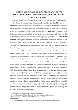
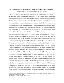
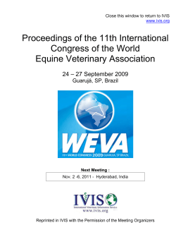
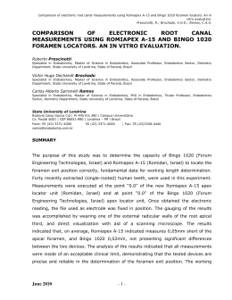
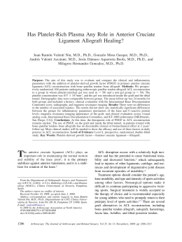
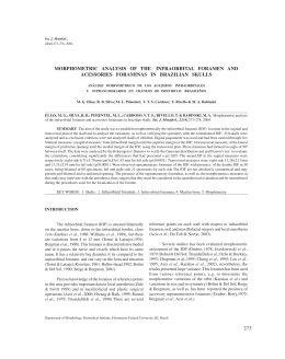
ACTA-1-2010-PELICULAS:1-2010
10/06/2010
03:58 p.m.
Página 63
63
RADIOGRAPHIC STUDY OF OSSIFICATION OF THE PTERYGOSPINOUS
AND PTERYGOALAR LIGAMENTS BY THE HIRTZ AXIAL TECHNIQUE
Rafaela R. Rosa1, Horácio Faig-Leite 2, Fabiola S. Faig-Leite3, Luiz C. Moraes1,
Mari E.L. Moraes1, Edmundo M. Filho1
Department of Diagnosis and Surgery, and 2Department of Biosciences and Diagnosis,
Faculty of Dentistry, São Paulo State University, UNESP, São José dos Campos, SP, Brazil.
3
Faculty of Medicine of Botucatu, UNESP, Botucatu, SP, Brazil.
1
ABSTRACT
The correct radiographic identification of ossification of the
pterygospinous and pterygoalar ligaments plays an important
role in surgical procedures for the treatment of trigeminal neuralgia. Most of these procedures are performed through the
foramen ovale, a site where these ligaments can be found to be
partially or completely ossified. We studied the radiographic
features of these ossified ligaments and their location in relation to the foramen ovale by the Hirtz axial technique. For this
purpose, 93 dry skulls from the Discipline of Anatomy, São José
dos Campos Dental School, UNESP, which presented partial or
complete ossification of these ligaments, were radiographed.
The pterygospinous ligament was detected on 27.97% of radiographs and was partially ossified in 19.36% of cases and
completely ossified in 8.61%. The pterygoalar ligament was
present in 62.35% of radiographs, being partially ossified in
49.44% and completely ossified in 12.91%. The pterygospinous
ligaments was found to be partially and completely ossified on
the same radiograph in 3.23% of cases, whereas the pterygoalar
ligament appeared partially and completely ossified on the same
radiograph in 6.45%. Furthermore, the pterygospinous ligament was thinner than the pterygoalar ligament and located
more medially in relation to the foramen ovale. The pterygoalar
ligament formed a large bone bar lateral to the foramen ovale,
often obliterating the lumen of the latter. The Hirtz axial technique is an excellent tool for the observation of complete or
partial ossification of the pterygospinous and pterygoalar ligaments in surgical procedures for the treatment of trigeminal
neuralgia performed through the foramen ovale.
Key words: pterygospinous ligament, pterygoalar ligament;
radiology.
ESTUDO RADIOGRÁFICO DA OSSIFICAÇÃO DOS LIGAMENTOS PTERIGOESPINHOSO
E PTERIGOALAR PELA TÉCNICA AXIAL DE HIRTZ
RESUMO
A correta identificação radiográfica da ossificação dos ligamentos pterigoespinhoso e pterigoalar é de grande importância nos
procedimentos cirúrgicos no tratamento da nevralgia trigeminal.
A maioria desses procedimentos é feita via forame oval, local onde
podemos encontrar esses ligamentos ossificados, parcial ou totalmente. Estudamos, pela técnica axial de Hirtz, as características
radiográficas desses ligamentos ossificados e sua localização em
relação ao forame oval. Para isso foram radiografados 93 crânios secos, pertencentes à Disciplina de Anatomia da Faculdade
de Odontologia de São José dos Campos - UNESP, que apresentavam a ossificação parcial ou total dos referidos ligamentos.
Encontramos o ligamento pterigoespinhoso em 27,97% das radiografias, sendo parcialmente ossificado em 19,36% e totalmente
em 8,61%. O ligamento pterigoalar estava presente em 62,35%
das radiografias, estando parcialmente ossificado em 49,44% e
totalmente em 12,91%. O ligamento pterigoespinhoso foi encontrado parcial e totalmente ossificado, numa mesma radiografia,
em 3,23% dos casos, enquanto o ligamento pterigoalar apareceu
parcial e totalmente ossificado, na mesma radiografia, em 6,45%
dos casos. Observamos ainda que o ligamento pterigoespinhoso
era menos espesso, em relação ao ligamento pterigoalar, e localizado mais para medial, em relação ao forame oval, enquanto que
o ligamento pterigoalar formava uma larga barra óssea lateralmente ao forame oval, obliterando muitas vezes o lúmen do
mesmo. A técnica axial de Hirtz é um excelente meio para a observação da ossificação total ou parcial destes ligamentos, quando
de procedimentos cirúrgicos que utilizam o forame oval para o
tratamento da nevralgia trigeminal.
INTRODUCTION
Various ligaments present in the skull base are of
clinical and surgical importance. Among them, the
pterygospinous and pterygoalar ligaments are located close to the foramen ovale and maintain an
important clinical relationship with this structure.
The pterygospinous ligament, described by Civinini
in 1829, connects the spinous process of the sphenoid
bone to Civinini’s spine located in the posterior margin of the lateral pterygoid lamina of the sphenoid
bone. In contrast, the pterygoalar ligament, described
by Hyrtl in 1862, extends from the undersurface of
Vol. 23 Nº 1 / 2010 / 63-67
Palavras chaves: ligamento pterigoespinhoso; ligamento pterigoalar; radiología.
ISSN 0326-4815
Acta Odontol. Latinoam. 2010
ACTA-1-2010-PELICULAS:1-2010
64
10/06/2010
03:58 p.m.
Página 64
Rafaela R. Rosa, Horácio Faig-Leite, Fabiola S. Faig-Leite, Luiz C. Moraes, et al.
the greater wing of the sphenoid bone to the lateral
pterygoid lamina of the sphenoid bone1,9,10.
Complete ossification of the pterygospinous ligament is known as the pterygospinous bar1,18,23,24,27,30
and complete ossification of the pterygoalar ligament as the pterygoalar bar1,4,19,24. When completely
ossified, the pterygospinous and pterygoalar ligaments form the pterygospinous foramen and porus
crotaphytico-buccinatorius, respectively18,24,27. Partial or complete ossification of these ligaments is
important from an anatomical, anthropologic and
clinical point of view, especially with respect to
treatment of trigeminal neuralgia1,18,23,24,27,30.
Trigeminal neuralgia is caused by nerve or microvascular compression in 80% of cases and by other factors
such as bone anomalies in the skull base in the remaining 20%9,18. Among these bone anomalies, ossified
pterygospinous and pterygoalar ligaments were the subject of this study. Various types of surgery are used for
the treatment of trigeminal neuralgia, including the
injection of alcohol into the trigeminal ganglion (alcoholization), vascular microdecompression2,3,9,16,23,26,28,29
and percutaneous procedures such as trigeminal
radiofrequency rhizotomy, glycerol rhizotomy and Fogarty balloon compression2,3,9,11,12,17,28,29. The completely
ossified pterygoalar ligament is of greater clinical
importance than the completely ossified pterygospinous
ligament since the latter generally does not represent an
obstacle to needle penetration through the foramen
ovale. In contrast, the completely ossified pterygoalar
ligament is more exuberant and may form a bony bar
that obliterates the foramen ovale, often impairing penetration of a needle through this foramen4,15,19,23-25.
Conventional radiography is an important tool for
the observation of these ossified ligaments. The
most recommended technique is the Hirtz axial
method5,18,19,23 or submentovertex projection, which
permits a clear observation of the anatomical struc-
Fig. 1: Lateral view of a skull showing the
completely ossified pterygospinous ligament.
Acta Odontol. Latinoam. 2010
tures of the skull base13,25. On Hirtz axial images, the
pterygospinous ligament appears obliterating the
foramen ovale and shows partial eclipse of its
lumen, whereas the completely ossified pterygoalar
ligament does not hide any radiopacity of the lumen
of the foramen ovale4,18,23,25.
The Hirtz axial radiograph performed in this study
is essential to the observation of the pterygospinous
ligament, ossified pterygoalar and foramen ovale.
This radiograph makes possible to detect these
anatomical structures considered of great relevance
and clinical applicability in individuals having
trigeminal neuralgia.
The objective of the present study was to demonstrate the importance of the Hirtz axial technique
for the analysis of the partially or completely ossified pterygospinous and pterygoalar ligaments in
terms of their relationship with the foramen ovale
and clinical significance.
MATERIAL AND METHODS
Ninety-three dry skulls of unknown age and gender, obtained from the Discipline of Anatomy, São
José dos Campos Dental School, UNESP, which
presented partial or complete ossification of the
pterygospinous (Fig. 1) and pterygoalar (Fig. 2) ligaments, were radiographed.
Conventional radiographs were obtained from skulls
by the Hirtz axial technique which is indicated for
the observation of skull base structures, such as foramen ovale, foramen spinosum, lateral pterygoid
lamina of the sphenoid bone, spinous process of
sphenoid bone, Civinini’s spine, and ossified pterygospinous and pterygoalar ligaments14,23,24,27.
The study was approved by the Ethics Committee on
Human Research of the São José dos Campos Dental
School, UNESP (protocol 073-2006-PH/CEP).
The radiographs were taken with a Funk Orbital X-15®
X-ray apparatus (Funk,
Ribeirão Preto, SP, Brazil)
at 80 kV, 5 mA and an
exposure time of 0.1 s.
Agfa Ortho CP-G Plus®
films measuring 18 x 24
cm mounted in EMB film
holders (Elétrica Médica
Brasileira, São Paulo,
Brazil) and Kodak Lanex
Regular®
intensifying
screens (Eastman Kodak
Company, Rochester, NY,
Fig. 2: Lateral view of a skull showing the
completely ossified pterygoalar ligament.
USA) were used.
ISSN 0326-4815
Vol. 23 Nº 1 / 2010 / 63-67
ACTA-1-2010-PELICULAS:1-2010
10/06/2010
03:58 p.m.
Página 65
Radiographic study of ossification ligaments
To align the auriculo-orbital plane (Frankfurt plane) of
the skulls and the film holders, a line was drawn perpendicularly to the horizontal plane of the cephalostat of the
X-ray apparatus. The adequate position of the median
sagittal plane in relation to the film plane was guaranteed by introduction of the ear rods of the cephalostat
into the external acoustic pores of the skull. A gauge was
used to transfer the height of the pore point from one
side to the other. The correct alignment between pore
points was confirmed by agreement between their
heights determined with the gauge. The two pores were
found to be aligned and parallel to the horizontal plane
in relation to the left orbital point (Fig. 3).
The images were analyzed by two examiners in a dark
room using a cold light negatoscope and a magnifying glass. The following radiographic characteristics
of ossification of the pterygospinous and pterygoalar
ligaments were recorded: skull identification, type of
ligament (pterygospinous or pterygoalar), type of
ossification (partial or complete), and side of ossified
ligament (right, left and bilateral).
The results were submitted to descriptive and inferential analysis using the chi-square test for uniform
distribution, with the level of significance set at 5%.
65
Fig. 3: Line indicating parallelism between the Frankfurt plane and
film holders and use of a gauge for alignment of the pore points.
RESULTS
Table 1 and Fig. 4 and 5 show the radiographic
results regarding the type of ossification of the
pterygospinous and pterygoalar ligaments.
Fig. 4: Radiograph showing
the pterygospinous ligament
which is completely ossified on
the left side and partially ossified on the right side (arrows).
Fig. 5: Radiograph showing
the pterygoalar ligament which
is completely ossified on the
right side and partially ossified
on the left side (arrows).
DISCUSSION
The pterygospinous and pterygoalar ligaments are
located close to the foramen ovale and are of anatomical, clinical and surgical importance8 because these
ossified ligaments may compress vascular-nervous
structures present in the region of the foramen ovale,
causing trigeminal neuralgia. Many studies in the literature only describe anatomical characteristics of the
Table 1: Distribution of the pterygospinous and pterygoalar ligaments according to ossification (partial or
complete) and side (right, left and bilateral) seen on radiographs
Pterygospinous
Type of ossification/side
Partially ossified
Pterygoalar
n
%
n
%
18
19.36
46
49.44
Right side
3
3.23
22
23.63
Left side
6
6.45
17
18.28
Bilateral
9
9.68
7
7.53
Completely ossified
8
8.61
12
12.91
Right side
3
3.23
3
3.23
Left side
0
absent
2
2.15
Bilateral
5
5.38
7
7.53
Partially and completely ossified
Total number of radiographs
Vol. 23 Nº 1 / 2010 / 63-67
3
3.23
6
6.45
29
31.20
64
68.80
ISSN 0326-4815
Acta Odontol. Latinoam. 2010
ACTA-1-2010-PELICULAS:1-2010
66
10/06/2010
03:58 p.m.
Página 66
Rafaela R. Rosa, Horácio Faig-Leite, Fabiola S. Faig-Leite, Luiz C. Moraes, et al.
partial or complete ossification of these ligaments, without a detailed analysis of the radiographic technique
used for the observation of these structures in the skull
base1,4,5,7,8,10,13,15,18,19,21,23-25,27,30. However, complete radiographic study of the ossified pterygospinous and
pterygoalar ligaments, as done in this investigation, is
very important because of the clinical significance of
these structures and their relationship with the foramen
ovale, and also for a better identification of the radiographic aspects of these ossified ligaments and other
adjacent structures of the skull base, thus contributing
to the treatment of trigeminal neuralgia.
In the present study, the radiographic location of the
ossified pterygospinous ligament was similar to its
anatomical location4,5,8,15,23. Radiographically, in our
findings, the ligament appeared as a thin radiopaque
structure located medial in relation to the foramen
ovale. According to the literature, the anatomical identification of the pterygospinous ligament is relatively
easy. This ligament generally consists of a thin bony
structure connecting Civinini’s spine to the spinous
process of the sphenoid bone and is located medially
to the foramen spinosum, a reference point for the
identification of the pterygospinous ligament4,13,27,30.
We observed in this radiographic study a higher frequency of partial ossification of the pterygospinous
ligaments (19.36%) when compared to complete ossification (8.61%). This percentage of partially ossified
pterygospinous ligament is higher than those reported in the anatomoradiographic studies of Priman and
Etter23 who observed partial ossification in 8% of
cases, and of Nayak et al.18 who reported a percentage of 3.84%. With respect to side, a partially ossified
pterygospinous ligament was detected on the right
side in 3.23% of skulls, on the left side in 6.45%, and
on both sides in 9.68%, with an average frequency of
unilateral occurrence of 4.84%. Our radiographics
results agree with those reported in the anatomical
study of Kapur et al.13, but differ from the findings of
Antonopoulou et al.1. Radiographically, in our study,
the pterygospinous foramen was present in 8.61% of
skulls, being bilateral in 5.38% of them, and was
located more medial in relation to the foramen ovale.
No comparison of our radiographics findings with the
literature is possible since only the anatomy of these
ligaments has been described in detail.
Analysis of the radiographs in our study showed the
presence of the porus crotaphytico-buccinatorius in
12 (12.91%) skulls, in agreement with Chouké and
Hodes6. Lower percentages were reported by
Chouké5, Priman and Etter23 and Kapur et al.13. In
contrast, Jovanovic et al.10, studying 30 skulls,
Acta Odontol. Latinoam. 2010
detected the porus crotaphytico-buccinatorius in a
higher percentage (43.4%) than that found in the
present study. Investigating 50 skulls, Antonopoulou
et al.1 detected the porus crotaphytico-buccinatorius
in only one skull unilaterally.
In the present radiographic study, the porus crotaphytico-buccinatorius was observed unilaterally on
five radiographs (5.68%). Comparison of these radiographic findings was not possible because of the lack
of radiographic studies on this subject. However, in
an anatomical study, Shaw24 detected the porus crotaphytico-buccinatorius on one side in 0.67% of cases,
a proportion lower than that reported by Priman and
Etter23 (2.6%). This difference might be explained by
differences in age and race. Faig-Leite et al.8 observed
the porus crotaphytico-buccinatorius in 19 (4.75%)
skulls, being unilateral in 12 of them (3%).
In our radiographic study, the pterygoalar ligament
was, at the same time, partially ossified on one side
and completely ossified on the other in six skulls
(6.45%). These results are similar to the anatomoradiographic findings of Priman and Etter23 who
identified different stages of ossification of the
pterygoalar ligament in 36 skulls (14.4%), with
ossification of this ligament being partial on one
side and complete on the other in 6 of them.
In their anatomoradiographic studies, Priman and
Etter23 and Chouké and Hodes6 found no case in
which the completely ossified pterygoalar ligament
branched into two Hyrtl foramina bilaterally. We also
observed only one porus crotaphytico-buccinatorius,
which was present bilaterally on seven radiographs
(7.53%). However, Patnaik et al.19 found two Hyrtl
foramina on the same side in the same skull.
Hirtz axial or submentovertex projection is the best
radiographic incidence for the observation of skull
base structures4,5,6,10,15,18. In our study, this technique
permitted a better observation of the pterygospinous
and pterygoalar ligaments, as well as the perfect
visualization of adjacent structures that were of
interest in this study.
The Hirtz axial radiograph performed in this study
made possible the perfect observation of the pterygospinous ligament, ossified pterygoalar and foramen
ovale with great clinical relevance and clinical applicability in individuals having trigeminal neuralgia.
CONCLUSIONS
The Hirtz axial technique permits good observation
of the partially or completely ossified pterygospinous
and pterygoalar ligaments. Radiographically, the ossified pterygospinous ligament was thinner and was
ISSN 0326-4815
Vol. 23 Nº 1 / 2010 / 63-67
ACTA-1-2010-PELICULAS:1-2010
10/06/2010
03:58 p.m.
Página 67
Radiographic study of ossification ligaments
normally located medially to the foramen ovale. In
contrast, the pterygoalar ligament was thicker, form-
67
ing a bone bar that obliterated the foramen ovale, and
was located more lateral in relation to the latter.
CORRESPONDENCE
Rafaela Rangel Rosa
e-mail: [email protected]
Faculdade de Odontologia do Campus
de São José dos Campos – UNESP
Department of Diagnosis and Surgery,
Discipline of Dental Radiology.
REFERENCES
1. Antonopoulou M, Piagou M, Anagnostopoulou S. An
anatomical study of the pterygospinous and pterygoalar bars
and foramina – their clinical relevance. J Craniomaxillofac
Surg 2008;36:104-108.
2. Ashkan K, Marsh H. Microvascular decompression for
trigeminal neuralgia in the elderly: a review of the safety
and efficacy. Neurosurgery 2004;55:840-850.
3. Bennetto L, Patel NK, Fuller G. Trigeminal neuralgia and
its management. BMJ 2007;334:201-205.
4. Chouké KS. On the incidence of the foramen of Civinini
and the porus crotaphitico-buccinatorius in American
whites and negroes I. Observations on 1544 skulls. Am J
Phys Anthropol 1946;4:203-225.
5. Chouké KS. On the incidence of the foramen of Civinini
and the porus crotaphytico-buccinatorius in American
whites and negroes II. Observations on 2745 additional
skulls. Am J Phys Anthropol 1947;5:79-86.
6. Chouké KS, Hodes PJ. The pterygoalar bar and its recognition by Roentgen methods in trigeminal neuralgia. Am J
Roentgenol 1951;65:10-12.
7. Das S, Paul S. Ossified pterygospinous ligament and its
clinical implications. Bratisl Lek Listy 2007;108:141-143.
8. Faig-Leite H, Faig-Leite FS, Fernandes RG. Anatomia do
ligamento pterigoalar e do forame crotafítico bucinatório
[resumo]. Int J Morphol 2007;25:15.
9. Hai J, Li ST, Pan QG. Treatment of atypical trigeminal neuralgia
with microvascular decompression. Neurol India 2006;54:1-4.
10. Jovanovic I, Vasovic L, Ugrenovic S, Zdravkovic D, Vlajkovic S, Dakovic-Bjelakovic M et al. Variable foramen of
Hyrtl of the human skull [abstract]. Acta Medica Medianae.
2003;42:1-5.
11. Kanpolat Y, Savas A, Bekar A, Berk C. Percutaneous controlled radiofrequency trigeminal rhizotomy for the treatment
of idiopathic trigeminal neuralgia: 25-year experience with
1.600 patients. Neurosurgery 2001;48:524-533.
12. Kaplan M, Erol FS, Ozveren MF, Topzakal C, Sam B,
Tekdemir I. Review of complications due to foramen ovale
puncture. J Clin Neurosci 2007;14:563-568.
13. Kapur E, Dilberovic F, Redzepagic S, Berhamovic E. Variation in the lateral plate of pterygoid process and the lateral
subzygomatic approach to the mandibular nerve [abstract].
Med Arh 2000;54:133-137.
14. Krmpotic-Nemanic J, Vinter I, Hat J, Jalsovec D. Mandibular
neuralgia due to anatomical variations. Eur Arch Otorhinolaryngol 1999;256:205-208.
15. Lepp FH, Sandner O. Anatomic–radiographic study of ossified pterygospinous and ‘‘innominate’’ ligaments. Oral Surg
Oral Med Oral Pathol 1968;26:244-260.
Vol. 23 Nº 1 / 2010 / 63-67
16. Liu HB, Ma Yi, Zou JJ, Li XG. Percutaneous microballoon
compression for trigeminal neuralgia. Chin Med J. 2007;
120:228-230.
17. Lopez BC, Hamlyn PJ, Zakrzewska JM. Stereotactic radiosurgery for primary trigeminal neuralgia: state of the
evidence and recommendations for futures reports. J Neurol Neurosurg Psychiatr 2004;75:1019-1024.
18. Nayak SR, Saralaya V, Prabhu LV, Pai MM, Vadgaonkar R,
D’Costa S. Pterygospinous bar and foramina in Indian
skulls: incidence and phylogenetic significance. Surg Radiol Anat. 2007;29:5-7.
19. Patnaik VVG, Rajan SK, Sanju B. Bilateral pterygoalar bar
and porus crotaphitico-buccinatorius: a case report. J Anat
Soc India 2001;50:161-162.
20. Peker T, Karaköse M, Anil A, Turgut HB, Gülekon N. The
incidence of basal sphenoid bony bridges in dried crania
and cadavers: their anthropological and clinical relevance.
Eur J Morphol 2002;40:171-180.
21. Peuker ET, Fischer, G Filler TJ. Entrapment of the lingual
nerve due to an ossified pterygospinous ligament. Clin Anat
2001;14:82-84.
22. Pollock BE. 754 percutaneous retrogasserian glycerol rhizotomy for patients with idiopathic trigeminal neuralgia: a
prospective analysis of factors related to pain relief
[abstract]. Neurosurgery 2004;55:470.
23. Priman J, Etter LE. The pterygospinous and pterygoalar
bars. Med Radiogr Photogr 1959;35:2-6.
24. Shaw JP. Pterygospinous and pterygoalar foramina: a role
in the etiology of trigeminal neuralgia? Clin Anat 1993;
6:173-178.
25. Skrzat J, Walocha J, Srodek R. An anatomical study of the
pterygoalar bar and the pterygoalar foramen. Folia Morphol
2005;64:92-96.
26. Spatz AL, Zakrzewska JM, Kay EJ. Decision analysis of
medical and surgical treatments for trigeminal neuralgia:
how patient evaluations of benefits and risks affect the utility of treatment decisions. Pain 2007;131:302-310.
27. Tebo HG. The pterygospinous bar in panoramic roentgenography. Oral Surg Oral Med Oral Pathol 1968;26:
654-657.
28. Toda K. Etiology of trigeminal neuralgia. Oral Science Int
2007;4:10-18.
29. Tun K, Celikmez R, Gürcan O, Gürcay AG, Türkoglu F,
Kaptanoglun E. Idiopathic bilateral trigeminal neuralgia
treated by bilateral microvascular decompression. Turkish
Neurosurg 2007;17:294-296.
30. von Lüdinghausen M, Kageyama I, Miura M, Alkhatib M.
Morphological peculiarities of the deep infratemporal fossa
in advanced age. Surg Radiol Anat 2006;28:284-292.
ISSN 0326-4815
Acta Odontol. Latinoam. 2010
Download