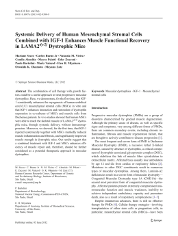
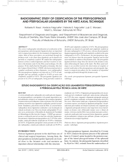
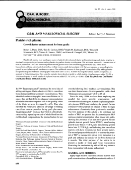
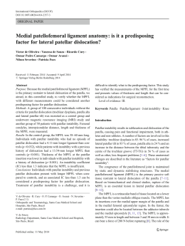
Knee Surg Sports Traumatol Arthrosc (2007) 15:1219–1227
DOI 10.1007/s00167-007-0385-x
KNEE
ACL reconstruction in a rabbit model using irradiated Achilles
allograft seeded with mesenchymal stem cells or PDGF-B
gene-transfected mesenchymal stem cells
Feng Li Æ Hongti Jia Æ Changlong Yu
Received: 12 March 2007 / Accepted: 26 June 2007 / Published online: 9 August 2007
! Springer-Verlag 2007
Abstract The present study was conducted to develop a
new strategy to accelerate reconstruction of the anterior
cruciate ligament (ACL) by modifying the Achilles allograft with autogenous mesenchymal stem cells (MSCs) or
PDGF-B transfected MSCs in a rabbit model. The allografts were first irradiated with Co60, stored at –80"C, and
then seeded with cells for implantation. Bilateral ACL
reconstructions were performed. On the left, the allograft
was either seeded with MSCs or PDGF-B transfected
MSCs and acted as the experimental group. On the right,
the graft without any cells seeded acted as control. At 3, 6
and 12 weeks after surgery, histological observation found
that implatation of MSCs or PDGF-B transfected MSCs
accelerated cellular infiltration into the ACL and enhanced
collagen deposition in the wound. PDGF-B transfected
MSCs could also lead to an initial promotion of angiogenesis. This gene transfer technique or cell implantion
may be a potentially useful tool for improving ligament
remodeling.
Keywords Mesenchymal stem cells ! Allograft !
Anterior cruciate ligament ! PDGF-BB ! Retrovirus
F. Li ! C. Yu (&)
Institute of Sports Medicine, Peking University Third Hospital,
No. 49, North Garden Road, Haidian District, Beijing 100083,
People’s Republic of China
e-mail: [email protected]
H. Jia
Department of Biochemistry and Molecular Biology,
School of Basic Medical Sciences, Peking University Health
Science Center, Beijing 100083, People’s Republic of China
Introduction
Anterior cruciate ligament (ACL) injuries are common, and
surgical reconstruction is desirable for patients who participate in vigorous sports activities. There are two kinds of
biological substitutes, autograft and allograft, which are
commonly used for ligament reconstruction. The use of
allogeneic tendons for ligament reconstruction has many
advantages [22], including the retention of normal tissues,
wider choice of graft size, and reduction in surgical and
anesthetic time. In our study, we used Achilles allograft for
ACL reconstruction.
Tendon grafts which stay in an intra-articular synovial
environment proceed through some phases of biologic
incorporation, such as necrosis, angiogenesis, cell repopulation and final maturation. Angiogenesis is an essential
step in the process of tendon healing and tendon graft
remodeling, in which neovascularization prompts delivery
of inflammatory cells, fibroblasts and growth factors to the
wound site. Therefore, we tried to develop a new strategy
by enhancing angiogenesis to accelerate the remodeling of
tendon graft.
With the application of knowledge gained from basic
science and clinical research, we tried to use autogenous
mesenchymal stem cells (MSCs) and PDGF-B gene to
modify the allograft to accelerate angiogenesis. MSCs are
pluripotential cells that are being investigated extensively
in a wide variety of settings, including enhancement of
healing of tendon injuries [2, 3, 30, 33]. PDGF-BB has
been shown to play essential roles in wound healing. Recent investigations [7] suggest that PDGF-BB mediates
many processes required for tissue repair, including chemotaxis (monocytes, neutrophils, fibroblast), proliferation
(fibroblasts, smooth muscle cells, capillary endothelial
cells), induction of several matrix molecules (fibronectin,
123
1220
hyaluronic acid) and the production and secretion of collagenase by fibroblasts. Furthermore, in vitro and in vivo
[4, 8, 12, 14, 16, 25, 27] application of PDGF to the tendon
cells and ligament has been demonstrated to enhance the
proliferation of fibroblasts and deposition of extracellular
matrix.
Based on those findings, we investigated the biological
effect of the introduction of PDGF-B gene and MSCs on
the healing process of the rabbit anterior cruciate
ligament.
Materials and methods
Experimental design
Bilateral ACL reconstructions with Achilles allografts
were performed by a single surgeon in 36 skeletally
mature female New Zealand white rabbits weighing 2.0–
2.5 kg each. Autologous bone marrow-derived MSCs
were harvested 3–5 weeks before ligament reconstruction
surgery. During bilateral ACL reconstruction, on the left
knee, the allograft was either seeded with MSCs or
PDGF-B transfected MSCs and acted as the treatment
group; on the right, the graft without any cells seeded
acted as control. The animals were euthanized at 3, 6 and
12 weeks postoperatively for histologic analysis. At each
time they were killed, 12 animals (six for MSCs group
and six for MSCs-PDGF-BB group) were used for analysis. A further two rabbits were used as normal control.
The experimental protocol, animal care and use procedures were in accordance with the policies of Peking
University Health Science Center and the National Institute of Health.
Knee Surg Sports Traumatol Arthrosc (2007) 15:1219–1227
Preparation of MSCs
Bone marrow samples were collected from the iliac crest of
New Zealand white rabbits by adopting previously published methods [3, 5]. A volume of 1 ml of bone marrow
was aspirated and put in complete Dulbecco’s Modified
Eagle Medium low glucose (DMEM-lg, Gibco, USA)
containing 10% fetal bovine serum (FBS), penicillin
100 lg/ml, streptomycin 100 lg/ml (pH 7.35). Samples
were washed twice with DMEM-lg and centrifuged at
2,000 rpm for 6 min. The supernatant was discarded and
the precipitating cell pellet was suspended in 10 ml of
MSCs growth media. Cells were counted and then plated at
17 · 106 to 22 · 106 cells per 100 mm dish. MSCs
adhered to the plates and proliferated to form colonies
between 5 and 8 days into primary culture. Once the
colonies of MSCs reached confluency, the adherent MSCs
were retrieved and subcultured again.
Transduction of cells
MSCs of Passage 1 were plated in six-well dishes. When
the cells reached 50% confluence, transductions were
performed by adding 1.6 ml fresh DMEM-lg, supplemented with 8lg/ml polybrene (Sigma, USA) to each well.
A total of 400 ll retrovirus supernatant was added to the
cells and they were incubated overnight at 37"C, the following morning the medium was replaced with 2 ml fresh
DMEM-lg supplemented with 200lg/ml G418. After
10–14 days of selection in G418, the clonal cells
(MSCs-PDGF-BB) were formed. They were trypsinized,
transferred to T-75 flasks and cultured in DMEM-lg. When
the cells became confluent, they were seeded onto graft for
later implantation.
Construction of retroviral vector
Graft preservation
The human PDGF-B fragment was cut from psv7dPDGF-B plasmid (kindly provided by Christer Betsholtz,
Institute of Medical Biochemistry, University of Gothenburg, Sweden) and inserted into a retrovirus vector
PLXRN (Clontech, USA) to get PLXRN-PDGF-B. The
Rous sarcoma virus promoter controls expression of the
Neor gene.
A total of 36 Achilles tendons were harvested from donor
New Zealand white rabbits and used as grafts. Each tendon
was separated into at least two bundles of ligament and
each was cut 2 mm wide and 3 cm long. Then these two
ligaments were used in the ACL reconstruction of one
animal. All the grafts were enclosed in tubes and placed in
dry ice in a container for gamma irradiation by Co60. The
exposure time of 9 h yielded a calculated dose of 2.5 Mrad.
Then the tendon graft was stored at –80"C for 2 to 5 weeks
before use.
Production of retroviral vector particles
The PLXRN-PDGF-B plasmids were used to transfect PT67 cells (Clontech, USA) to get retroviral particles. Clonal
amphotropic packaging cells were derived as described in
Clontech protocol. The retrovirus supernatants were filtered through a 0.45 lm syringe filter (Sartorius, German)
and stored at –80"C before use.
123
Seeding MSCs or MSCs-PDGF-BB on the Achilles
graft
At the time of transplantation, the frozen grafts were
thawed in saline gauze at room temperature. In the
Knee Surg Sports Traumatol Arthrosc (2007) 15:1219–1227
experiment group, MSCs or MSCs-PDGF-BB were
embedded in fibrin glue (Beixiu Company, China) by
mixing the cell pellet with the fibrin part and then mixing
this solution with the thrombin part. The final concentration
was approximately 1.5 · 106 cells/ml. Of this mixture, a
0.5 ml sample was placed in an Achilles graft before it
became gel and allowed to gel in a culture dish in a CO2
incubator for 10 min at 37"C. In the control group, 0.5 ml
of fibrin glue without cells was incubated in the same
manner. The graft was washed gently three times in PBS
solution, 20 min before implantation.
Surgical protocol
Bilateral ACL reconstructions were performed. A lateral
parapatellar arthrotomy was used to expose the knee joint.
The native ACL was divided and tibial and femoral tunnels
were created with a 2 mm drill. The combined length of the
tunnel from the anterior femoral surface, across the joint, to
the anterior tibial surface was recorded. The graft was then
inserted via the holding suture using a Beath pin from the
tibial and femoral tunnels at the same time slowly. We
tethered each end of the graft with the suture to a screw
inserted into the bone, manually applying minimal graft
tension in a direction aligned with the long axis of the bone
tunnel with the knee flexed at 90". The gap between the
tunnel and the graft was filled with an additional 100ll of
fibrin glue. Then the wound was closed in layers. After
surgery, all the animals were allowed to move freely in
their cages.
1221
ELISA
The level of human PDGF-BB in cell culture medium and
lavaged fluid were measured with ELISA kits (mouse antihPDGF neutralizing antibody, R&D Systems, USA)
according to the manufacturer’s instructions.
Histologic analysis
The femur and tibia were removed; the knee joints were
left intact, and the specimens fixed in 4% paraformaldehyde. The samples of ACL were embedded in paraffin and
cut into 5lm thick sections longitudinal to the bony tunnels. The slides were stained using hematoxylin and eosin,
toluidine blue staining and immunohistochemical stain for
collagens type I (Sigma, USA) and III (Oncogene, USA)
and PDGF-BB (rabbit anti-serum, Lab Vision Corporation,
USA). Four knees from two rabbits with normal ACLs
were harvested and prepared in a similar manner for histologic analysis.
Results
RT-PCR
As described in Materials and methods, the MSCs-PDGFBB contain the entire coding sequence of human PDGFBB, while the MSCs do not (Fig. 1).
Analysis of PDGF-B mRNA and protein production
ELISA
RT-PCR
MSCs-PDGF-BB were found to secrete PDGF-BB at a
steady-state level of 1,100 ng/106cells/24 h, whereas the
MSCs did not. The concentration of PDGF-BB from the
perfusion of joint cavity in the gene treatment group is as
shown in Table 1
Total RNA was extracted from confluent tissue culture
flasks of MSCs or MSCs- PDGF-BB using Trizol (Invitrogen, USA), according to the manufacturer’s instructions.
5-TCTGCTGCTACCTGCGTCTG-3 and 5-GCGTTGGT
GCGGTCTATG-3 were used as primers for RT-PCR.
Obtained PCR products were separated on 1% agarose gels
and stained with ethidium bromide.
Gross observations
The intra-articular knee gross examination showed intact
grafts in all cases.
Arthrosis fluid puncture
Histologic findings in the 3-week group
Number 7 needles were introduced from the superior aspect into the upper compartment of the joint cavity of the
knee. The knee joints on both sides were perfused with
1 ml saline and the perfusate samples were collected. Then
they were centrifuged at 6,000 rpm at 4"C for 10 min to
remove cells and other debris. Thereafter, the supernatant
was transferred to another tube and was immediately frozen at –80"C for ELISA.
HE staining showed that 3 weeks after surgery, in the
control group, host cells had already invaded into the graft
and most of them were inflammatory cells; while in the
graft seeded with MSCs, granulation was formed and
vascularization could be observed. In the gene-transfected
group, significantly more cells and hypervascularity could
be found (Fig. 2).
123
1222
Knee Surg Sports Traumatol Arthrosc (2007) 15:1219–1227
Immunohistochemical staining of collagen type I
and III
After transplantation, collagen type III increased in the
grafts along with the growth of cells. Collagen deposition
increased significantly in the PDGF-B gene-introduced
wound at 3 weeks. After 6–12 weeks, collagen type I
gradually increased to replace type III in the MSCs group
and gene-transfected group (data not shown).
Immunohistochemical staining of PDGF-BB
The graft treated with MSCs-PDGF-BB demonstrated intense staining cells at 3 weeks. After 6–12 weeks, the
staining was gradually decreased (Fig. 5), whereas the
wounds treated with cells or without cells had virtually no
staining (data not shown).
Discussion
Fig. 1 Detection of mRNA of human PDGF-BB by RT-PCR in
MSCs and MSCs-PDGF-BB. Lane 1, molecular weight markers;
lane2, MSCs; lane 3, MSCs-PDGF-BB
Histologic Findings in the 6w-group
In the control group cells invaded further into the graft;
while in both the MSCs group and gene-transfected group
some chondrocyte-like cells appeared (Fig. 3).
Histologic findings in the 12-week group
In the control group, a large amount of fibrocytes were
found in the graft; while in the MSCs group and genetransfected group, the grafts had structure similar to normal
ACL, especially in the MSCs group. However, the number
of cells was still more than normal (Fig. 4).
Table 1 The PDGF-BB expression of perfusate sample from knee
joint in the gene-transfected group (mean deviation)
Group
0
PDGF-BB (pg/ml)
0
3 weeks
522.76
6 weeks
12 weeks
728.848
412.09
123
This is the first report known to us that evaluates the effect
of cell therapy and gene therapy technique on ACL allograft reconstruction. The aim of our experiment was to
develop a new strategy to accelerate the remodeling of
allograft and to build an allograft tendon bank for ligament
reconstruction.
Although ACL reconstruction with allogeneic tendon
has many advantages, its use has not been chosen widely.
There are many reasons, such as marked inflammatory
rejection responses and the risks of disease transmission,
which prevent its use. As for the rejection response, Minami et al. [17] showed that normal MHC antigens are in
the tendon cell component of the graft and numerous
studies [1, 6, 10, 11, 17, 21, 28] have demonstrated that
deep freezing at –80"C, by destroying these cells, can reduce graft antigencity. To reduce the risks of disease
transmission, gamma irradiation [22] is now widely used as
a safe and effective secondary sterilization technique. It
can inhibit HIV transmission in T lymphocytes. Graft
sterilization by gamma irradiation was also reported to
produce less deleterious effects on healing.
After dealing with the allograft to reduce its side effect, we
tried to find a way to modify the allograft to accelerate its
ligamentation. For this, we used the MSCs and hPDGF-B
gene. Many researches [5, 9, 15, 29] were done to find suitable plant cells to accelerate the remodeling of the allograft.
In vitro study on MSCs in tendon healing by Van et al. [29]
found that MSCs seemed to be the most suitable candidate
for the development of tissue-engineered ligament, with the
highest cell proliferation and highest collagen production,
compared with ACL cells or skin fibroblasts, when the cells
were seeded onto a resorbable suture material, respectively.
Knee Surg Sports Traumatol Arthrosc (2007) 15:1219–1227
1223
Fig. 2 Photomicrographs
of 3-week ACL reconstruction.
a–c H&E stain, original
magnification ·100; d–f
toluidine blue stain, original
magnification ·100; a, d control
group; b, e MSCs group; c, f
gene-transfected group. In the
control group, 3 weeks after
surgery, host cells had already
invaded into the graft, while in
the MSCs group, granulation
was formed and vascularization
could be observed. In the genetransfected group, significantly
more cells and hypervascularity
could be found. b, c The arrows
show blood vessels)
Histological findings of our study suggested that
implantation of autologous cells accelerated the cellular
infiltration into the ACL and enhanced the maturation of
collagen. We noticed that administration of cultured
autologous cells in MSCs group and gene-transfected
group caused an influx of cells into the ACL graft. But we
are not sure where the cells were from: the MSCs applied
to the surface of tendon grafts may have served as a source
of additional recruitable fibroblast-like cells for tendon
repopulation, or they may have been involved in activation
and recruitment of local fibroblast precursors, and bone
marrow-derived cells may have contributed to the graft that
was placed in a bone tunnel.
MSCs also have the potential to migrate and transform
into certain cells. In another study [19], MSCs, which were
obtained from green fluorescent protein (GFP) transgenic
SD rat and cultivated, were injected into normal SD rats in
which multiple tissues had been injured including the
anterior cruciate ligament. GFP-positive cells could
mobilize into the injured ACL. Watanabe et al. [31] also
demonstrated that when the autogenous MSCs were explanted into the injured MCL of the animal, the cells could
migrate and survive for a certain time.
In our study, some fibroblast-like cells, fibrocartilagelike cells and endothelium of blood vessels were also found
from 3 to 12 week during the remodeling process in cell
plant groups. It was hypothesized that MSCs from bone
marrow would differentiated into certain cells in the
healing ligament following adaption to the specific environmental conditions of the tendon, and it may be reasonable to expect that MSCs could differentiate as well as
promote the regeneration and maturing of the graft.
Many studies [4, 8, 12, 14, 16, 25, 27] have also attempted to determine the effects of PDGF-BB on a ligament engineering system. It has been demonstrated that it
promoted fibroblast proliferation, matrix synthesis, neovascularization, and mechanical properties.
In our studies in the gene-transfected group, more cells
and blood vessels could be found at 3 weeks, and more
collagen was synthesized in the ACL as compared with the
control and MSCs group (data not shown). Similar to our
findings were the results of a study by Nakamura et al. [20],
which described an increased vascularity and enhanced
collagen deposition in the wound of a patellar ligament
after PDGF-B gene transfer in rats.
The mechanisms by which PDGF affect ligament healing are complex. Kuroda et al. [24] studied immunohistochemically the presence and the level of bFGF, TGF-ß,
PDGF AA and PDGF-BB expression in a model of ACL
reconstruction using a free patellar tendon autograft. They
123
1224
Knee Surg Sports Traumatol Arthrosc (2007) 15:1219–1227
Fig. 3 Photomicrographs
of 6-week ACL reconstruction.
a–c H&E stain, original
magnification ·100; d, e, f
toluidine blue stain, original
magnification ·100; a, d control
group; b, e MSCs group; c, f
gene-transfected group. In the
control group, cells invaded
further into the graft; while in
both the MSCs group and in the
gene-transfected group, some
chondrocyte-like cells appeared.
b The arrows show
chondrocyte-like cells; e, f the
arrows show extracellular
matrix around the cells positive
with toluidine blue stain.)
found that all tested growth factors were upregulated with a
maximum expression at 3 weeks and up to 60% of all cells
were stained PDGF positive. PDGF is one of the most
effective growth factors during tendon graft remodeling.
Tendon grafts are exposed to the reduced PO2 of the
intra-articular environment, Petersen et al. [23] showed that
PDGF as well as hypoxia strongly enhanced VEGF
secretion from tenocytes. Besides this VEGF-mediated
angiogenetic effect, PDGF furthermore induces the synthesis of other growth factors, including IGF, and regulates
the presence of other receptors [32]. Therefore, it could be
concluded that the expression of PDGF-BB by the small
number of transfected cells may activate a cascade of
PDGF-BB throughout the wound.
Some researches [18] showed that there are some
growth factors receptors such as PDGFR on the surface of
MSCs. MSCs from bone marrow might have a better
response to PDGF as compared with those from the ACL
or MCL regarding proliferation and migration. Thus, it
might be suggested that a tendon graft seeded with MSCsPDGF-BB is more likely to promote MSCs or fibroblast
proliferation and migration and accelerate graft tissue
remodeling.
The observations of Kuroda et al. [24] also imply that if
a growth factor is administered to the tendon graft, its
123
tissue concentration should be highest around the third
week to enhance the effect of the other intrinsical growth
factors. In our study at 3 weeks, we found significantly
higher vascularity in the PDGF-transfected grafts. The
concentration of PDGF-BB in articular fluid from genetransfected rabbit increased at 3 week, got to the highest
point at 6 week and then dropped down. This rapid
reduction in the level of their localization indicates that
once the extrinsic cells infiltrate to the graft and revascularization is complete, these growth factors may have less
significance for subsequent remodeling. Therefore, it is
very important to find the appropriate time point for the
administration of growth factors to promote healing.
In vivo study by Hildebrand et al. [12] demonstrated that
the improvements in the MCL structural properties were
dose-dependent to PDGF-BB. That is, a higher dose of
PDGF-BB improved more structural properties of the femur–MCL–tibia complex than a lower dose of PDGF-BB
did. In both the MSCs group and gene-transfected group,
although there are much more differences in morphology at
the early stages, the structure of the ACL in the two groups
have less significant differences at the later stages, especially at 12 weeks. For an ACL reconstruction model, we
don’t know if the dosage we used was appropriate to
maximally enhance graft remodeling, or this indicates that
Knee Surg Sports Traumatol Arthrosc (2007) 15:1219–1227
1225
Fig. 4 Photomicrographs of
12-week ACL reconstruction
and normal ACL. a–c, g H&E
stain, original magnification
·100; d–f, h toluidine blue
stain, original magnification
·100; a, d control group;
b, e MSCs group; c, f genetransfected group; g, h normal
ACL. In the control group, a
large amount of fibrocytes were
found in the graft; while in the
MSCs group and genetransfected group grafts were
similar to normal ACL
structure. The normal ACL
(g, h) demonstrated more
heterogenous cell types and
arrangements. Most cells were
enclosed within lacunae. The
ACL (h) is positive to toluidine
blue stain; the nucleus is blue
while the extracellular matrix is
red-purple, especially adjacent
to the cells. b, c, g The arrows
show cell arrangements within
lacunae. e, f, h The arrows show
the extracellular matrix around
cells positive to toluidine blue
stain)
the growth factor PDGF may have early effect on the ACL
reconstruction and have less significance for subsequent
remodeling. It is essential to find an appropriate dosage for
ACL reconstruction.
As we know, there are limitations of topically applied
growth factor proteins, including rapid degradation of the
protein and inefficient action at the cellular level. Delivery
of PDGF by gene therapy is potentially a much more
efficient method of delivering active proteins with resultant
sustained activity; From the perspective of clinical utility,
BMSCs can be easily accessed in most patients without
significant additional surgery or the risk of immune
reaction, and these cells can be rapidly expanded in a cell
culture media. Therefore, such combined therapy of gene
therapy and cell therapy may be an effective approach for
ACL allograft reconstruction.
We chose to use a rabbit model for ACL reconstruction.
This is a model that has been validated in previous reports.
Although we have designed special tools for the surgery in
order to minimize injury to tissues during the operation, it
is necessary to recognize some limitations concerning the
experimental model used in this study. Rabbit is too small
for surgery and the model is not similar to a human being.
There were also significant differences between experi-
123
1226
Knee Surg Sports Traumatol Arthrosc (2007) 15:1219–1227
Fig. 5 Photomicrographs of
ACL reconstruction in genetransfected group in
immunohistochemical staining
of PDGF-BB (original
magnification ·200). The graft
treated with MSCs-PDGF-BB
demonstrated intense staining
cells at 3 weeks (a), after 6–
12 weeks the staining was
gradually decreased (b, c)
mental and control limbs in histology and other tests. More
accurate and reliable experiment such as biomechanical
testing is needed with a large animal model, such as sheep
or dog, to obtain the results for judging the possibility of
the use of gene therapy to enhance the process of ligament
reconstruction in human beings.
Conclusions
We have demonstrated that the application of PDGF-B
gene to a tendon graft is effective in changing certain
structural properties during the early phase of tendon graft
remodeling after ACL reconstruction. PDGF-B gene leads
to an initial promotion of angiogenesis and subsequent
enhanced collagen deposition in the wound. Therefore, this
gene transfer technique may be a potentially useful tool for
improving tendon repair.
In our study, we have also shown that MSCs can be
transplanted to serve as a source for healing tissues or used
as a molecular vehicle for therapeutic purposes. The
application of MSCs is effective in enhancing collagen
deposition and changing certain structural properties.
We have only used one growth factor to improve the
structural properties of remodeling ligament. On finding an
appropriate dosage, or combining with other growth factors, this effect might further be improved. This should
encourage further research.
Acknowledgments We would like to thank Professor Carl-Henrik
Heldin, Ludwig Institute for Cancer Research, Sweden and Christer
Betsholtz, Institute of Medical Biochemistry, University of Gothenburg, Sweden for helping us in our study.
References
1. Arnoczky SP, Warren RF, Ashlock MA (1986) Replacement of
the anterior cruciate ligament using a patellar tendon allograft: an
experimental study. J Bone Joint Surg Am 68A:376–385
123
2. Awad HA, Butler DL, Boivin GP, Smith FN, Malaviya P,
Huibregtse B, Caplan AI (1999) Autologous mesenchymal
stem cell-mediated repair of tendon. Tissue Eng 5:267–277
3. Awad HA, Butler DL, Dressler MR, Smith F, Boivin GP, Young
RG (2003) Repair of patellar tendon injuries using mesenchymal
stem cells and collagen scaffolds. J Orthop Res 21:420–431
4. Batten ML, Hansen JC, Dahners LE (1996) Influence of dosage
and timing of application of platelet-derived growth factor on
early healing of the rat medial collateral ligament. J Orthop Res
14:736–741
5. Bellincampi LD, Closkey RF, Prasad R, Zawadsky JP, Dunn MG
(1998) Viability of fibroblast-seeded ligament analogs after
autogenous implantation. J Orthop Res 16:414–420
6. Bos GD, Goldberg VM, Powell AE, Heiple KG, Zika JM (1984)
The effect of histocompatibility matching on canine frozen bone
allografts. J Bone Joint Surg Am 65A:89–96
7. Heldin C-H, Westermark B (1999) Mechanism of action and
in vivo role of Platelet-derived growth factor. Phys Rev 79:1283–
1316
8. DesRosiers EA, Yahia L, Rivard CH (1996) Proliferative and
matrix synthesis response of canine anterior cruciate ligament
fibroblasts submitted to combined growth factors. J Orthop Res
14:200–208
9. Dunn MG, Liesch JB, Tiku ML, Zawadsky JP (1995) Development of fibroblast-seeded ligament analogs for ACL reconstruction. J Biomed Mater Res 29:1363–1371
10. Friedlaender GE, Strong DM, Sell KW (1976) Studies on the
antigenicity of bone: freeze-dried and deep-frozen bone allografts
in rabbits. J. Bone Joint Surg Am 58A:854–858
11. Graham WC, Smith PA, McGuire MP (1955) The use of frozen
stored tendons for grafting: an experimental study. J Bone Joint
Surg Am 37A:624
12. Hildebrand KA, Woo SL, Smith DW, Allen CR, Deie M, Taylor
BJ, Schmidt CC (1998) The effects of platelet-derived growth
factor-BB on healing of the rabbit medial collateral ligament: an
in vivo study. Am J Sports Med 26:549–554
13. Kang HJ, Kang ES (1999) Ideal concentration of growth factors
in rabbit’s flexor tendon culture. Yonsei Med J 40:26–29
14. Letson AK, Dahners LE (1994) The effect of combinations of
growth factors on ligament healing. Clin Orthop Relat Res
308:207–212
15. Lin VS, Lee MC, O’Neal S, McKean J, Sung KL (1999) Ligament tissue engineering using synthetic biodegradable fiber
scaffolds. Tissue Eng 5:443–452
16. Meaney Murray M, Rice K, Wright RJ, Spector M (2003) The
effect of selected growth factors on human anterior cruciate ligament cell interactions with a three-dimensional collagen–GAG
scaffold. J Orthop Res 21:238–244
Knee Surg Sports Traumatol Arthrosc (2007) 15:1219–1227
17. Minami A, Ishii S, Ogino T (1982) The effect of the immunological antigenicity of the allogenic tendinosus tendon grafting.
Hand 14:111–119
18. Minguell JJ, Erices A, Conget P (2002) Mesenchymal stem cells.
Exp Biol Med 226:507–520
19. Agung M, Ochi M, Yanada S, Adachi N, Izuta Y, Yamasaki T,
Toda K (2006) Mobilization of bone marrow-derived mesenchymal stem cells into the injured tissues after intraarticular
injection and their contribution to tissue regeneration. Knee Surg
Sports Traumatol Arthrosc 14:1307–1314
20. Nakamura N, Shino K, Natsuume T, Horibe S, Matsumoto N,
Kaneda Y, Ochi T (1998) Early biological effect of in vivo gene
transfer of platelet-derived growth factor (PDGF)-B into healing
patellar ligament. Gene Ther 5:1165–1170
21. Peacock EE, Petty J (1960) Antigenicity of tendon. J Surg
Gynecol Obstet 110:187–192
22. Peter M, Prokopis, Schepsis AA (1999) Allograft use in ACL
reconstruction. Knee 6:75–85
23. Petersen W, Pufe T, Zantop T, Tillmann B, Mentlein R (2003)
Hypoxia and PDGF have a synergistic effect that increases the
expression of the angiogenetic peptide vascular endothelial
growth factor in Achilles tendon fibroblasts. Arch Orthop Trauma
Surg 123:485–488
24. Kuroda R, Kurosaka M, Yoshiya S, Mizuno K (2000) Localization of growth factors in the reconstructed anterior cruciate ligament: immunohistological study in dogs. Knee Surg Sports
Traumatol Arthrosc 8:120–126
25. Scherping SC Jr, Schmidt CC, Georgescu HI, Kwoh CK, Evans
CH, Woo SL (1997) Effect of growth factors on the proliferation
of ligament fibroblasts from skeletally mature rabbits. Connect
Tissue Res 36:1–8
1227
26. Schmidt CC, Georgescu HI, Kwoh CK, Blomstrom GL, Engle
CP, Larkin LA, Evans CH, Woo SL (1995) Effect of growth
factors on the proliferation of fibroblasts from the medial collateral and anterior cruciate ligaments. J Orthop Res 13:184–190
27. Spindler KP, Clark SW, Nanney LB, Davidson JM (1996)
Expression of collagen and matrix metalloproteinases in ruptured
human anterior cruciate ligament: an in situ hybridization study.
J Orthop Res 14:857–861
28. Stevenson S, Honn RB, Templeton JW (1983) Effects of tissue
antigen matching on the healing of fresh cancellous bone allografts in dogs. Am J Vet Res 44:204–206
29. Van Ei jk F, Saris DB, Riesle J, Willems WJ, Van Blitterswi jk
CA, Verbout AJ, Dhert WJ (2004) Tissue engineering of ligaments: a comparison of bone marrow stromal cells, anterior
cruciate ligament, and skin fibroblast as cell source. Tissue Eng
10:893–903
30. Wakitani S, Goto T, Pineda SJ, Young RG, Mansour JM, Caplan
AI, Goldberg VM (1994) Mesenchymal cell-based repair of large,
full-thickness defects of articular cartilage. J Bone Joint Surg Am
76:579–592
31. Watanabe N, Woo SL, Papageorgiou C, Celechovsky C, Takai S
(2002) Fate of donor bone marrow cells in medial collateral
ligament after simulated autologous transplantation. Microsc Res
Tech 58:39–44
32. Yasuhiro Y, Abrahamson S (2000) Dose-related effects of
platelet-derived growth factor-BB vary by different rabbit tendons in vitro. Transactions of the 46th annual meeting of the
Orthopaedic Research Society
33. Young RG, Butler DL, Weber W, Caplan AI, Gordon SL, Fink
DJ (1998) Use of mesenchymal stem cells in a collagen matrix
for Achilles tendon repair. J Orthop Res 16:406–413
123
Download