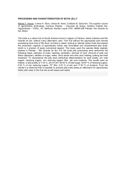
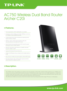
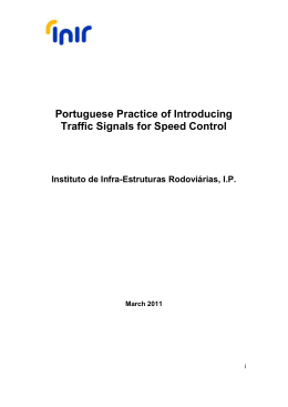
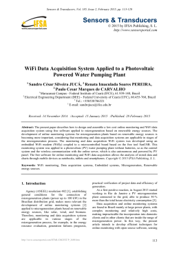
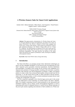
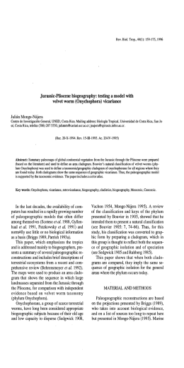
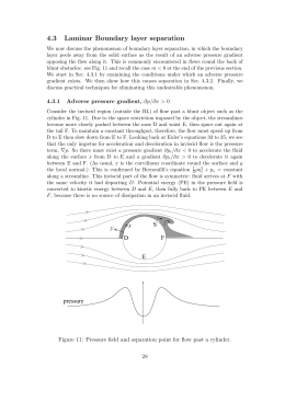
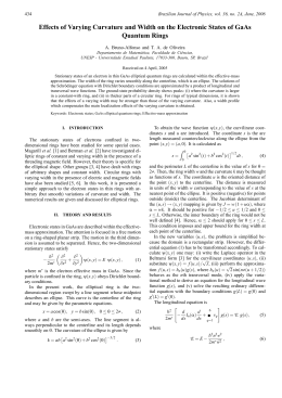
Mobile Remote Monitoring Of Biological Signals
Murilo F. da Rocha, Dario F. G. de Azevedo, Thais Russomano, Márcio V. Figueira and Sérgio Helegda
Abstract— This research purposes the development of a
telemedicine system, which is capable of remote monitoring and
digitalization of the patients biological signals. It includes a
mobile device which transmits the patient electroencephalogram
(EEG) and electrocardiogram (ECG) to a monitoring host using
the wireless communication, allowing mobility to the patient in
hospital or in his dally routine.
I.
SENSORS
AMPLIFIERS
INTRODUCTION
Patients searching for medical care in hospitals often face
several problems, such as no vacancy, high cost, risk of
infection, lack of mobility, adaptation to a strange
environment and the food, lack of productivity, isolation
from professional environment, among others. This work
reports the development of a telemedicine system that is
capable of remotely monitoring ECG and EEG signals. A
device is attached to the patient so that the acquisition,
processing, analysis and transmission of the biological data
are automatically performed. The user end device of the
telemedicine system, named User Terminal (UT), is
connected to a wireless transmitter. This arrangement allows
the UT to send information through the wireless network, so
that the biological data information can be received in a
Monitoring Host (MH), where the medical doctors analyze
the data through internet. The UT sends the biological data
to the MH on a real-time basis. This feature allows the
patients to stay in their houses or offices and move around
freely.
II. MATERIALS AND METHODS:
The following blockdiagram (Fig. 1) shows the several
protocols and ways of communications between the parts of
this solution. Each link performs one or more different
possibilities of communication.
DSP
PROCESSOR
WIRELESS
WIFI
INFRARED
WIRELESS
BLUETOOTH
Fig. 1. Blockdiagram of this system.
Sensors are attached to the patient and are connected to a
microcontrolled device for filtering, A/D conversion, digital
signal processing, primary analysis, data encoding and
transmission to the MH via a wireless transmitter connected
to the UT (Fig. 2).
Sensor
Cables
Analog
Hardware
Wireless
transmitter
Digital
Hardware
Fig. 2. Blockdiagram of the UT.
A differential amplifier [9] (DA) (Fig. 3) has been used to
amplify the electrical signal from the heart
M. F. da Rocha is with the Pontifical Catholic University of Rio Grande
do Sul, Brazil (email:[email protected] ).
D. F. G. de Azevedo is with the Pontifical Catholic University of Rio
Grande do Sul, Brazil (email:[email protected] ).
T. Russomano is with the Pontifical Catholic University of Rio Grande
do Sul, Brazil (email:[email protected] ).
M. V. Figueira is with the Pontifical Catholic University of Rio Grande
do Sul, Brazil (email:[email protected] ).
S. Helegda is with the Pontifical University Catholic of the Rio Grande
do Sul. Porto Alegre, RS, Brazil. (e-mail: [email protected] ).
Sensor
AD
N
Cables
Fig. 3. Blockdiagram of the analog hardware.
LP
Digital
Hardware
The interference from the AC electric power system is
quite high so that the UT includes a notch filter (N) in the
frequency of 60 Hz [1].
Before sending this signal to the digital module of the UT,
it is filtered and amplified. A low-pass filter (LP) reduces the
noise level as well the highest frequencies from the ECG and
EEG.
The UT digital module (Fig. 4) is controlled by a iMX
microcontroller [7], [8]. It is a dual core hybrid processor.
The first core is based on ARM technology, common used
on PDAs due to its connectivity. The second core is a DSP
(digital signal processor) which rapidly process digital data
(for real time systems). The iMX have an ideal combination
for portable applications wireless:
low price, good
performance and low consumption of power.
fibrillation, skipped beat, PVC (premature ventricular
contraction), R-on-T, Bigeminy, Trigeminy, Interpolated
PVC and APB (atrial premature beats) [2], [3], [16].
If the parameters are within the programmed limits [2],
[3], the device sends the statistics information of these
parameters using the wireless network. This happens on
determined time intervals to ensure the communication link
between the patient and the MH [4]. These informations are
transmitted using communication protocol wireless [5].
All the information about the patient and monitored data
are available on the Internet. The patient and the operator
can access the status data. The doctor can access the data and
change the monitoring configurations and verify the ECG or
EEG waveform patient data. Both the waveform and the
parameters are presented in the patient’s homepage.
The web pages were developed using HTML [6],
JavaScript [11] languages and MySQL [9] data base.
Clock
Generation
III. RESULTS:
Analog
Hardware
iMX
wireless transmitter
Fig. 4. Blockdiagram of the digital hardware.
The biologic signals are digitalized by a high quantization
and high digitalization ADC connected to the DSP
processor.
Normal
Bradycardia
Tachycardia
Asystole
Skipped Beat
The UT and MH functions were fully tested in laboratory:
sensor interface, amplifiers, A/D converter, signal processing
algorithms, user interface, communication protocols and MC
software. Transmission delays were measured [3], [12].
Simulation of the several programmed arrhythmia was
performed and validated by medical doctors.
The new technique proposed in this work allows detection
of short time arrhythmia or waves altered brain [14], [15]
that occur on specific situations of the patient daily life.
Once the cardiac parameters and waves of the brain are
transmitted to and recorded at the MH in a daily basis, there
is a great increase in the monitoring of the patient cardiac
and brain system. In a busy hospital environment, short time
arrhythmia and waves altered brain may not even be
detected.
In most cases, the patients who have used the telemedicine
system proposed in this work, have successfully lived
normally with his relatives and friends within his family and
work environment.
PVC
R-on-T
Bigeminy
Trigeminy
Interpolated
PVC
APB
Fig. 5. Arrhythmias.
The UT monitors the heart rate and the interval times
between the R pulses of the ECG against the specified limits.
This allows the MC to identify several cardiac arrhythmias
[1] (Fig. 5): bradycardia, tachycardia, asystole, ventricular
IV. CONCLUSION
The telemedicine system proposed in this work was
successfully designed, implemented and tested in laboratory.
The system functions were implemented and validated.
Validation with patients remains to be performed.
Automatic analysis of other medical parameters is under
development and should improve the whole system
The telemedicine system presented in this paper aims to
identify cardiac arrhythmias and altered brain waves.
However, the proposed system also allows the measurement
and recording of several other important parameters, such as
blood pressure, body temperature, etc.
The use of the wireless network imply real-time operation,
becoming a great differential of this system: the monitoring
of the heart and brain in real time.
For commercial purposes, it is necessary the development
of security levels, like password checks and cryptography.
These applications were not implemented yet.
The application Location Based Service (LBS) may
empower this solution delivering the information about the
location of the patient reducing the time to his rescue.
.
REFERENCES
[1] W. Tompkins, J. Webster, “Design of MicrocomputerBased Medical Instrumentation”, 1981.
[2] D. Azevedo, “Iniciação à Eletrocardiografia”, 1999.
[3] W. Deccache, "ECG de Bolso", 2001.
[4] Telecommunications Industry Association
“ANSI/TIA/EIA-136-350-A-2000”, 2000.
2000,
[5] IEEE - Institute of Electrical and Electronics Engineers
- Normas técnicas dos protocolos BlueTooh e Wi-Fi.
http://www.ieee.org/, 2005.
[6] J. A. Ramalho, “HTML4: teoria e prática”, 2000.
[7] Freescale, “iMX xx Family - User’s Guide”, 2005.
[8] Analog Devices, “AD623”, 1999.
[9] R. Prates, “MySQL - Guia de Consulta Rápida”, 2000.
[10] J. Evaristo, “Programando em Linguagem C”, 2001.
[11] J. A. Ramalho, “JavaScript : prático e rápido”, 2002.
[12] A. C. Guyton, “Neurociência Básica”, 1991.
[13] A. C. Guyton, “Anatomia e Fisiologia do Sistema
Nervoso”, 1976.
[14] J. Delay, G. Verdeaux, “Electroencefalografia Clínica”,
1967.
[15] L. R. P. Junior, “Eletroencefalogramas Básicos”, 1990.
[16] D. F. G. de Azevedo, E. P. de Moura, M.C. F. de
Castro, F.C.C. de Castro, T. Russomano, “Telemedicine:
Remote Monitoring of Cardiac Patients,” in Proc. 25th Ann.
Intl. Conf. IEEE Eng. In Méd. and Biol. Soc., EMBC’03,
Cancun, Mexico.
Download