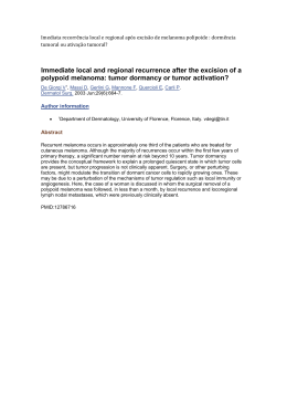
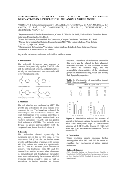
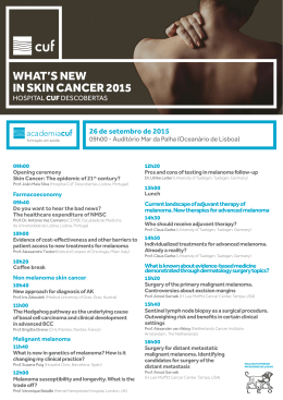
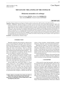
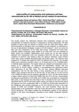
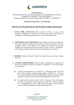
Case report of a surgical procedure in pacient
with Acral Lentiginous Melanoma
Carolina Pompermaier1, Clarissa Pieresan Winkelmann1, Fernando
de Marco dos Santos1, Isnard Elman Litvin1, Rochele Barcelos Prá1,
Rafael Fontana1
1University
of Caxias do Sul
Introduction
Acral lentiginous melanoma is the least
common variant of radial growth phase
melanomas. It arises most commonly on
palmar, plantar, subungual, and mucosal
surfaces.
Objectives
Report a surgical procedure accomplished
in a patient with letiginous acral
melanoma.
Methods
Patient V.F., 48 years old, male, caucasian,
recognized pigmented ulcerated blackned
lesion in the right subungual region after
non-traumatic loss of the nail in 2014. The
medical aid sugg-
ested trauma or fungal damage. In 2015,
it was observed appearance of right
inguinal lymphadenophaty associated
with growth. The PET-CT showed
hypermetabolic lymphadenophaty in the
right external iliac chain, which the
biggest was 1,4 cm. The patient
underwent amputation of the hallux and
inguinal lymph node biopsy. Pathology
indicated the dignosis of acral lentiginous
melanoma Clark IV, Breslow 8,0 mm, with
metastatic lymph nodal disease. Thus, it
was made iliac and retroperitoneal
lymphadenectomy.
Results
Cutaneous malignant melanoma is the
most lethal form of skin cancer. Of its
histologic subtypes, acral lentiginous
melanoma is the least common, more
prevalence in dark-skinned individuals.
The incidence increases in age, and it's
associated with poorer survival rates,
which may be due to delay in diagnosis.
Wide local excision is the treatment of
choice. The regional lymphadenectomy is
indicated to lymph nodes that have 1 to 4
cm. Intraoperative lymphatic mapping
and sentinel node biopsy are effective
techniques in identifying suspicious
nodes.
Image 1. Acral lentiginous melanoma, 40x, HE
Conclusions
The early detection is the bottom for the
vanced disease. The resection of the first
site is indicated to prevent possible
metastasis. Thus, the physician should
investigate if the patient has other similar
skin lesions and orther tests that deviate
a possible neoplasia.
References
Image 2. Acral lentiginous melanoma, 400x, HE
patient who has ALM to have a better
prognosis. In this case, the differential
diagnosis was late, indicating a more ad-
1. Coleman WP, III, Loria PR, Reed RJ, Krementz ET. Acral
Lentiginous Melanoma. Arch Dermatol. 1980, p. 773-776.
Disponível
em:
<http://archderm.jamanetwork.com/article.aspx?articleid=54
1206>. Acesso em: 07 jul. 2015.
2. HARMELIN, Eric S. et al. Acral lentiginous melanoma: an
illustrative case report and brief review of the literature. The
Journal Of Foot And Ankle Surgery. Miami, p. 540-545. dez.
1998.
Disponível
em:
<http://www.sciencedirect.com/science/article/pii/S1067251
698800331>. Acesso em: 03 jul. 2015.
3. RIDGEWAY, Calvin A. et al. Acral Lentiginous Melanoma.
Jama Surgury. Illinois, p. 88-92. jan. 1995. Disponível em:
<http://archsurg.jamanetwork.com/article.aspx?articleid=5961
14#References>. Acesso em: 07 jul. 2015.
4. SUTHERLAND, Carl M. et al. Acral lentiginous melanoma. The
American Journal Of Surgery. New York, jul. 1993. p. 64-67.
Disponível
em:
<http://www.sciencedirect.com/science/article/pii/S00029610
05805860>. Acesso em: 07 jul. 2015.
5. WARD, D.j. et al. Acral lentiginous melanoma: an illustrative
case report and brief review of the literature. British Journal
Of Plastic Surgery. Brighton, p. 619-623. out. 1984. Disponível
em:
<http://www.sciencedirect.com/science/article/pii/000712268
4901619>. Acesso em: 03 jul. 2015.
Image 3. Melanocitic lesion in the ungeal region
Autor a ser contatado:
Rochele Barcelos Prá
[email protected]
Hospital Geral de Caxias do Sul
Rua Prof. Antônio Vignoli, 255
Caxias do Sul, RS
Brasil
Download