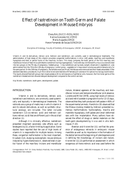
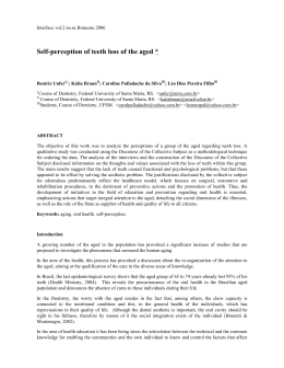
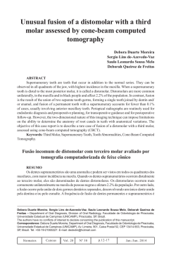
IMPACTED TEETH PREVALENCE IN THE CITY OF CURITIBA, PARANÁ, BRAZIL
190
IMPACTED TEETH PREVALENCE IN THE CITY OF
CURITIBA, PARANA, BRAZIL
PREVALÊNCIA DE DENTES RETIDOS NA CIDADE
DE CURITIBA, PARANÁ, BRASIL
Rodrigo SANDRIN **
Daniel Luiz Gaertner ZORZETTO ***
Clóvis MARZOLA ****
João Lopes TOLEDO-FILHO *****
José Lucas BARBOSA ******
Ivonete Barreto HAAGSMA *******
_________________________________________
* Monograph presented for conclusion of Specialization Course in Oral and Traummatology
Maxillofacial Surgery in 2008.
** Ex Resident of the Course in Oral and Traummatology Maxillofacial Surgery by the Brazilian
College of Buco Maxillofacial Surgery and Traummatology and Base Hospital of
Bauru, São Paulo State, Brazil. Student that concluded the Specialization Course in
Oral and Traummatology BMF Surgery APCD - Bauru, 2007 and author of the
monograph.
*** Professor of the Specialization Course of Oral and Traummatology Buco Maxillofacial
Surgery, APCD – Bauru, São Paulo State, Brazil. Effective member of the Base
Hospital of Bauru, São Paulo, Brazil. Titular member of the Brazilian College of Buco
Maxillofacial Surgery and Traummatology.
**** Retired Titular Professor of Surgery of School of Dentistry of the University of São Paulo –
FOB – USP. Professor of the Specialization Course of Oral and Traummatology Buco
Maxillofacial Surgery, APCD – Bauru, São Paulo State, Brazil. Effective member of
the Base Hospital of Bauru, São Paulo, Brazil. Titular member of the Brazilian College
of Buco Maxillofacial Surgery and Traummatology. Credenciated Professor of the
Albert Einstein Israelite Hospital, São Paulo, SP, Brazil. Co-adviser of this report.
***** Titular Professor of Anatomy of School of Dentistry of the University of São Paulo – FOB USP. Professor of the Specialization Course of Oral and Traummatology Buco
Maxillofacial Surgery, APCD – Bauru, São Paulo State, Brazil. Effective member of
the Base Hospital of Bauru, São Paulo, Brazil. Titular member of the Brazilian College
of Buco Maxillofacial Surgery and Traummatology.
****** Specialist in Radiology and Orthodonty and proprietor of the “All Doc Radiology Clinic”
in the Curitiba City, in Paraná state in Brazil.
******* Specialist in Radiology and proprietor of the “All Doc Radiology Clinic” in the Curitiba
City, in Paraná state in Brazil.
IMPACTED TEETH PREVALENCE IN THE CITY OF CURITIBA, PARANÁ, BRAZIL
191
ABSTRACT
This research reports a radiographic review of 9028 panoramic
radiographs from the digital archives of a Radiologic Institute of Curitiba city,
state of Paraná, “ALL DOC” with the purpose of evaluating the prevalence of
dental retention and their characteristics such as gender, location, age group and
teeth more frequently found retained. Three thousand and six hundred and thirty
six (3636) (40,27%) panoramic radiographs were found with retained teeth, being
female gender more affected (58,22%). The age group researched was from 13 to
95 years old. The posterior region of right mandible presented the major number
of retention cases (28,64%) and the teeth more frequently found retained were
lower third molars (54,65%), followed by upper third molars (40,09%) and upper
canines (1,61%).
RESUMO
Esta pesquisa apresenta uma revisão de 9028 radiografias
panorâmicas do arquivo digital do instituto de radiologia da cidade de Curitiba,
estado do Paraná, “ALL DOC” com a finalidade de avaliar a prevalência de
dentes retidos e suas características quanto ao gênero, localização, faixa etária e
dentes mais freqüentemente encontrados retidos. Foram encontradas 3636
(40,27%) radiografias panorâmicas com dentes retidos, sendo o gênero feminino o
mais acometido (58,22%). A faixa etária pesquisada variou dos 13 aos 95 anos.
A região posterior da mandíbula direita foi a que mais apresentou casos de
retenção (28,64%) e, os dentes mais freqüentemente encontrados retidos foram os
terceiros molares inferiores (54,65%), seguidos pelos terceiros molares superiores
(40,09%) e, caninos superiores (1,61%).
Uniterms: Dental retention; retained third molars; prevalence.
Unitermos: Retenção dental; terceiros molares retidos; prevalência.
INTRODUCTION
Impacted teeth are defined when a tooth is embedded totally or
partly inside the bone by the time of normal eruption, with or without
maintenance of follicular sac integrity. (MARZOLA, 1995 e 2005).
Impaction can occur with any type of teeth, nevertheless impaction
of third molars and maxillary canines seem to occur more frequently than for
other type of teeth. (MARZOLA; MADEIRA; CASTRO, 1968).
One of the reasons for the higher impacted third molar frequency is
the fact of being the last tooth to erupt (OLASOJI; ODUSANYA, 2000).
As to this line of thought, it has been said that the higher
prevalence of mandibular third molar impaction is due to mandibular bone
thickness and compact tissue and either for the space for this teeth were limited by
one side for the mandibular second molar and at the other for the ascending ramus
IMPACTED TEETH PREVALENCE IN THE CITY OF CURITIBA, PARANÁ, BRAZIL
192
of mandible, acting both as an obstacle for its eruption (MARTINHÃO;
BARROS; CAMPOS et al., 1992).
Impacted teeth are potentially related to periodontal disease,
pericoronitis, caries and adjacent tooth reabsorption (NICODEMO; RANGEL;
BAZZARELLA, 1982).
Those facts justify the value of this study, emphasizing the early
diagnose and treatment planning for these anomalies that if left untreated may
cause patient health issues.
With this purpose, we propose to evaluate the prevalence of
impacted teeth in the City of Curitiba, the gender prevalence and the teeth more
founded in this condition.
LITERATURE REVIEW
There are two forms of dental impaction: teeth completely bone
embedded (endosseous impaction) or partly or totally gingival mucosa embedded
(subgingival impaction). It can be partly impacted with rupture of follicular sac
(semi-impaction) (MARZOLA, 1995 e 2005).
The frequency of dental impaction hasn’t been addressed until
recent years. BLUM (1923) was the first to report mandibular third molars (86%),
maxillary canines (51%) and maxillary third molars (33%) as the most frequently
impacted teeth.
In a study of 6389 cases, 1432 of them with full mouth radiographs,
MEAD (apud MARZOLA, 2005) reported that 461 of 581 impacted teeth
(nearly 80%) were third molars, being 248 mandibular and 213 maxillary; another
teeth reported with high frequency was the maxillary canine(23).
FONSECA (1956) evaluated 1000 patients clinic and
radiographically, finding 174 (17,4%) patients with impacted teeth, 93 (18,6%)
male e 81 (15,4%) female. The author could prove the higher prevalence of
impacted mandibular third molars (66,6%), followed by maxillary third molars
(33,3%), maxillary canines (14,3%), mandibular premolars (2,8%), mandibular
canines (1,7%); supernumerary teeth (1,4%, 8 maxillary fourth molar and 1
mandibular) and two cases of mesiodens (0,2%).
In a study with 3874 radiographs, the prevalence of at least one
impacted teeth was 16,7%. The most frequently impacted teeth were maxillary
third molars (21,9%) and mandibular ones (17,5%), followed by maxillary
canines and mandibular premolars. No significant statisticcal difference was
found by gender to third molar impaction. Nevertheless, maxillary canine
impaction was more frequent in female (1,17% versus 0,51% male) (DACHI;
HOWELL, 1961).
In the City of Ribeirão Preto (SP, Brazil) it was noticed 1000
radiographs, with 63 (6,3%) showing 99 impacted teeth, among them mandibular
third molars with high prevalence (34,3%); maxillary canines (18,2%); maxillary
third molars (17,2%); mandibular second premolars (10,1%); mandibular first
premolars (4%); maxillary second premolars (4%); maxillary central incisor (3%);
supernumerary (3%); mandibular canines (2%); maxillary lateral incisor (1%) and
mandibular first and second molars (1% each). By gender: 6.6% female and 5.9%
male (GRANDINI; VERRI; STIVANIN, 1966).
IMPACTED TEETH PREVALENCE IN THE CITY OF CURITIBA, PARANÁ, BRAZIL
193
In a study with 1760 patients in the city of Araçatuba (SP, Brazil) it
was observed that impaction was present in 17.9% of the patients and agenesis
occurred in 7.6%. Mandibular and maxillary third molars were the most
impaction-affected teeth (49.35 % each). Maxillary canine’s impaction was seen
in 1.4% of cases. Thus, in gender, the prevalence was similar (45.3% male and
41.1% female) (MARZOLA; MADEIRA; CASTRO, 1968).
It was found 6.4% prevalence of impacted teeth in 1000 patients
evaluated clinically and radiographically, 6,68% female and 6,05% male.
Nevertheless, supernumerary teeth suffered more impaction (26,02%); although
difference between them and mandibular third molars was lower (23,29%). Other
teeth suffering from impaction were maxillary canines (24,66%); maxillary third
molars (9,59%); mandibular canines (5,48%); maxillary second premolars
(4,11%); maxillary central incisor (4,11%); mandibular second premolars (1,37%)
and mandibular first premolars (1,37%). Of supernumerary teeth, all were
maxillary, 57,90% posterior and 42,10% anterior (5 mesiodens) (SALOMÃO;
SENI, 1970).
A study at Harlem Hospital (USA) where 95% of the patients were
afro-Americans, evaluated 3745 panoramic radiographies. The authors reported
18,20% patients with one or more impaction, male and female with equal
prevalence. Maxillary third molars were most frequently impacted than
mandibular (58,87% and 41,13% respectively). Other impaction reported was
canine (3,94%); second molar (0,67%); first and second premolar (0,49% each);
first molar (0,25%) e lateral incisor (0,08%) (KRAMER; WILLIAMS, 1970).
Following the same line of research, a radiograph evaluation was
performed with 3000 patients. In 245 (8,16%) of 3000 patients it was found 360
impacted teeth: mandibular third molar (37,78%); maxillary third molar (20,28%);
maxillary canine (16,94%); supernumerary (11,39%) and different teeth with
lower percentage. Impaction were more frequent in female (8,26%) than male
(8,06%) (VERRI; OLIVEIRA; GRANDINI et al., 1973).
A research conducted among Dentistry students in the City of São
José dos Campos between 18 and 23 years old showed that 40% of the sample
with third molar impaction, 45,2% female and 31.2% male. Impaction prevalence
in decreasing order was: right mandibular third molar (39 cases); left mandibular
third molar (34 cases); right maxillary third molar (33 cases) and left maxillary
third molar (31 cases) (NICODEMO; RANGEL; BAZZARELLA, 1982).
Prevalence and distribution of impacted teeth in 1895 radiographs
was 583 (30,8%) with one or more impaction at the following order: mandibular
third molar (48,1%); maxillary third molar (29,6%); maxillary canine (11,9%);
mandibular canine (3,5%); mandibular second premolar (3,3%); maxillary second
premolar (2,2%); maxillary second molar (0,5%); mandibular first premolar
(0,4%); maxillary central incisors (0,2%); mandibular second molar (0,1%);
maxillary first premolar (0,1%); other (0,3%). There was no significant statisticcal
difference in gender (BROWN; BERKMAN; COHEN et al., 1982).
Swedish student’s radiographs were evaluated for prevalence of
impaction of third molars. Analyzing 113 female and 144 male, it was reported
that 33% of the sample showed one or more third molars impacted. The
prevalence of third molar impaction was higher in mandible (66%) than in
maxillae (34%) (SCHERSTÉN; LYSELL; ROHLIN, 1989).
IMPACTED TEETH PREVALENCE IN THE CITY OF CURITIBA, PARANÁ, BRAZIL
194
A study of 334 panoramic radiographies in the City of Maringa
(PR) showed 58 third molars impacted in 36 (10,78%) radiographies. The
minimal age criteria of inclusion in this study were 25 years old. 34 mandibular
third molars were observed (58,6%) and 24 maxillary third molars (41,4%).
Distribution of impacted teeth ranged from 67,2% female to 32,8% male
(MARTINHÃO; BARROS; CAMPOS et al., 1992).
A study of dental anomalies was designed through the analysis of
934 panoramic radiographies of patients aging from 3 to 80 years old. It was
found some kind of anomaly in 550 (58,9%) of the radiographs, such as impacted
teeth (21,2%) and supernumerary teeth (2,3%) (CARVALHO; SIMI;
ABDALLA et al., 1997).
A comparison between the prevalence in third molar impaction in
an urban population (city of Lagos, Nigeria) and a rural one was made.
Evaluating 1200 patients clinically and radiographically in each group, a higher
prevalence was observed in the urban population (22.8%) than in the rural one
(3.1%). Mandibular third molar impaction was numbered in 15,1% of impaction
in urban cases versus 1.9% at rural population, as for maxillary third molar, 6.2%
were urban versus 0.3% rural. Authors reported that the difference in impacted
teeth may be due to changes in eating habits occurred in the urban population,
such as the addition of less rigid food, therefore demanding less maxillary
function. It was also reported that dental friction during mastigatory movements
of rigid food requires a high muscle activity stimulating mandibular growth
(OLASOJI; ODUSANYA, 2000).
In the years 2001 and 2002 the impaction prevalence in 88
panoramic radiographies of surgery attending patient’s of the Dentistry school of
the City of Feira de Santana (BA) was evaluated. As a result 59,1% of the sample
was female and 40.9% male. It was reported that the majority of impacted teeth
were mandibular third molars (49,3%), followed by maxillary third molars
(36,9%); supernumerary (6,7%); maxillary canines (3,8%); incisors (1,9%) and
mandibular premolars (1,4%) (FARIAS; SANTOS; CAMPOS et al., 2003).
A research designed in Singapore, China, with 1000 panoramic
radiographies of patients ranging from 20 to 40 years old found out 68,6% of the
population researched with impacted third molar teeth. It was observed higher
prevalence in mandible (90%) than maxillae (28%) and female suffered in higher
number (56%) than male (44%) (QUEK; TAY; TAY et al., 2003).
Aiming to analyze third molar position prevalence in private
practice patients in the Cities of Cunha Porã, Maravilha and Palmitos (SC, Brazil),
585 panoramic radiographies were observed, being 210 male and 375 female.
1815 impacted third molars were recorded, 465 teeth number 28; 453 teeth
number 38; 450 teeth number 18 and 447 teeth number 48 (MARZOLA;
COMPARIN; TOLEDO-FILHO, 2006).
2651 panoramic radiographies of a digital data center in the City of
Curitiba (PR) were analyzed for the prevalence of impacted teeth (third molar,
molar, premolar, canine and incisor). 425 radiographies showed impacted teeth,
185 male (43,53%) and 240 female (56,47%), ages ranging from 15 to 88 years.
971 impacted teeth were observed: 267 (teeth number 38); 242 (teeth number 48);
217 (teeth number 18); 187 (teeth number 28); 14 (teeth number 23); 12 (teeth
number 13) and other in lower percentage (TOLEDO, 2007).
IMPACTED TEETH PREVALENCE IN THE CITY OF CURITIBA, PARANÁ, BRAZIL
195
At the same digital data center, 3000 panoramic radiographies were
also evaluated for the prevalence of impacted canines finding 2,23% of impaction.
Higher prevalence in female (58,2%) than male (41,8%) was observed. The
frequency of impaction was: teeth number 23 (44,8%); teeth number 13 (41,8%);
teeth number 33 (7,4%) and teeth number 43 (6%) (OLIVEIRA, 2007).
Authors studying 848 panoramic radiographies found 64
supernumerary impacted teeth, most of them in permanent dentition. As to
location, the maxilla anterior region had the most occurrences, with 57,8%. Other
regions in maxilla were molar (10,9%); premolar (9,4%); canine (7,8%).
Mandible: premolar (9,4%). Other areas, 4,8%. In gender, there was higher
prevalence in male (8,4%) then female (5,2%) (CUNHA-FILHO; PURICELLI;
HENNIGEN et al., 2002).
It was also observed that in supernumerary teeth the most common
was mesiodens, being related one case of a 5 year-old male, with 2 impacted
palatal mesiodens, one of them inverted (ABREU-E-LIMA; MOTISUKI;
BORDIN, 2002).
MATERIAL AND METHOD
9028 panoramic radiographies were examined from a radiological
digital institute arquive called “ALL DOC” in the city of Curitiba, PR, Brazil,
taken for different therapeutic reasons. The study protocol was evaluated by the
‘Hospital de Reabilitação de Anomalias Craniofaciais’ of São Paulo University,
Bauru, Brazil ethical committee. The required inclusion criteria were based on
permanent tooth, discarding those with deciduous dentition and permanentdeciduous dentition.
Minimal age consider for inclusion was 13 years, due to the fact
that at this age maxillary canines have already erupted and because at this phase,
we can predict the impaction or not of third molars. Radiographies researched in
this study were from both male and female patients and age ranged from 13 to 95
years.
Prior to the beginning of collect data, the examiner was adjusted.
Digital radiographies were analyzed clockwise counting impacted teeth after
careful anatomic region consideration.
Images were analyzing using a LG 17” monitor and desktop
computer; light in the room was adequate, during one hour daily for about four
months, by a buco-maxillo-facial surgery specialized dentist.
Data were manually classified in proper cards in the end of each
evaluation. In the final evaluation stage these info were condensed in a computer
file using Microsoft Excel® software in order to facilitate percentage study of the
data collected.
RESULTS
From 9028 panoramic radiographies, 3685 (40,82%) were male and
5343 (59,18%) female. Age ranged from 13 to 95 years. From this amount, 3636
(40,27%) radiographies showed impacted teeth and 59,73% did not showed any
signals of impaction (Graph 1).
IMPACTED TEETH PREVALENCE IN THE CITY OF CURITIBA, PARANÁ, BRAZIL
196
40,27%
impacted teeth
no impacted teeth
59,73%
Graph 1 – Prevalence of impacted teeth in the population studied.
From the total of impacted teeth, 2117 (58,22%) were female and
1519 (41,78%) male (Graph 2).
41,78%
male
female
58,22%
Graph 2 – Prevalence of impacted teeth according to genre.
IMPACTED TEETH PREVALENCE IN THE CITY OF CURITIBA, PARANÁ, BRAZIL
197
Most frequently impacted teeth was 48 (27,79%); 38 (26,86%); 28
(20,18%); 18 (19,91%); 13 (0,81%) and 23 (0,80%). Other type’s equals 3,65%
(Graph 3).
30,00%
27,79%
26,86%
25,00%
20,18% 19,91%
20,00%
15,00%
10,00%
3,65%
5,00%
0,81%
0,80%
Teeth
13
Teeth
23
0,00%
Teeth
48
Teeth
38
Teeth
28
Teeth
18
Other
tooth
Graph 3 – Prevalence according to the frequency of impacted teeth type.
There were 65 impacted supernumerary cases (0,65%), maxillary
fourth molars (0,43%) in higher range, followed by mandibular premolars
(0,12%); mandibular fourth molars (0,05%); maxillary premolars (0,02%);
maxillary incisor (0,02%) and mesiodens (0,01%) (Graph 4).
0,01%
0,02%
4º MS
0,02%
PMI
0,05%
4º MI
0,12%
PMS
IS
0,43%
Graph 4 – Supernumerary prevalence.
Mesiodens
IMPACTED TEETH PREVALENCE IN THE CITY OF CURITIBA, PARANÁ, BRAZIL
198
DISCUSSION
The dental impaction theme has been reckoned by several authors
throughout the years.
The amount of impacted teeth found in this study seem to
correspond to those reported before (NICODEMO; RANGEL;
BAZZARELLA, 1982 (40%); BROWN; BERKMAN; COHEN et al., 1982
(30,8%) and SCHERSTÉN; LYSELL; ROHLIN 1989 (33%)), opposing from
other outcomes (FONSECA, 1956 (17,4%); DACHI; HOWELL, 1961 (16,7%);
GRANDINI; VERRI; STIVANIN, 1966 (6,3%); MARZOLA; MADEIRA;
CASTRO, 1968 (17,9%); SALOMÃO; SENI, 1970 (6,4%); KRAMER;
WILLIAMS, 1970 (18,20%); VERRI; OLIVEIRA; GRANDINI et al., 1973
(8,16%); MARTINHÃO; BARROS; CAMPOS et al., 1992 (10,78%);
CARVALHO; SIMI; ABDALLA et al., 1997 (21,2%); OLASOJI;
ODUSANYA, 2000 (22,8% and 3,1% in two different population); QUEK;
TAY; TAY et al., 2003 (68,6%); TOLEDO, 2007 (16,03%) and OLIVEIRA,
2007 (2,23%) (Fig. 1).
Regarding the most frequent gender affected by impacted teeth,
some authors described higher prevalence in females, (GRANDINI; VERRI;
STIVANIN, 1966; SALOMÃO; SENI, 1970; VERRI; OLIVEIRA;
GRANDINI et al., 1973; NICODEMO; RANGEL; BAZZARELLA, 1982;
MARTINHÃO; BARROS; CAMPOS et al., 1992; FARIAS; SANTOS;
CAMPOS et al., 2003; QUEK; TAY; TAY et al., 2003; MARZOLA;
COMPARIN; TOLEDO-FILHO, 2006; TOLEDO, 2007 and OLIVEIRA,
2007), this was also seen in this study. Others may disagree describing males
with better prevalence (FONSECA, 1956; MARZOLA; MADEIRA; CASTRO,
1968; SCHERSTÉN; LYSELL; ROHLIN, 1989 and CUNHA-FILHO;
PURICELLI; HENNIGEN et al., 2002). Nevertheless, researches showed
similar prevalence in both gender (DACHI; HOWELL, 1961; KRAMER;
WILLIAMS, 1970 and BROWN; BERKMAN; COHEN et al., 1982).
For the most impacted tooth type findings, present investigation
outcome are in agreement with several authors (BLUM, 1923; MEAD apud
MARZOLA, 2005; FONSECA, 1956; GRANDINI; VERRI; STIVANIN,
1966; VERRI; OLIVEIRA; GRANDINI et al., 1973; NICODEMO;
RANGEL; BAZZARELLA, 1982; BROWN; BERKMAN; COHEN et al.,
1982; SCHERSTÉN; LYSELL; ROHLIN, 1989; MARTINHÃO; BARROS;
CAMPOS et al., 1992; OLASOJI; ODUSANYA, 2000; FARIAS; SANTOS;
CAMPOS et al., 2003; QUEK; TAY; TAY et al., 2003 and TOLEDO, 2007),
who reported mandibular third molars with high prevalence than other teeth.
Some studies showed lower prevalence of mandibular third molars when
compared with other teeth (DACHI; HOWELL; 1961; SALOMÃO; SENI,
1970; KRAMER; WILLIAMS, 1970 e MARZOLA; COMPARIN;
TOLEDO-FILHO, 2006).
Approaching impacted maxillary canine, the outcome was similar
to other authors (MARZOLA; MADEIRA; CASTRO, 1968; KRAMER;
WILLIAMS, 1970; FARIAS; SANTOS; CAMPOS et al., 2003; TOLEDO,
2007 and OLIVEIRA, 2007), opposing to contemporaneous and previous
researches (BLUM 1923; FONSECA, 1956; GRANDINI; VERRI;
IMPACTED TEETH PREVALENCE IN THE CITY OF CURITIBA, PARANÁ, BRAZIL
199
STIVANIN, 1966; SALOMÃO; SENI, 1970; VERRI; OLIVEIRA;
GRANDINI et al., 1973 and BROWN; BERKMAN; COHEN et al., 1982).
The supernumerary prevalence was similar to several studies
(FONSECA, 1956; GRANDINI; VERRI; STIVANIN, 1966 and
CARVALHO; SIMI; ABDALLA et al., 1997). However, different of some
studies (SALOMÃO; SENI, 1970; VERRI; OLIVEIRA; GRANDINI et al.,
1973 and FARIAS; SANTOS; CAMPOS et al., 2003), where the
supernumerary prevalence was higher.
Maxillary fourth molars were the most common finding among
supernumerary teeth, despite the statement of most common being mesiodens
(ABREU-E-LIMA; MOTISUKI; BORDIN, 2002), appearing just one case at
the present study, as in other researches (FONSECA, 1956 and MARZOLA;
FREITAS; ALVAREZ et al., 1968).
Discussing location of supernumerary teeth the outcome of the this
study does not agree with some of the literature (CUNHA-FILHO;
PURICELLI; HENNIGEN et al., 2002) where the anterior maxillae had more
prevalence opposing to posterior maxillae, as showed at the present study and
other (SALOMÃO; SENI, 1970).
Great part of supernumerary teeth occurence was in patients with
multiple impacted supernumerary teeth (MARZOLA, 1995 and 2005).
CONCLUSIONS
Within the analysis of 9028 radiographies in this study:
1. Impacted teeth prevalence in Curitiba, PR, Brazil was 40,27%.
2. Gender prevalence showed that female are most likely in 58,22%
to present dental impaction.
3. Mandibular third molars has the higher impaction rate (54,65%),
followed by maxillary third molars (40,09%) and maxillary canines (1,61%).
Fig. 1 – Some factors prevalence research about impacted teeth.
Authors
Impaction
Prevalence
BLUM, 1923
----------------
MEAD apud
MARZOLA, 2005
----------------
---------------
248
213
23
17,4%
16,7%
Male
18,6%
---------------
66,6%
17,5%
33,3%
21,9%
14,3%
-------------
34,3%
17,2%
18,2%
49,3%
49,3%
1,4%
23,29%
9,59%
24,66%
FONSECA,
1956
DACHI; HOWELL,
1961
GRANDINI; VERRI;
STIVANIN, 1966
MARZOLA;
MADEIRA; CASTRO,
1968
SALOMÃO; SENI,
1970
6,3%
17,9%
6,4%
Gender
Third Lower Third Upper
Molar
Molar
Prevalence
Prevalence
--------------86%
33%
-
Female
6,6%
Male
45,3%
Female
6,68%
Upper
Canine
Prevalence
51%
IMPACTED TEETH PREVALENCE IN THE CITY OF CURITIBA, PARANÁ, BRAZIL
KRAMER; WILLIANS,
1970
AITASALO;
LEHTINEN; OKSALA
apud MARZOLA,
2005
VERRI; OLIVEIRA;
GRANDINI et al.,
1973
NICODEMO;
RANGEL;
BAZZARELLA, 1982
BROWN; BERKMAN;
COHEN et al., 1982
SCHERSTÉN;
LYSELL; ROHLIN,
1989
200
18,20%
---------------
41,13%
58,87%
--------------
---------------
---------------
39,2%
36,9%
-------------
8,16%
Female
8,26%
37,78%
20,28%
16,94%
40%
Female
45,2%
73
64
--------------
48,1%
29,6%
11,9%
66%
34%
------------
33%
--------------Male
56,03%
--------------
Feminino
60,6%
-------------
--------------
--------------
10,78%
Female
67,2%
58,6%
41,4%
-------------
----------------
Female
56%
417
601
---------------
21,2%
--------------
---------------
-------------
---------------
22,8%/3,1%
---------------
15,1%/1,9%
6,2%/0,3%
--------------
---------------
Female
59,1%
49,3%
36,9%
3,8%
68,6%
Female
56%
90%
28%
---------------
---------------
Female
375
900
915
---------------
TOLEDO, 2007
16,03%
Female
56,47%
509
404
26
OLIVEIRA,
2007
---------------
Female
58,2%
----------------
-----------------
54,65%
40,09%
MARZOLA; NARYFILHO; KAWAKAMI
et al., 1990
MARTINHÃO;
BARROS; CAMPOS
et al., 1992
OLIVEIRA; SPOHR;
ZENI et al., 1996
CARVALHO; SIMI;
ABDALLA et al.,
1997
OLASOJI;
ODUSANYA, 2000
FARIAS; SANTOS;
CAMPOS et al.,
2003
QUEK; TAY; TAY
et al., 2003
MARZOLA;
COMPARIN;
TOLEDO-FILHO,
2006
PRESENT
RESEARCH
30,8%
40, 27%
Female
58,22%
2,23%
1,61%
REFERENCES *
ABREU-E-LIMA; F.; MOTISUKI, C.; BORDIN, M. M. Mesiodens: Detecção e
intervenção cirúrgica precoce. Rev. gaúcha Odont., v. 50, n. 2, p. 69-73, 2002.
IMPACTED TEETH PREVALENCE IN THE CITY OF CURITIBA, PARANÁ, BRAZIL
201
AITASALO, K.; LEHTINEN, R.; OKSALA, E. An orthopantomographic study
of prevalence of impacted teeth. J. oral Surg. v. 1, p. 117-20, 1972. In:
MARZOLA, C. Fundamentos de Cirurgia Buco Maxilo Facial. CDR. Bauru: Ed.
Independente, 2005.
AZENHA, M. R.; MARZOLA, C.; BRANDT-FILHO, S. H. O. et al., Multiple
impacted supernumerary teeth in a not sindromic patient – Surgical clinical case
report. Revista de Odontologia da Academia Tiradentes de Odontologia, Bauru, v.
5, n. 3, p. 374-83, 2007.
AZENHA, M. R.; PEREIRA, L. C.; SQUILLACE, J. M. et al., Quartos molares
superiores bilaterais retidos – Relato de caso clínico-cirúrgico. Revista da
Academia Tiradentes de Odontologia, Revista eletrônica, Bauru, v. 4, n. 2, p. 28491, 2007.
BLUM, T. Malposed teeth: their classification pathology and treatment. Int. J.
Orthodont. Oral Surg. Radiol., Lakewood, v. 9, p. 122-137, 1923.
BROWN, L. H.; BERKMAN, S.; COHEN, D. et al., A radiological study of the
frequency and distribution of impacted teeth. J. dent. Assoc. S. Afr., v. 37, p. 62730, 1982.
CARVALHO, P. L.; SIMI, R.; ABDALLA, C. A. et al., Estudo da prevalência
das anomalias dentais por meio das radiografias panorâmicas. Rev. Odont. Univ.
St. Amaro, São Paulo, v. 2, n. 3, p. 28-30, 1997.
CUNHA-FILHO, J. J.; PURICELLI, E.; HENNIGEN, T. W. et al., Ocorrência de
dentes supranumerários em pacientes do serviço de Cirurgia e Traumatologia
Buco-Maxilo-Facial, Faculdade de Odontologia da UFRGS, no período de 1998 a
2001. Rev. Fac. Odont. Porto Alegre, Porto Alegre, v. 43, n. 2, p. 27-34, 2002.
DACHI, S. F.; HOWELL, F. V. A survey of 3874 routine full-mouth radiographs
II. A study of impacted teeth. Oral Surg. Oral Pathol. Oral Med., St. Louis, v. 14,
n. 10, p. 1165-69, 1961.
FARIAS, J. G.; SANTOS, F. A. P.; CAMPOS, P. S. F. et al., Prevalência de
dentes inclusos em pacientes atendidos na Disciplina de Cirurgia do Curso de
Odontologia da Universidade Estadual de Feira de Santana. Rev. Pesq. Bras.
Odontoped. Clín. Integr., João Pessoa, v. 3, n. 2, p. 15-9, 2003.
FONSECA, J. B. Incidência da inclusão dentária em 1000 pacientes com exame
radiográfico completo. Sel. Odont., São Paulo, v. 11, p. 21-8, 1956.
GRANDINI, S. A.; VERRI, R. A.; STIVANIN, D. Estudo da incidência dos
dentes inclusos: pesquisa através do exame radiográfico em 1000 pacientes. Rev.
Assoc. paul. Cir. Dent., São Paulo, v. 20, p. 90-8, 1966.
HOPCRAFT, M. Multiple supernumerary teeth: Case report. Aust. dent. J.,
Sidney, v. 43, n. 1, p. 17-9, 1998.
KIM, T.; ARTUN, J.; BEHBEHANI, F. et al., Prevalence of third molar
impaction in orthodontic patients treated nonextraction and with extraction of 4
premolars. Am. J. Orthod. Dentofacial Orthop., St. Louis, v. 123, n. 2, p. 138-45,
2003.
KRAMER, R. M.; WILLIAMS, A. C. The incidence of impacted teeth. A survey
at Harlem Hospital. Oral Surg., St. Louis, v. 29, n. 2, p. 237-41, 1970.
_____________________________________________
* According the ABNT norms.
IMPACTED TEETH PREVALENCE IN THE CITY OF CURITIBA, PARANÁ, BRAZIL
202
MARTINHÃO, Z. G.; BARROS, W. M. R.; CAMPOS, G. M. et al., Estudo da
incidência de terceiros molares inclusos, por meio de radiografias panorâmicas, e
aplicação da informática na computação dos dados. Odont. Mod., Rio de Janeiro,
v. 19, n. 6, p. 6-12, 1992.
MARZOLA, C.; MADEIRA, M. C.; CASTRO, A. L. Ocorrência de retenção e
agenesia dental em 1760 indivíduos. Arq. Cent. Estud. Fac. Odont. Univ. Fed. M.
Gerais, Belo Horizonte, v. 5, n. 1, p. 33-46, 1968.
MARZOLA, C.; FREITAS, J. A. S.; ALVAREZ, L. C. et al., Mesiodens e
retenção do incisivo medial superior: relato de um caso. Rev. bras. Odont., Rio de
Janeiro, v. 25, n. 149, p. 3-8, 1968.
MARZOLA, C.; CAMPANELLA, E. J. Terceiro molar retido no processo
coronóide da mandíbula. Rev. gaúcha Odont., Porto Alegre, v. 33, p. 127-33,
1985.
MARZOLA, C.; DAMANTE, J. H. Odontodisplasia total. Múltiplos dentes não
irrompidos. Rev. gaúcha Odont., Porto Alegre, v. 34, n. 2, p. 140-3, 1986.
MARZOLA, C.; NARY-FILHO, H.; KAWAKAMI, R. et al., Retenção de
terceiros molares inferiores: etiologia, acidentes de irrupção, classificação e
técnica cirúrgica. Rev. Odonto Ciênc., Porto Alegre, v. 5, n. 10, p. 9-25, 1990-92.
MARZOLA, C. Retenção Dental, 2ª ed. São Paulo: Pancast, 1995, p. 15-242.
MARZOLA, C. Fundamentos de Cirurgia Buco Maxilo Facial. CDR. Bauru: Ed.
Independente, 2005.
MARZOLA, C.; COMPARIN, E.; TOLEDO-FILHO, J. L. Prevalência das
posições de terceiros molares nos municípios de Cunha Porã, Maravilha e
Palmitos, no Extremo oeste de Santa Catarina. Rev. bras. Cir. Traumatol. BucoMaxilo-Fac., São Paulo, v. 3, n. 1, p. 2-14, 2006.
MEAD, S. V. Incidence of impacted teeth. Int. Orthodont. Oral Surg. Radiol. v.
16, p. 885-90, 1930. In: MARZOLA, C. Fundamentos de Cirurgia Buco Maxilo
Facial. CDR. Bauru: Ed. Independente, 2005.
NICODEMO, R. A.; RANGEL, F. J. C.; BAZZARELLA, C. B. Prevalência de
terceiros molares inclusos entre estudantes da Faculdade de Odontologia de São
José dos Campos. Ars. Curandi, Rio de Janeiro, v. 8, n. 4, p. 13-15, 1982.
OLASOJI, H. O.; ODUSANYA, S. A. Comparative study of third molar
impaction in rural and urban areas of South–Western Nigeria. Odontostomatol.
Trop., Dakar, v. 23, n. 90, p. 25-8, 2000.
OLIVEIRA, D. L. Prevalência de caninos retidos na cidade de Curitiba – PR.
Monografia apresentada ao curso de Especialização em Cirurgia e Traumatologia
BMF na cidade de Bauru, SP, Brasil. Associação Paulista de Cirurgiões Dentistas,
2007.
OLIVEIRA; M. G.; SPOHR, A. M.; ZENI, E. L. et al., Radiografia panorâmica
na complementação diagnóstica de inclusões de terceiros molares. Rev. Odonto
Ciênc., Porto Alegre, v. 11, n. 22, p. 83-91, 1996.
QUEK, S. L.; TAY, C. K.; TAY, K. I. et al., Pattern of third molar impaction in a
Singapore Chinese population: a retrospective radiographic survey. Int. J. oral
Maxillofac. Surg., v. 32, p. 548-52, 2003.
SALOMÃO, J. I. S.; SENI; S. M. T. Estudo clínico-radiográfico da incidência dos
dentes inclusos em mil pacientes. Rev. gaúcha Odont., Porto Alegre, v. 18, n. 2, p.
83-9, 1970.
SCHERSTÉN, E.; LYSELL, L.; ROHLIN, M. Prevalence of impacted third
molars in dental students. Swed. dent. J., Malmo, v. 13, n. 4, p. 7-13, 1989.
IMPACTED TEETH PREVALENCE IN THE CITY OF CURITIBA, PARANÁ, BRAZIL
203
TOLEDO, G. L. Estudo da prevalência de dentes retidos através de radiografias
panorâmicas digitais no município de Curitiba, PR, Brasil. Monografia
apresentada ao curso de Especialização em Cirurgia e Traumatologia BMF na
cidade de Bauru, SP, Brasil. Associação Paulista de Cirurgiões Dentistas, 2007.
VERRI, R. A.; OLIVEIRA, M. A.; GRANDINI, S. A. et. al., Estudo clínico
radiográfico da incidência dos dentes inclusos em 3000 indivíduos. Rev. Assoc.
paul. Cir. Dent., São Paulo, v. 27, n. 5, p. 274-79, 1973.
ZORZETTO, D. L. G.; MARZOLA, C.; TOLEDO-FILHO, J. L. et al., Cirurgia
de terceiros molares inferiores retidos: complicações pós-operatórias –
observações clínicas. Rev. gaúcha Odont., Porto Alegre, v. 48, n. 2, p. 102-8,
2000.
o0o
Download