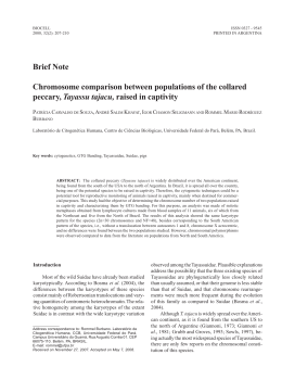
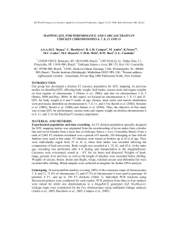
Case Report Interrupted Aortic Arch Type B in A Patient with Cat Eye Syndrome Sintia Iole Nogueira Belangero1, Fernanda Teixeira da Silva Bellucco1, Mirlene C. S. P. Cernach1, April M. Hacker2, Beverly S. Emanuel2, Maria Isabel Melaragno1 Universidade Federal de São Paulo1, São Paulo, SP - Brasil; Children´s Hospital of Philadelphia (University of Pennsylvania)2, Filadélfia, Pensilvânia - United States We report a patient with cat eye syndrome and interrupted aortic arch type B, a typical finding in the 22q11.2 deletion syndrome. Chromosomal analysis and fluorescent in situ hybridization (FISH) showed a supernumerary bisatellited isodicentric marker chromosome derived from chromosome 22. The segment from 22pter to 22q11.2 in the supernumerary chromosome found in our patient does not overlap with the region deleted in patients with the 22q11.2 deletion syndrome. However, the finding of an interrupted aortic arch type B is unusual in CES, although it is a frequent heart defect in the 22q11 deletion syndrome. Introduction Cat eye syndrome (CES; OMIM #115470) is a rare syndrome, clinically characterized by downslanting palpebral fissures, ocular coloboma, anorectal, heart and renal malformations, ears with preauricular tags and/or pits and mental retardation1,2. CES is often associated with significant phenotypic variability, ranging from patients with almost normal phenotype to those with severe abnormalities1. None of the features is consistently present. Only 41% of CES patients have the classical combination of iris coloboma, anal anomalies and pre-auricular anomalies1. Thus, many patients cannot be identified as having CES by phenotype alone. The diagnosis is based on the presence of a supernumerary marker chromosome derived from chromosome 22. This marker chromosome is usually isodicentric, contains a single active centromere and often exhibits satellites on both ends. Therefore, patients have partial tetrasomy from 22 pter to 22q11.23. The molecular breakpoints that originate the CES chromosome (CEC) are usually clustered in two intervals. The proximal one is the most common breakpoint and corresponds to the proximal deletion breakpoint interval found for the 22q11 deletion. The more distal breakpoint overlaps with the common distal deletion breakpoint of the 3 Mb deletion seen in the 22q11 deletion syndrome4. Thus, based on the location of the two breakpoints, CEC can be classified into three types. They can be isodicentric Key words Aorta, thoracic; Cat Eye Syndrome; chromosome deletion; heart defects, congenital. Mailing address: Sintia Iole Nogueira Belangero• Rua Botucatu, 740 - Vila Clementino – 04023-900 - São Paulo, SP - Brasil E-mail: [email protected] Manuscript received September 21, 2007, revisd manuscript received December 05, 2007; accepted February 12, 2008. e29 with both proximal or both distal breakpoints, resulting in a smaller or a larger symmetric chromosome, respectively. CEC can also be originated by one proximal and one distal breakpoint, resulting in an asymmetric CEC. In the latter case, there is one additional copy of the DiGeorge chromosomal region (DGCR), whereas in the larger isodicentric CEC there are two, and there is no extra DGCR copy in the smaller CEC. The distal boundary of the CES chromosomal region is proximal to the DGCR5. It covers approximately 2 Mb of 22q11.2 with its distal boundary. No obvious correlation between the severity of the associated phenotype and the size of the duplicated region has been found in CES4,5. In the present study, a patient with a supernumerary marker chromosome, cat eye syndrome and interrupted aortic arch type B is described. Case report Patient The Research Ethics Committee of UNIFESP approved this study, and informed consent was obtained from the patient’s parents. The proband was female, and the first child born to young parents (mother 22 yrs, father 26 yrs). The birth was by Caesarian section at 39 weeks of gestation. The birth weight was 2760 g, birth length was 47 cm, and cephalic circumference was 34 cm. The mother observed that the child looked tired after feeding. At 3 months of age an echocardiogram was performed and revealed interventricular communication (IVC), moderate hypoplasia of the aortic valve and ring, moderate dilation of the pulmonary trunk, left-turning aortic arch, presence of a B-type interruption of the aortic arch, and presence of a good-caliber arterial channel with aorta-pulmonary artery shunt. These findings were subsequently confirmed by other echocardiograms. At 4 months of age (Figure 1), her weight was 4410 g (<3%ile), length was 61.5 cm (50%ile) and head circumference was 41 cm (50%ile). The child presented with a broad and prominent forehead, downslanting palpebral fissures, hypertelorism, inner epicanthic folds, bilateral preauricular pits, abnormal ears, flat nasal bridge, anteverted nares, long philtrum, carp-shaped mouth and slight malar hypoplasia. The child died at 5 months of age. The cause of death was an infection (bronchopneumonia) that did not allow the surgical correction of the cardiopathy. Cytogenetic analysis Chromosomal analysis revealed a supernumerary bisatellited marker chromosome in all metaphase spreads studied. Parental karyotypes were normal. FISH using Belangero et al Interrupted aortic arch type B and Cat Eye syndrome Case Report Figure 1 - Patient at 4 months of age. The arrow shows a preauricular pit. the centromeric 14,22 (D14Z1/D22Z1) probe (Vysis ) was positive in the marker chromosome. Hybridization with DiGeorge/VCFS-Tuple1 probe (Cytocell ) showed absence of DGS/VCFS chromosomal region in the marker chromosome. FISH with cosmid probes revealed that the marker chromosome hybridized with the c106E4 (proximal), but not with the c103A2 probe (distal). This fact identified the marker as an isodicentric chromosome 22 with breakpoint in the region of the low copy repeat A (LCR A) (Figure 2). Discussion Considering the child’s phenotype and that approximately 50% of cases with interrupted aortic arch type B are associated with a microdeletion of 22q11.2, the initial diagnosis for the patient was the 22q11.2 deletion syndrome6-8. Nonetheless, the patient presented a karyotype with a supernumerary chromosome and was diagnosed as having CES. A review of 74 patients with CES showed heart defects in 37, of which 16 had a total anomalous pulmonary venous return and three had Tetralogy of Fallot. Other heart anomalies were atrial septal defect, hypoplasia of the mitral valve, atrium or ventricle, monoventricle and persistence of the left superior vena cava1. In another study, 51 patients with CES were referred as having congenital heart defects2. Eighteen of them had ventricular septal defect, 15 total anomalous pulmonary venous connection, 16 atrial septal defect, seven Tetralogy of Fallot, eight patent ductus arteriosus, five aortic malformations, five pulmonary stenosis, three tricuspid atresia and three hypoplastic left heart syndrome. Our patient presented a symmetric CES chromosome that did not contain the region deleted in patients with the 22q11.2 deletion syndrome. Previous studies indicated that the majority (6/9) of CES chromosome duplications did not extend to the commonly deleted region Figure 2 - Scheme of chromosome 22, not in scale. Centromeres are represented by dark circles and NORs by squares. (a) 22pter to 22q11.2 segment of a normal 22 chromosome. The lines above the scheme show the location of the CES and the DiGeorge chromosomal Region (DGCR). The boxes represent the LCRs A to D that flank DGCR9. Regions corresponding to FISH probes are represented by vertical lines. (b) Scheme of the isodicentric bisatellited chromosome found in the CES patient with breakpoint in the LCR A between c106e4 and c103a2 probes. Arq Bras Cardiol 2009;92(5):e29-e31 e30 Belangero et al Interrupted aortic arch type B and Cat Eye syndrome Case Report of patients with 22q11.2 deletion syndrome5. Nonetheless, our patient presented interrupted aortic arch type B, a frequent heart defect in the 22q11.2 deletion syndrome, but which had not been described in CES. Thus, although the etiology for this heart lesion is likely the result of chromosomal abnormality, other potential etiologies, such as other involved genomic regions, environmental etiologies or interactions between genetic and environmental factors will have to be explored. Acknowledgements This work was supported by CNPq and CAPES, Brazil and grants DC02027, CA39926 from the NIH (BSE) and funds from the Charles E.H. Upham endowed chair (BSE). Potential Conflict of Interest No potential conflict of interest relevant to this article was reported. Sources of Funding This study was funded by CNPq, CAPES e DC02027, CA39926 (Grants) de NIH (BSE) e de Charles E. H. Uphan endowed chair. Study Association This article is part of the thesis of doctoral submitted by Sintia Iole Nogueira Belangero, from Universidade Federal de São Paulo. References 1. Berends MJ, Tan-Sindhunata G, Leegte B, van Essen AJ. Phenotypic variability of Cat-Eye syndrome. Genet Couns. 2001; 12: 23-34. 2. Rosias PR, Sijstermans JM, Theunissen PM, Pulles-Heintzberger CF, De DieSmulders CE, Engelen JJ, et al. Phenotypic variability of the cat eye syndrome: case report and review of the literature. Genet Couns. 2001; 12: 273-82. 3. Mears AJ, el-Shanti H, Murray JC, McDermid HE, Patil SR. Minute supernumerary ring chromosome 22 associated with cat eye syndrome: further delineation of the critical region. Am J Hum Genet. 1995; 57: 66773. 4. McTaggart KE, Budarf ML, Driscoll DA, Emanuel BS, Ferreira P, McDermid HE. Cat eye syndrome chromosome breakpoint clustering: identification of two intervals also associated with 22q11 deletion syndrome breakpoints. Cytogenet Cell Genet. 1998; 81: 222-8. 5. Mears AJ, Duncan AM, Budarf ML, Emanuel BS, Sellinger B, Siegel-Burtelt J, et al. Molecular characterization of the marker chromosome associated with e31 Arq Bras Cardiol 2009;92(5):e29-e31 cat eye syndrome. Am J Hum Genet. 1994; 55: 134-42. 6. Botto LD, May K, Fernhoff PM, Correa A, Coleman K, Rasmussen SA, et al. A population-based study of the 22q11.2 deletion: phenotype, incidence, and contribution to major birth defects in the population. Pediatrics. 2003; 112: 101-7. 7. Takahashi K, Kuwahara T, Nagatsu M. Interruption of the aortic arch at the isthmus with DiGeorge syndrome and 22q11.2 deletion. Cardiol Young. 1999; 9: 516-8. 8. Goldmuntz E, Clark B, Mitchell L, Jawad AF, Cuneo BF, Reed L. Frequency of 22q11 deletions in patients with conotruncal defects. J Am Coll Cardiol. 1998; 32: 492-8. 9. Shaikh TH, Kurahashi H, Saitta SC, O’Hare AM, Hu P, Roe BA. Chromosome 22-specific low copy repeats and the 22q11.2 deletion syndrome: genomic organization and deletion endpoint analysis. Hum Mol Genet. 2000; 9: 489501.
Download