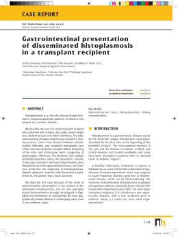
375
Microepidemia de histoplasmose em Blumenau, Santa Catarina
Case Report
An outbreak of histoplasmosis in the city of
Blumenau, Santa Catarina*
FLÁVIO DE MATTOS OLIVEIRA1, GISELA UNIS2, LUIZ CARLOS SEVERO3
ABSTRACT
Acute pulmonary histoplasmosis is rarely diagnosed and is often confused with tuberculosis. Most knowledge of the
disease has been derived from descriptions of epidemics in which a number of individuals were exposed to the same
source of infection. Isolation of Histoplasma capsulatum var. capsulatum from soil samples is conclusive evidence of an
epidemic focus. This is the first report of an outbreak of histoplasmosis, in which two cases were reported and the fungus
was isolated at the focus of the epidemic, in the state of Santa Catarina. Further epidemiological studies are needed in
order to determine the prevalence of the infection statewide.
Keywords: Histoplasmosis/diagnosis; Histoplasmosis/epidemiology; Histoplasma/isolation & purification; Lung
disases, fungal; Disease outbreaks; Case reports [publication type]
INTRODUCTION
The thermal dimorphic fungus Histoplasma
capsulatum var. capsulatum causes various clinical
manifestations, depending on the anatomical and
immunological status of the host and on the quantity
of fungal inoculum. Acute pulmonary histoplasmosis
occurs when an otherwise healthy individual inhales
a large quantity of fungal propagules.(1)
In previously healthy individuals, acute
pulmonary histoplasmosis manifests as cough, fever,
dyspnea, and asthenia. In primary infections, these
symptoms appear three weeks after exposure to the
fungus, whereas they appear more rapidly (within
one week after exposure) in cases of re-infection.
The course of the disease is self-limited, with
spontaneous regression of the symptoms. Clinical
and laboratory findings vary depending on
whether or not the host has been previously
infected and on the quantity of inoculum.(2) The
fungus is found in the upper layers of the soil
and is propagated through airborne dispersion.(3)
Length of exposure determines disease severity.(4)
Most knowledge of the disease has been
* Study carried out in the Santa Casa Hospital Mycology Laboratory, Porto Alegre, Rio Grande do Sul, Brazil.
1. Santa Casa Hospital Mycology Laboratory
2. PhD in Internal Medicine from the Universidade Federal do Rio Grande do Sul (UFRGS, Federal University of Rio
Grande do Sul)
3. Researcher for the Conselho Nacional de Desenvolvimento Científico e Tecnológico (CNPq, National Council for
Scientific and Technological Development)
Correspondence to: Luiz Carlos Severo. Laboratório de Micologia, Hospital Santa Rita, Santa Casa-Complexo Hospitalar.
R. Annes Dias, 285 - CEP: 90020 090, Porto Alegre, RS, Brasil
Tel: 55 51 3214-8435. E-mail: [email protected]
Submitted: 25 February 2005. Accepted, after review: 14 September 2005.
J Bras Pneumol. 2006;32(4):375-8
376
Oliveira FM, Unis G, Severo LC
derived from descriptions of epidemics in which a
number of individuals were exposed to the same
source of infection,(2) such epidemics being more
easily recognized, since they affected an entire
group of people simultaneously. The simultaneous
onset of symptoms in more than one patient should
raise the suspicion of histoplasmosis and can be
used to rule out the possibility of tuberculosis.
Histoplasmosis mimics tuberculosis in its clinical,
radiological, and histopathological aspects. It is
therefore necessary to carry out specific tests, using
cultures to identify the etiologic agent.
Since 1958, 26 outbreaks of histoplasmosis
have been reported, all occurring in one of eight
Brazilian states (Rio de Janeiro, Rio Grande do Sul,
São Paulo, Distrito Federal, Minas Gerais, Paraíba,
Amazonas and Bahia), and the fungus was isolated
from soil in five of those states (Rio de Janeiro,
Rio Grande do Sul, São Paulo, Distrito Federal and
Paraíba).(5) Herein we describe the first outbreak
of acute pulmonary histoplasmosis in which H.
capsulatum was isolated at the focus of the
epidemic in the state of Santa Catarina.
The presence of pulmonary nodules on the chest
X-ray and tomography of the chest (Figures 1A and
1B), together with the absence of expectoration,
justified the performance of a biopsy to evaluate
the pulmonary nodule.
The microbiological evaluation revealed
negative double-immunodiffusion for H.
capsulatum Hematoxylin and eosin staining of the
lung biopsy revealed tuberculous granuloma with
caseous necrosis, sputum smear microscopy was
negative, and the results of the Grocott-Gomori
methenamine silver (GMS) staining of the sections
were negative. In view of these findings, empirical
treatment for tuberculosis was initiated.
Due to the knowledge that the disease is related
to contact with bat guano, the material was sent
to the Santa Casa Hospital Mycology Laboratory,
in the city of Porto Alegre, in the state of Rio Grande
do Sul, for mycological re-evaluation. This reevaluation revealed small, single-budding yeastlike organisms suggestive of H. capsulatum in the
GMS staining of new tissue sections. The patient
was started on itraconazole (200 mg/day) and
presented favorable evolution.
CASE REPORT
Case 1
A 67-year-old white male, resident of the city
of Blumenau, in the state of Santa Catarina, and a
sales representative, presented with dyspnea,
asthenia, chest pain, and sweats ten days after
having cleaned his attic, where there are water
tanks. He was in contact with bat guano for a limited
period of time when the area was being swept.
A
Case 2
A 50-year-old white male, resident of the city
of Blumenau and a stonemason, presented a
clinical profile similar and simultaneous to that
described in Case 1 ten days after the cleaning of
the same area, in which there was bat guano. The
patient was exposed to a large quantity of dust in
an enclosed space. The patient was hospitalized
for seven days. Two chest X-rays, one presenting
B
Figure 1 - A and B: Computed tomography of the chest revealing a right-sided pulmonary nodule and a triangular
opacity in the lower left lobe with its base angled toward the pleura
J Bras Pneumol. 2006;32(4):375-8
377
Microepidemia de histoplasmose em Blumenau, Santa Catarina
A
B
Figure 2 - A and B: Chest X-ray revealing diffusely disseminated micronodules in both lungs and subsequent control of
the condition with regression of the lesions
diffusely disseminated micronodules in both lungs
and one showing the subsequent control of the
condition, are shown in Figures 2A and 2B.
The microbiological evaluation revealed
negative double-immunodiffusion for H.
capsulatum. Hematoxylin and eosin staining of the
transbronchial biopsy sample revealed a
granulomatous chronic inflammatory process,
sputum smear microscopy was negative, and the
results of the GMS staining of the section were
negative. In view of these results, empirical
treatment for tuberculosis was initiated.
Mycological re-evaluation of the material was
carried out in the same laboratory used for the
Case 1 samples, and the GMS staining of new
sections also revealed small, single-budding yeastlike organisms suggestive of H. capsulatum. The
patient was started on itraconazole (200 mg/day)
and presented clinical improvement one month
after the onset of symptoms.
DISCUSSION
In Brazil, 26 outbreaks of histoplasmosis,
involving 184 patients, have been reported since
1958(5) (Table 1). The number of cases per outbreak
ranged from two to thirteen. The main sources of
infection were visits to caves where there was bat
guano (ten outbreaks), followed by visits to
abandoned mines (five), and contact with droppings
from chicken coops (three outbreaks). In 11 of those
outbreaks, H. capsulatum was isolated from soil.
Epidemiological history can raise diagnostic
suspicion. Knowledge of clinical syndromes,
together with clinical suspicion, can avoid the use
of empirical treatment. Radiological findings,
negative test results, and the high frequency of
tuberculosis in Brazil were the motivations for
prescribing tuberculosis treatment for both patients.
Immunodiffusion test results, although negative
for these two patients, have high sensitivity and
TABLE 1
Outbreaks of histoplasmosis reported in Brazilian states
State
RJ
RS
SP
MG
AM
PB
BA
GO
SC
Isolation from soil
+
+
+
+
+
+
Histoplasmin
skin test*
93
89
86
64
50
32
32
22
6,3
APH
n
15
3
3
1
1
1
1
1
1**
*Maximum value found (%); **Present study
RJ: Rio de Janeiro; RS: Rio Grande do Sul; SP: São Paulo;
MG: Minas Gerais; AM: Amazonas; PB: Paraíba; BA: Bahia;
GO: Goiás; SC: Santa Catarina. APH: acute pulmonary
histoplasmosis
J Bras Pneumol. 2006;32(4):375-8
378
Oliveira FM, Unis G, Severo LC
are positive in approximately 75% of cases. (6)
Length of exposure determines disease severity.
This explains the fact that the second patient, who
was the one who swept the area, presented a more
severe form of the disease, with diffuse pulmonary
alterations on the chest X-ray, and required
hospitalization. However, a short exposure time
results in focal pulmonary lesions, as in Case 1.
The diagnosis is confirmed by the presence of
H. capsulatum in the lung tissue biopsy. The tissue
section must be stained according to the GMS
technique.(6) In both cases, the diagnosis of the
outbreak was made through GMS staining of lung
biopsy samples, which revealed small, oval-shaped,
single-budding yeast-like organisms suggestive of
H. capsulatum.
Treatment is not usually indicated for
immunocompromised patients with acute
pulmonary histoplasmosis, since the disease is selflimiting and presents minimal morbidity. Treatment
should be considered for those who remain
symptomatic for more than a month or who present
diffuse radiological alterations or hypoxemia.(7)
When amphotericin B was the only antifungal drug
available, treatment was not indicated due to the
toxicity of the drug.(8) With the advent of the azole
antifungals, which have the advantage of easy oral
administration and good tolerability, the indications
increased. The treatment of choice is the use of
itraconazole (200 mg/day) from six to twelve weeks.(7)
In the cases described herein, we opted for
treatment because the patients had been
symptomatic for more than a month, and because,
in Case 2, hospitalization was required.
Isolation of the fungus from soil demands
meticulous work and collection of various samples
from multiple locations, since H. capsulatum
concentrates in small microfoci presenting ideal
growth conditions, such as temperature between 22ºC
and 29ºC and relative humidity between 67% and
87% or more, at certain times of year.(3) The technique
used to identify the fungus is intraperitoneal
J Bras Pneumol. 2006;32(4):375-8
inoculation in mice, which are killed four weeks later.(9)
The present study represents the first time that it
was possible to isolate H. capsulatum from soil in
the state of Santa Catarina, this being conclusive
evidence of an epidemic focus. (4) The only
epidemiologic survey in the state of Santa Catarina
was carried out in 1955, involving 110 soldiers in
several cities and employing various dilutions of
histoplasmin (1:10, 1:100, 1:1000). The rate of
positivity was found to be 6.3%, which does not
reflect the current situation. (10) Outbreaks of
histoplasmosis are usually more frequent in areas of
higher endemicity. Further epidemiological studies
are needed in order to determine the prevalence of
the infection statewide.
REFERENCES
1. Goodwin RA, Loyd JE, Des Prez RM. Histoplasmosis in
normal hosts. Medicine (Baltimore). 1981;60(4):231-66.
2. Goodwin RA Jr, Des Prez RM. State of the art:
histoplasmosis. Am Rev Respir Dis. 1978;117(5):929-56.
3. Wheat LJ, Conces D, Allen SD, Blue-Hnidy D, Loyd J.
Pulmonary histoplasmosis syndromes: recognition,
diagnosis, and management. Semin Respir Crit Care
Med. 2004;25(2):129-44.
4. Larsh HW. Histoplamosis. In: Di Salvo AF. Occupational
mycoses. Philadelphia: Lea & Febiger; 1983. p.29-42.
5. Unis G, Roesch EW, Severo LC. Histoplasmose pulmonar
aguda no Rio Grande do Sul. J Bras Pneumol.
2004;31(1):52-9.
6. Wheat LJ. Laboratory diagnosis of histoplasmosis:
update 2000. Semin Respir Infect. 2001;16(2):131-40.
7. Wheat J, Sarosi G, McKinsey D, Hamill R, Bradsher R,
Johnson P, et al. Practice guidelines for the management
of patients with histoplasmosis. Infectious Diseases
Society of America. Clin Infect Dis. 2000;30(4):688-95.
8. Severo LC, Petrillo VF, Camargo JJ, Geyer GR, Porto NS.
Acute pulmonary histoplasmosis and first isolation of
Histoplasma capsulatum from soil of Rio Grande do
Sul, Brasil. Rev Inst Med Trop S Paulo. 1986;28(1):51-5.
9. Ajello L, Zeidberg LD. Isolation of Histoplasma
capsulatum and Allescheria boydii from soil. Science.
1951;113(2945):662-3.
10. Fava SC, Fava Netto C. Epidemiologic surveys of
histoplasmin and paracoccidioidin sensitivity in Brazil.
Rev Inst Med Trop S Paulo. 1998;40(3):155-64.
Download