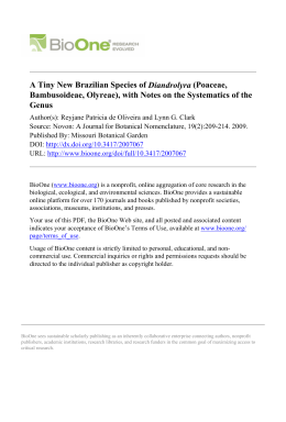
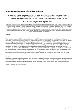
Neodiversity 8: 50–54 www.neodiversity.org Published online 17th June 2015 IMPROVING MICROTECHNIQUES FOR PROCESSING LEAF BLADES OF GRASSES USING ICHNANTHUS PALLENS (SW.) MUNRO EX BENTH. AS A MODEL SPECIES L.A.JESUS JUNIOR1,3, R.P.OLIVEIRA1, K.R.B.LEITE2 1Programa de Pósgraduação em Botânica, Departamento de Ciências Biológicas, Universidade Estadual de Feira de Santana, Av. Transnordestina, s.n., 44036900, Feira de Santana, BA, Brazil. 2Departamento de Botânica, Instituto de Biologia, Universidade Federal da Bahia, Rua Barão de Jeremoabo, s.n., Campus Universitário, 40170115, Salvador, BA, Brazil 3Author for correspondence: [email protected] AB ST R AC T Poaceae materials are usually difficult to include in historesin, and in order to solve this problem we present in this paper some modifications on leaf section microtechniques to improve sample processing in specimens of this family. The sections obtained here present higher quality than those obtained by traditional protocols, which can provide more precise anatomical descriptions in the future. The modifications here suggested are also advantageous by providing the reduction in the total time of processing the materials. Anatomical studies are widely used in Poaceae, being an important tool for both taxonomic and evolutionary studies (Prat 1936, Ellis 1976, 1979, Renvoize 1987, GPWG 2001, Oliveira et al. 2008a, 2008b, Guglieri et al. 2008, Pelegrin et al., 2009, Jesus Junior et al. 2012). However the microtechniques that have been traditionally used often do not provide good materials for the optical analysis in certain taxonomic groups in this family. This situation requires great investment for protocol optimization but no guarantee of success. Among the microtechniques most widely used, historesin embedding has been proved to be a viable alternative for anatomical studies in Poaceae, especially for leaf cross sections. One of the main reasons for the popularity of this technique is the smaller quantity of material required for the analysis. Allied to this, this technique is a faster process, provides an ideal thickness of the materials for the analyses, and additionally presents the possibility of using herbarium samples, which is impracticable in hand cut techniques (Meira & Martins 2003). Despite this, some Poaceae materials have been presenting problems for infiltration in the historesin, and adaptations in the protocols found in the literature have been necessary, such as those described by Sass (1940). Considering this, in the current paper we propose modifications in the technique of historesin embedding used in the leaf samples of this family. This can improve the processing of samples from representatives of Poaceae and contribute for anatomical studies in this family. MATERIAL AND METHODS The materials were obtained in field trips throughout Brazil. Voucher specimens are deposited in the herbarium of the Universidade Estadual de Feira de Santana (HUEFS): C.Silva 745 (Petrópolis, Rio de Janeiro), C.Silva 841 (Angra dos Reis, Rio de aneiro) and K.M.Pimenta 165 (Igrapiúna, 50 JESUS JUNIOR ET AL. IMPROVED TECNIQUES FOR POACEAE ANATOMY Rio de Janeiro). All samples correspond to specimens of Ichnanthus pallens (Sw.) Munro ex Benth., which belongs to subfamily Panicoideae and was selected as the model for this study. The first two samples were fixed in a solution of 70% formalin:acetic acid: alcohol (FAA70) and later transferred to a 70% ethanol solution (Johansen 1940) until processing. These materials correspond to herbarium specimens and were rehydrated by following the protocol of reversal of herborization indicated by Smith & Smith (1942), and finally kept in a 70% ethanol solution until the preparation of leaf sections. For the analysis of all materials we selected the median portion of the second leaf blade below the inflorescence. Tree replications per specimen were embedded in historesin (Leica@). Before the inclusion, the materials were dehydrated in a graded ethanol series (80%, 90% and 100%; 2 hours per changing). The materials were then embedded in a 1:1 mix of 100% ethanol and historesin for 2 hours, followed by embedding in pure historesin for a further 2 hours, and then kept in a refrigerator for 72 hours in 2.0 mL microtubes with activated historesin (prepared following the manufac turer's instructions). After this period the samples were included in polyethylene plates according to the manufacturer's instructions and maintained in an oven at 40 °C for 6 hours. Finally, the blocks were glued with cyanoacrylate in wood bases and placed in silica gel until the moment of use. Then, the blocks were sectioned with disposable blades (Leica 818) in a rotative microtome (Micron), with 8, 9 and 10 µm. The cuts were subsequently transferred to Petri dishes with distilled water at 45 ºC for dilatation and finally stained with 0.25% toluidine blue in 0.1 M phosphate buffer pH 6.8 (Kraus & Arduin 1997), in a similar procedure used for freehand cuts. The process also includes washing and mounting of the cuts on slides with coverslip using Entelan (Merck) (adapted from Sass 1940). All the procedures were carried out at the Laboratory of Plant Micromorphology (LAMIV) at Universidade Estadual de Feira de Santana, using the Zeiss optical microscope Primo Star with the cuts and sample processing. Photomicrographs were taken using an Olympus BX51 microscope with an attached DP25 camera and the software “Imaging Software Cellsens” version 2.3 build 7045. RESULTS AND DISCUSSION The materials analyzed (Fig. 1) exhibited anatomical patterns similar to those described by Metcalfe (1960) for tribe Paniceae s.l., group to which I. pallens belongs. The anatomical patterns include: radiated chlorenchyma, simple sheath surrounding the vascular the bundles in the mesophyll, and double sheath associated with the midrib, where the parenchymatous sheath lacks chloroplasts, which resembles a sheath of the Kranz type and indicates that the I. pallens is a C3 species. The epidermis has bulliform cells on both sides of the blade, and substomatic chambers commonly associated with vascular bundles of second and third orders. When infiltration with historesin in a sample was insufficient, a tissue retraction occurred, resulting in cuts with indistinct cell boundaries (Fig. 1 AB). However, using the new protocol described here the cuts are sharper (Fig. 1CE) than those obtained by the manufacturer's standard protocol (Fig. 1AB). The quality of the latter do no allow accurate anatomical descriptions. The protocol modifications proposed here for Poaceae materials proved to be advantageous in terms of reducing the total time of processing (see Table 1), which in turn, enables the study of larger quantities of samples in a shorter time. This improvement is possible because some steps of the standard protocol become unnecessary, such as the 51 JESUS JUNIOR ET AL. IMPROVED TECNIQUES FOR POACEAE ANATOMY arrangement of the sections in an excess of historesin on the blade, and the washing that follows the coloration process in a glass container. The new protocol also avoids occasional losses of sectioned materials in the washing step. Figure 1. A–E. Transversal sections of leaves of Ichnanthus pallens. A–B. Sections obtained using the standard protocol as described in the literature. A. General view of the mesophyll. B. Midrib. C–E. Sections obtained using the new protocol. C. Midrib with inflated parenchymatous leaf sheath. D. Detail of third order vascular bundle with associated substomatic chambers. E. View of the mesophyll with the distribution of bulliform cells. D–E. Captions: bp – Inflated parenchymatous leaf sheath; cse – substomatic chambers; cb – bulliform cell; pr – radiate parenchyma. Bars = 50 µm. 52 JESUS JUNIOR ET AL. IMPROVED TECNIQUES FOR POACEAE ANATOMY Other advantages of this new protocol are its potential to be used also for other graminoid groups such as Cyperaceae, and its relative low cost to be implemented, providing, in turn, faster answers to taxonomic and evolutionary questions. 20105) and a Productivity Research Grant (PQ1D) to RPO; and to the Fundação de Amparo à Pesquisa do Estado da Bahia for additional financial support (PNE 0020/ 2011). LITERATURE CITED ACKNOWLEDGMENTS Ellis, R.P. 1976. A procedure for standardizing comparative leaf anatomy in the Poaceae I. The leafblade as viewed in transverse section. Bothalia 12:65–109. Ellis, R.P. 1979. A procedure for standardizing comparative leaf anatomy in the Poaceae II. The epidermis as seen in surface view. Bothalia 12: 641–671. GPWG (Grass Phylogeny Working Group) 2001. Phylogeny and subfamilial classification of the grasses (Poaceae). Annals of the Missouri Botanical Garden 88: 373–457. Guglieri, A.; LonghiWagner, H.M.; Zuloaga, F.O. 2008. Anatomia foliar das espécies de Panicum L. subg. Panicum (Poaceae: Panicoideae: Paniceae) The authors thank the Center for the Study of Biodiversity of the Michelin Ecological Reserve, in special Dr. Kevin Flesher for the financial support and infrastructure during the field trips; Christian da Silva for the collected materials; Marlon Machado for English revision; the Coordenação de Aperfeiçoamento de Pessoal de Nível Superior for scholarship grant for LAJJ; the Conselho Nacional de Desenvolvimento Científico e Tecnológico (CNPq) for financial support (grants 562349/20103 e 563558/ 53 JESUS JUNIOR ET AL. IMPROVED TECNIQUES FOR POACEAE ANATOMY ocorrentes no Brasil. Iheringia, Série Botânica 63: 279–293. Jesus Junior, L.A.; Oliveira, R.P.; Leite, K.R.B.; Silva, L.B. 2012. Comparative analysis of the leaf anatomy in two Parodiolyra species (Poaceae: Olyreae) occurring on forests in Eastern Brazil. Brazilian Journal of Biology 72: 205–210. Johansen, D.A. 1940. Plant microtechnique. McGrawHill Book Co. Inc., New York. Kraus, J.E. & Arduin, M. 1997. Manual básico de métodos em morfologia vegetal. EDUR, Seropédica, Rio de Janeiro. Meira, R.M.S.A. & Martins, F.M. 2003. Inclusão de material herborizado em metacrilato para estudos de anatomia vegetal. Revista Árvore 27: 109–112. Metcalfe, C.R. 1960. Anatomy of the Monocotyledons. Claredon Press, Oxford. Oliveira, R.P.; LonghiWagner, H.M.; Leite, K.R.B. 2008. A contribuição da anatomia foliar para a taxonomia de Raddia Bertol. (Poaceae: bambusoideae). Acta Botanica Brasilica 22: 1–19. Oliveira, R.P.; LonghiWagner, H.M.; Leite, K.R.B. Hollowell, V.C. 2008. Pariana multiflora (Poaceae: Bambusoideae: Olyreae): a new species from Brazil and notes on the leaf anatomy of this genus in Eastern Coast Brazil. Systematic Botany 33: 262–266. Pelegrin, C.M.G.; LonghiWagner, H.M.; Oliveira, P.L. 2009. Anatomia foliar como subsídio à taxonomia de espécies do Complexo Briza L. (Poaceae: Pooideae: Poeae). Acta Botanica Brasilica 23: 666–680. Prat, H. 1936. La Systématique des Graminées. Annales des Sciences Naturelles; Botanique 6.10: 165–258. Renvoize, S.A. 1987. A survey of leaf blade anatomy in grasses XI. Paniceae. Kew Bulletin 42: 739–768. Sass, J. 1940. Elements of Botanical Micro technique. New York and London. Smith, F.H. & Smith, E.C. 1942. Anatomy of the inferior ovary of Darbya. American Journal of Botany 29: 464–471. ISSN 18095348 (print), ISSN 23582847 (online) 54
Baixar