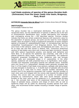
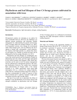
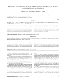
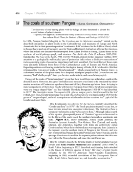
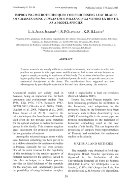
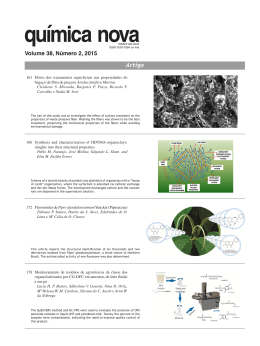
BIOCELL
2009, 33(3): 155-165
ISSN 0327 - 9545
PRINTED IN ARGENTINA
Leaf blade anatomy and ultrastructure of six Simira species
(Rubiaceae) from the Atlantic Rain Forest, Brazil
TARSILA MARIA DA SILVA MORAES1, CLAUDIA FRANCA BARROS2, SEBASTIÃO JOSÉ DA SILVA NETO3, VALDIRENE
MOREIRA GOMES4 AND MAURA DA CUNHA1*
1.
2.
3.
4.
Laboratório de Biologia Celular e Tecidual, Centro de Biociências e Biotecnologia, Universidade Estadual do Norte
Fluminense, Campos dos Goytacazes, RJ, Brazil.
Laboratório de Botânica Estrutural, Programa Mata Atlântica, Diretoria de Pesquisas, Instituto de Pesquisas Jardim Botânico
do Rio de Janeiro, RJ, Brazil.
Programa Mata Atlântica – Diretoria de Pesquisas, Instituto de Pesquisas Jardim Botânico do Rio de Janeiro, RJ, Brazil.
Laboratório de Fisiologia e Bioquímica de Microrganismos, Centro de Biociências e Biotecnologia, Universidade Estadual
do Norte Fluminense, Campos dos Goytacazes, RJ, Brazil.
Key words: leaf anatomy, structure, morph-anatomy
ABSTRACT: Simira is a predominantly woody Neotropical genus comprising 41 taxa, 16 of which occur in
Brazil and eight of them in the southeastern region of Brazil. Leaf blades of Simira eliezeriana Peixoto, S.
glaziovii (K. Schum.) Steyerm., S. grazielae Peixoto, S. pikia (K. Schum.) Steyerm., S. rubra (Mart.) Steyerm.,
S. sampaioana (Standl.) Steyerm. were collected in the southeastern region of Brazil and fixed according to
usual methods for light and electron microscopy. The leaf blades show typical characteristics of the Rubiaceae
family as dorsiventral mesophyll and paracytic stomata. The presence of two bundle sheaths that extend to the
upper epidermal layer, prismatic crystal and crystal-sand, alkaloids in the mesophyll and the organization
micromorphological of the outer periclinal wall are considered characteristics representative for the genus.
This study also demonstrates some leaf blade characteristics that can be used to Simira species identification
(leaf surface, domatia types, epicuticular wax types and patterns of epidermis anticlinal cell walls).
Introduction
The Atlantic Rain Forest constitutes the second most
threatened ecosystem in the world, and has been reduced
over the years (Myers et al., 2000). This biome presents
a high diversity of plant families and Rubiaceae is one
of the most important families in several remnant forests in Rio de Janeiro state/Brazil (Guedes-Bruni, 1998).
The Rubiaceae family has ca. 650 genera and 13,000
species with a wide geographical distribution in the
tropical and subtropical areas (Struwe, 2002). Among
*Address correspondence to: Maura Da Cunha.
E-mail: [email protected]
Received: September 10, 2008. Revised version received: July
13, 2009. Acepted: July 19, 2009.
these, Simira Aubl. is a predominantly woody Neotropical genus consisted of 41 species, 16 of which occur
in Brazil, and eight from southeastern Brazil (Silva Neto,
2000). This genus is important due to its economic value,
e.g. production of handmade pieces, urban landscaping
and dyestuff.
The Rubiaceae family is a monophyletic group (Judd
et al., 1999) and belongs to the Gentianales order (Struwe,
2002). Robbrecht (1988) divided this family into four
subfamilies: Antirheoideae, Cinchonoideae, Ixoroideae
and Rubioideae based on morphological characters. Phylogenetic studies based on molecular data alone or in
combination with morphological data have drastically
changed the Rubiaceae classification at subfamily (e.g.
Bremer et al., 1995; Bremer, 1996), tribal (e.g. Bremer
156
and Thulin, 1998; Rova et al., 2002), generic (e.g.
Nepokroeff et al., 1999; Andersson, 2001), and species
level (e.g. McDowell and Bremer, 1998; Persson, 2000).
The molecular studies proposed a new classification in
three subfamilies - Rubioideae, Cinchonoideae and
Ixoroideae (Bremer et al., 1995; Rova et al., 2002). Together, macromolecular data, morphological and anatomical characters have contributed to characterize several
subgroups of this family (Piesschaert et al., 2000; Dessein
et al., 2001; Jansen et al., 2001, 2002). The genus Simira
has been classified in the Cinchonoideae subfamily and
Rondeletieae tribe (Robbrecht, 1988) based on morphological data. A recent phylogenetic study based on molecular data, however, placed Simira in the tribe Simireae
within the Ixoroideae subfamily (Rova et al., 2002). Because of this change in the Simira classification, it is necessary to reunite data of several study areas.
Anatomical and micromorphological characters of
the leaf blade with possible taxonomic significance have
been studied as a source for solving taxonomic problems.
The comparative leaf anatomy and micromorphology in
Psychotria (Da Cunha and Vieira, 1993/97; Gomes et
al., 1995; Vieira and Gomes, 1995), Rondeletia (Kocsis
et al., 2004), Rudgea (Mantovani and Vieira, 1993/97;
Leo et al., 1997), Rustia (Vieira et al., 2001), Coussarea
(Tavares and Vieira, 1994) and Bathysa (Nascimento et
al., 1996; Barros et al., 1997) have been studied as well
as the wood anatomy of several species of Rubiaceae of
the Atlantic Forest timber (Callado et al., 1997). In Simira
genus, the comparative structure of colleters (Klein et
al., 2004) and wood anatomy (Callado and Silva Neto,
2003) in several species has been studied.
The present study aims to characterize the leaf blade
anatomy and micromorphology of six Simira species
of Atlantic Rain Forest from Southeastern Brazil, and
to contributing to the knowledge the implications in survival strategies and taxonomic importance below the
genus level. We particularly want to identify anatomical and morphological variations, which may be useful
in species identification as well as to test the validity of
the sections currently recognized in Simira.
TARSILA MARIA DA SILVA MORAES et al.
Reserva Florestal da Companhia do Vale do Rio Doce
(19º 06' - 19º 18' S and 39º 45' - 40º 19' W), in the Municipal District of Linhares, Espírito Santo state, Brazil, at Barragem de Saracuruna (22º 12’ - 22º 17’ S and
43º 30’ – 43º 35’ W), in the Municipal District of Duque
de Caxias, and at Reserva Biológica de Tinguá (22º28’
– 22º 39’ S and 22º 35 – 43º 34’ W), in the Municipal
District of Duque de Caxias and Nova Iguaçu, in Rio de
Janeiro state, in the southeastern region of Brazil.
Light microscopy
Leaf fragments were fixed in a solution of 2.5%
glutaraldehyde and 4.0% paraformaldehyde in 0.05 M
cacodylate buffer at pH 7.2, for two hours. Subsequently,
the samples were rinsed three times with the buffer and
post-fixed for two hours at room temperature with 1.0%
osmium tetroxide in 0.05 M cacodylate buffer at pH
7.2. The post-fixed samples were dehydrated in a graded
series of acetone solutions. Subsequently, the material
was infiltrated and embedded in epoxi resin (Polybed).
Microtome sections (1.0 μm) were cut and stained with
toluidine blue (0.05% aqueous solution). The slides were
sealed with Entellan® (Merck) and examined with an
Axioplan ZEISS microscope. For epidermis, dissociation was achieved using a solution 1:1 of 10% nitric
acid and 10% chromium oxide (Jensen, 1962), and
stained with 10% safranine. Epidermis outer periclinal
cell walls visualization, images were obtained using an
ANALYSIS SIS LINK/OXFORD - ZEISS.
Histochemistry
Histochemical tests were carried out on freehand
sections of the collected material alkaloids were localized by the Dragendorff test described by Costa
(1982); and crystals were tested by the insolubility in
acetic acid and solubility in hydrochloric acid (McLean
and Cook, 1958), and examined with an Axioplan
ZEISS microscope.
Scanning electron microscopy
Material and Methods
Plants
Mature leaves of Simira eliezeriana Peixoto, S.
glaziovii (K. Schum.) Steyerm., S. grazielae Peixoto, S.
pikia (K. Schum.) Steyerm., S. rubra (Mart.) Steyerm.,
S. sampaioana (Standl.) Steyerm. were collected at
The fragments of leaf blades were fixed, post-fixed
and dehydrated as for light microscopy. The samples
were subsequently critically point dried in CO2, sputter
coated with 20 nm gold, and observed with a digital
scanning electron microscope (ZEISS DSEM 962). The
authenticity of epicuticular wax was verified by washing leaves in chloroform for one minute and comparing
to the unwashed specimens under SEM.
LEAF BLADE STRUCTURE OF SIX SIMIRA SPECIES
Transmission electron microscopy
The samples were fixed, post-fixed, dehydrated and
embedded as described previously. The ultrathin sections (80 nm) were collected in copper grids (300 mesh),
stained with acetate with 1.0% uranyl followed by 5.0%
lead citrate for routine observation. Three other stains
were used to elucidate the cytochemical of the outer
periclinal cell walls. (1) The preservation and contrast
of lipids were enhanced and observed using imidazolebuffered osmium tetroxide (Angermüller and Fahimi,
1982). (2) 0.5 mg.ml-1 Ruthenium red was used to detect negatively charged components of the outer
periclinal cell walls (Luft, 1971). (3) Periodic acidthiocarbohydrazide (THC)-silver proteinate (PATAg)
was used to detect polysaccharides containing 1,2-glycol groups. For the latter technique, sections were stained
with periodic acid and solutions not containing peri-
157
odic acid and THC as control (Thiéry, 1967). Sections
were observed at 80 kV using a transmission electron
microscope (ZEISS EM 900).
Extraction and determination of leaf proteins
The protein extraction was performed as described
by Granier (1988) with some modifications. About 5 g
of leaf blades were dry crushed in a mortar cooled with
liquid nitrogen. The dry powder was resuspended in 5
mL of extraction buffer containing 100 mM Tris-HCl,
pH 8.7, 1 mM dithiothreitol (DTT), 1 mM ascorbic acid,
5 mM Mg Cl2 and 1% polyvinylpyrrolidone (PVP). The
extract was centrifuged at 20,000 g for 1 hour at 4ºC. The
supernatant was then mixed with 10% Trichlorile acetic,
0.07% 2-mercaptoethanol in cold acetone and kept at 18ºC for 1 h. After a 30 min centrifugation at 30,000 g,
the supernatant was removed and the pellet was recov-
FIGURES 1-4. Scanning electron microscopy of Simira Aubl. species. 1. Pocket domatia in S. grazielae
Peixoto. 2. Hair-tuft domatia in S. pikia (K. Schum.) Steyerm. 3. S. glaziovii (K. Schum.) Steyerm. adaxial
surface. 4. S. pikia (K. Schum.) Steyerm. abaxial surface. Scale bars= (1) 200 μm; (2) 500 μm; (3, 4) 50 μm.
158
TARSILA MARIA DA SILVA MORAES et al.
ered and submitted to further electrophoresis. The protein determination was performed by the method of
Bradford (1976), using bovine serum albumin as standard and SDS-polyacrylamide gel electrophoresis (SDSPAGE) was performed as described by Laemmli (1970).
Proteins utilized as molecular mass standards for PAGESDS were bovine serum albumin (66 kDa), ovalbumin
(45 kDa), glyceraldehyde-3-phosphate dehydrogenase (36
kDa), carbonic anhydrase (29 kDa), trypsinogen (24 kDa),
trypsin inhibitor (20 kDa) and β-lactoglobulin (14 kDa).
Results
The presence of domatia is observed in the abaxial
surface between the midrib and secondary veins. Simira
grazielae (Fig. 1) presented domatia in pockets, while
the other species presented domatia in tufts of hairs (Fig.
2). Cuticular ornamentation is observed on both surfaces
as in S. glaziovii (Fig. 3), except in S. pikia (Fig. 4), and
is restricted near the stomata of the other species studied.
The cuticle of epidermic cells is non-striated (Fig. 3) on
FIGURES 5-10. Scanning electron microscopy of Simira Aubl. species. 5. S. grazielae Peixoto abaxial
surface showing stomata with parallel cuticular striations. 6. S. rubra (Mart.) Steyerm. abaxial surface
showing stomata with perpendicular cuticular striations. 7. S. elizeriana Peixoto adaxial surface showing smooth layer of epicuticular wax. 8. S. grazielae Peixoto adaxial surface showing epicuticular wax in
granules. 9. S. sampaioana (Standl.) Steyerm. trichome with cuticular striations. 10. S. elizeriana Peixoto
trichome without cuticular striations. Scale bars= (5, 6, 7, 8, 10) 10 μm; (9) 5 μm.
LEAF BLADE STRUCTURE OF SIX SIMIRA SPECIES
both surfaces, except in S. pikia (Fig. 4). The cuticle ornamentations is observed only near the stomata, except
in S. elizeriana. In S. grazielae (Fig. 5), these cuticular
striations are parallel to the stomata axis, while in other
species they are perpendicular (Fig. 6). Epicuticular wax
is observed in all species studied. Simira glaziovii, S. pikia,
S. rubra, and S. sampaioana present a fine film; however, a smooth layer is seen S. elizeriana (Fig. 7) and
granules in S. grazielae (Fig. 8).
159
The species studied exhibit unicellular, pluricellular
(Fig. 12) and stellate (Fig. 14) trichomes on both surfaces, except in S. grazielae that has a glabrous surface.
Trichomes of S. pikia, S. rubra, and S. sampaioana (Fig.
9) show surface ornamentations, not observed in S.
elizeriana (Fig. 10) and S. glaziovii.
In a transverse section of the leaf blade, the adaxial
and abaxial epidermis present a single layer covered with
a fine cuticle in all species as can observed in S. rubra
FIGURES 11-16. Light microscopy of Simira Aubl. species.
11. Transverse section of S.
rubra (Mart.) Steyerm. leaf
blade showing dorsiventral
mesophyll. 12. Transverse
section of S. rubra (Mart.)
Steyerm. leaf blade showing
lateral projections along main
vein forming domatia (arrow).
13. Transverse section of S.
pikia (K. Schum.) Steyerm.
Note two layers of the bundle
sheath and parenchymatic
layer forming crystalliferous
series (stars). 14. Frontal view
of stellate trichome in abaxial
surface of S. rubra (Mart.)
Steyerm. 15. Straight anticlinal wall in S. sampaioana
(Standl.) Steyerm. leaf blade.
16. Strong sinuous anticlinal
wall in S. pikia (K. Schum.)
Steyerm. leaf blade. Scale
bars= (11) 30 μm (12, 14) 50
μm (13) 20 μm; (15, 16) 40 μm.
160
(Fig. 11, 12), and in S. pikia (Fig. 13). The Simira species studied have dorsiventral mesophyll. Palisade tissue comprises two layers in S. elizeriana and one or
two layers in the other species; and the number of spongy
tissue layers varies from three layers in S. sampaioana
to six in S. glaziovii. The species present prismatic crys-
TARSILA MARIA DA SILVA MORAES et al.
tals and crystal-sand, identified by the insolubility in
acetic acid and solubility in hydrochloric acid, as well
as alkaloids, identified by Dragendorff reagent, in the
vacuole of mesophyll cells.
Paracytic stomata slightly protrude above the level
of the other epidermal cells. Stomata are not usually
FIGURES 17-24. Transmission
electron microscopy of Simira Aubl.
outer periclinal cell wall. 17. S.
glaziovii (K. Schum.) Steyerm.
adaxial epidermis. 18. S. elizeriana
Peixoto abaxial epidermis. 19. S.
sampaioana (Standl.) Steyerm.
adaxial epidermis with Ruthenium
red treatment. 20. S. pikia (K.
Schum.) Steyerm. abaxial epidermis
with Ruthenium red treatment. 21. S.
glaziovii (K. Schum.) Steyerm. adaxial
epidermis with Thiéry treatment. 22.
Abaxial epidermis of S. rubra (Mart.)
Steyerm. with Thiéry treatment. 23. S.
elizeriana Peixoto adaxial epidermis
with imidazole treatment. 24. S.
grazielae Peixoto abaxial epidermis
with imidazole treatment. Scale Bars=
(17, 18, 20, 24) 0.25 μm; (21) 0.15 μm
(22) 0.9 μm (19, 23) 0.4 μm.
LEAF BLADE STRUCTURE OF SIX SIMIRA SPECIES
seen over primary and secondary veins. In transverse
sections, the guard cell presents a parietal thickening
that is uniform.
In epidermis, at both leaf surfaces, the pattern of
anticlinal cell walls varies from straight to sinuous:
straight anticlinal walls in S. sampaioana (Fig. 15);
slightly undulating walls forming an “S” in the S.
elizeriana, S. glaziovii and S. rubra; and S. grazielae
and S. pikia (Fig. 16) exhibit a strong sinuous. Anticlinal walls of epidermal cells are thin or moderately thick.
The vascular system shows a collateral arrangement
and two layers of the bundle sheath (Fig. 11). The inner
sheath is constituted by bundle perivascular fibers presenting an interruption in the phloematic tissue and parenchymatic cells in the outer bundle. The parenchymatic
cells of the S. pikia bundle sheath contain a large amount
of prismatic crystals, forming a crystalliferous series
(Fig. 13). The bundle sheaths are extended towards
adaxial epidermis in all species (Fig. 11, 13).
In the TEM cross sections, the outer periclinal cell
walls in both epidermis show a fine epicuticular wax
and three distinct layers: the inner polysaccharide-rich
layer, mainly composed of cellulose; the intermediate
cuticular layer, with a tree-like polysaccharide-rich network, immersed in a matrix of cutine; and a cuticle
proper layer (Fig. 17-24). The cuticle proper is homogenous and relatively electron lucent. The cuticular layer
is formed by a reticulated network layer and composed
of a cutine matrix and a tree-like layer composed of
polysaccharides and pectins, as observed with cytochemistry stains. The polysaccharide portion of the
cuticular layer reacts with PATAg for polysaccharides
and with ruthenium red, for pectin (Fig. 19-21). In contrast, the lipid portion of the cuticular membrane, the
cuticle proper and the matrix of the cuticular layer react
with imidazole (Fig. 23, 24). The species studied present
differences in cuticular layer organization showing the
tree-like layers more prominent in the adaxial surface
(Fig. 17, 19, 21, 23). In S. pikia that presents a cuticular
striated relief, the cuticular layer keeps up with a striated organization, mainly the tree-like layer (Fig. 20).
The preliminary analysis of the soluble proteins of
Simira and Bathysa species shows the presence of several specific proteins between 66 and 14.5 kDa. Two
bands of the same molecular weight of specific proteins between 66 and 45 kDa are observed for the Simira
and Bathysa species (Fig. 25, arrows). Two other proteins close to 36 kDa are also detected in the Bathysa
species (Fig. 25, arrows). A band of approximately 24
kDa is observed just in the lanes of the Simira species.
Our study also demonstrated proteins with low molecu-
161
lar masses, more specifically below 14.5 kDa, present
in all species of the Bathysa and Simira species analysed.
Discussion
Anatomical and ultrastructural studies have indicated important characteristics capable of distinguishing taxa, as well as relating structural characteristics
with the environment (Bredenkamp and Van Wyk, 2000;
Kong, 2001). The characteristics observed may be useful for identifying characters of genera, and to distinguish species and possible implications in survival strategies in the Atlantic Rain Forest.
The presence of domatia in the abaxial surface of
the leaf blades between the midrib and secondary veins
occur in the six species studied, and Robbrecht (1988)
relate that it is a common character in the Rubiaceae
family. Domatia are structures that frequently shelter
small predators or fungivore arthropods. These structures appear scattered among 277 families of angiosperms, occurring mostly in tropical and subtropical areas (Agrawal and Karban, 1997). The presence
FIGURE 25. SDS-polyacrylamide gel electrophoresis
of the proteins extracted from the leaf blades of the
Simira and Bathysa species. (1) Simira pikia; (2) S.
glaziovii; (3) S. rubra; (4) Bathysa gymnocarpa; (5) B.
stipulata. (M) Markers (kDa).
162
and type of domatia constitute a morphological character with useful systematic value to separate genera
and species (Adâmoli Barros, 1959). The domatia in
pockets are only found in Simira grazielae and is a
good character to separate this species from the others. Lateral projections in the midrib vein were also
observed forming wakes covered with hairs in S. rubra,
described as a domatia type in Psychotria velloziana
(Da Cunha and Vieira, 1993/97). The organisms that
living in domatia, are indirectly beneficial for the plant
because they reduce the herbivore arthropods or the
pathogenic fungi that commonly inhabit the surface
of the plant leaves (O’Dowd and Wilson, 1989;
Agrawal et al., 2000). These structures are possibly
associated with defense mechanisms against microorganisms in Atlantic Rain Forest. Further studies are
required to determine the possible role of these structures in plant protection.
The leaf micromorphology aspects can sometimes
reflect the adaptation of plants to their habitat (Juniper and Jeffree, 1983); however, several characters can
also provide conclusive data for taxa separation
(Barthlott, 1981). Only S. pikia exhibit cuticular ornamentation in both surfaces, distinguishing the others
species studied. In the species studied, the adaxial surfaces are smooth, except in Simira pikia that exhibit
cuticular ornamentation that distinguish this species
from the others. The cuticle of the plants is covered
with epicuticular waxes that possess considerable ultrastructural and chemical diversity. These waxes show
variable shapes that can be used in taxonomy (Barthlott
et al., 1998). In the species studied, three types of epicuticular waxes were observed according to Barthlott
et al. (1998): a smooth layer in Simira elizeriana; a
granular type in S. grazielae, and a fine film in the
other species. These different types were a complementary characteristic for separating S. elizeriana and
S. grazielae.
Structural aspects of the epidermis, such as the form
of the anticlinal cell walls, seem to be related to the light
intensity. According to Combes (1946), shaded leaves exhibit epidermis with sinuous anticlinal cell walls. The anticlinal cell wall of Simira sampaioana is straight in the
adaxial epidermis, as in Psychotria nuda and P. leiocarpa
(Vieira et al., 1992), and Coussarea meridionalis and C.
graciliflora (Tavares and Vieira, 1994). Other species of
Simira show variation in the sinuosity, such as S. grazielae
and S. pikia, suggesting that others factors, besides luminosity, may be related to the expression of this characteristic. According to Barthlott (1981), the form of the anticlinal cell wall can be useful as a taxonomic value. The
TARSILA MARIA DA SILVA MORAES et al.
straight anticlinal cell wall of S. sampaioana was used to
separate it from the other species.
The outer periclinal cell walls are organized forming layers of varied compositions that are observed by
TEM. Few studies have utilized technical cytochemistry to identify these components (Lyshede, 1978;
Tenberge, 1992; Barros and Miguens, 1998). In all species studied, three distinct layers were observed: the inner polysaccharide-rich layer mainly composed of cellulose, the intermediate cuticular layer, with a tree-like
polysaccharic-rich network immersed in a matrix of
cutine, and the cuticle proper in all species studied.
Comparative analyses in Simira leaf blades have shown
evidence of biochemical similarities that can be useful
for taxonomy.
Rubiaceae trichomes have a relatively simple
structure and present only minor diversity (Robbrecht,
1988). According to Metcalfe and Chalk (1950), they
can be unicellular, uniseriate, in tuff or rarely stellate.
In the species studied, trichomes were unicellular,
pluricellular and stellate at both surfaces, except in
Simira grazielae. Cuticular ornamentations were observed on the surface of the trichomes of S. pikia, S.
rubra and S. sampaioana as described for others
Rubiaceae, i.e. Bathysa australis and Psychotria
suterella (Barros et al., 1997).
The vascular system in the Simira species shows a
collateral arrangement described before in other
rubiaceous species (Robbrecht, 1988). The presence of
two layers of the bundle sheath extending until the
adaxial epidermis observed in Simira species can constitute a diagnostic characteristic of the genus. The
bundle sheath prevents the vascular terminations from
being exposed to the air contained in the intercellular
spaces; involved in the short distance transportation
between the bundle and the mesophyll; the sheath extensions have facilitated this function, taking the product of the bundle to the epidermis cells (Dickison, 2000).
The same author believes that these extensions provide
an additional mechanical support for the leaf blade.
Several types of crystalline inclusions are found
in Rubiaceae species and may be a taxonomic character. In the present investigation, prismatic crystals and
crystal-sand were observed in the mesophyll cells corroborating with the occurrence of crystalline inclusions
for this genus as described by Metcalfe and Chalk
(1950). These crystal types have been already observed
in wood of many genera of Cinchonoideae and
Ixoroideae (Jansen et al., 2002). In S. pikia, prismatic
crystals were also observed in the parenchymatic
bundle sheath. The presence of crystals in plants has
LEAF BLADE STRUCTURE OF SIX SIMIRA SPECIES
been attributed to a possible defense function against
herbivory, ion balance role or tissue support (Franceschi
and Horner Jr., 1980).
Robbrecht (1988) reported the presence of indolic,
quinolinic and isoquinolinic alkaloids in tribes of
Rubiaceae. Some benziliquinolinic alkaloids identified
have been mentioned as an important taxonomic character (Dahlgren, 1975; Cronquist, 1981). Chromatographic analyses have isolated alkaloids from the wood
and bark extract of the S. glaziovii (Alves et al., 2001;
Bastos et al., 2001), S. salvadorensis and S. rubra
(Arnason et al., 1983). In the species studied, alkaloids were identified in mesophyll cells through the
positive reaction to the Dragendorff reagent. Phytochemistry studies are necessary to determine their
chemical composition.
The accumulation of specific proteins has a taxonomic value and can be used to additionally characterize plant taxa (Chen et al. 1997). Several studies have
shown the importance of protein markers identification
present in some families as: Poaceae (Chen et al., 1997),
Brassicaceae (El Naggar, 2001; Marques et al., 1998),
Cucurbitaceae (Pasha and Sen, 1991; Castro et al.,
1999), Fabaceae (Misset and Fontenelle, 1992) and
Leguminosae (=Fabaceae) (Niknam et al., 2004). These
techniques have also been used in phylogenenetic studies in the Rubiaceae family. Through the preliminary
analysis of the present soluble proteins in the Rubiaceae
species, the presence of a great diversity of molecular
masses of several proteins can be observed. Some bands
seem to be characteristic of each species or each genus
and may come to represent specific markers to be used
for identification of Bathysa and Simira genus.
The results reported here as the presence of
domatia, “rubiaceous” stomata and dorsiventral mesophyll in Simira leaf blades confirms the leaf characteristics for the Rubiaceae family (Metcalfe and Chalk,
1950; Robbrecht, 1988). The presence of two layers
of the bundle sheath extending until the adaxial epidermis, as well as the presence of alkaloids may be
taxonomic value in Simira genus, helping to segregate
this genus. Micromorphological and anatomical aspects such as cuticle ornamentation, domatia type,
epicuticular wax type, the pattern of anticlinal cell
walls, and the presence or absence of trichomes, can
be used to segregate the Simira species studied. The
combination of anatomical and micromorphological
aspects of the leaf blade may be helpful in the systematic of these species. However, it is necessary more
information about others Simira species for the genus
comparative study.
163
Acknowledgements
We thank the Conselho Nacional de Desenvolvimento Científico e Tecnológico (CNPq), the Fundação
de Amparo a Pesquisa do Rio de Janeiro (FAPERJ),
Coordenação de Aperfeiçoando de Pessoal de Nível
Superior (CAPES), the Fundation Margareth Mee, and
the PETROBRÁS for their financial support. We thank
the technicians M.A.S.C. Dutra, B.F. Ribeiro, and G.A.
de Moraes of LBCT/CBB/UENF. Mr. Walter da Silva
for the help in plant collects. This study forms part of
the MSc degree thesis of T.M.S.M., carried out at the
Universidade Estadual do Norte Fluminense.
References
Adâmoli de Barros MA (1959). Ocorrência das domácias na
família Rubiácea. Anais da Escola Superior de Agricultura
“Luiz de Queiroz” 16: 311-337.
Agrawal AA, Karban R, Colfer RG (2000). How leaf domatia and
induced plant resistance affect herbivores, natural enemies
and plant performance. Oikos 89: 70-80.
Agrawal AA, Karban R (1997). Domatia mediate plant arthropods
mutualism. Nature 387: 562-563.
Alves CCF, Cranchi DC, Carvalho MG, Silva SJ (2001).
Triterpenos, esteróide glicosilado e alcalóides isolados de
Simira glaziovii. Floresta e Ambiente 8: 174-179.
Andersson L (2001). Margaritopsis (Rubiaceae, Psychotrieae) is
a pantropical genus. Systematics and Geography of Plants
71: 73-85.
Angermüller S, Fahimi DH (1982). Imidazole-buffered osmium
tetroxide: an excellent stain for visualization of lipids in
transmission electron microscopy. Histochemical Journal 14:
823-825.
Arnason T, Morand P, Salvador J, Reyes I, Lambert J, Towers N
(1983). Phototoxic substances from Flaveria trinervis and
Simira salvadorensis. Phytochemistry 22: 594-595.
Barros CF, Callado CH, Da Cunha M, Costa CG, Pugialli HRL,
Marquete O, Machado RD (1997). Anatomia ecológica e
micromorfologia foliar de espécies de floresta montana na
Reserva Ecológica de Macaé de Cima. In: Serra de Macaé
de Cima: florística e conservação em mata atlântica ( HC
Lima, RR Guedes-Bruni, eds.), p. 275-296. Instituto de
Pesquisa do Jardim Botânico do Rio de Janeiro Rio de
Janeiro, Rio de Janeiro.
Barros CF, Miguens FC (1998). Ultrastructure of the epidermal
cell of Beilshmeidia rigida (Mez) Kosterm (Lauraceae). Acta
Microscopica 7: 1-11.
Barthlott W (1981). Epidermal and seed surface characteristics
of plants: systematic applicability and some evolutionary aspects. Nordic Journal of Botany. 1: 345-354.
Barthlott W, Neinhuis C, Cutler D, Ditsch F, Meuse I, Theisen I,
Wilhelmi H (1998). Classification and terminology of plant
epicuticular waxes. Botanical Journal of the Linnean Society 126: 237-260.
Bastos ABFDO, Carvalho MG, Velandia JR, Braz-Filho R (2001).
Constituintes químicos isolados de Simira glaziovii (K.
Schum) Steyerm. e a distribuição dos deslocamentos
164
químicos dos átomos de carbono e hidrogênio do alcalóide
ofiorina e seus derivados. Química Nova 25: 241-245.
Bradford MM (1976). A rapid sensitive method for the quantification of microgram quantities of protein utilising the principle of dye binding. Biochemistry 72: 248-254.
Bredenkamp CL, Van Wyk AE (2000). The epidermis in Passarina
(Thymelaeaceae): structure, function and taxonomic significance. Bothalia 30: 69-86.
Bremer B (1996). Phylogenetic studies within Rubiaceae and relationships to other families based on molecular data. Opera Botanica Belgica 7: 33-50.
Bremer B, Andreasen K, Olsson D (1995). Subfamilial and tribal
relationships in the Rubiaceae based on rbcL sequence data.
Annals of the Missouri Botanical Garden 82: 383-397.
Bremer B, Thulin M (1998). Collapse of Isertieae, re-establishment of Mussaendae, and a new genus of Sabiceeae
(Rubiaceae); phylogenetic relationships based on rbcL data.
Plant Systematics and Evolution 211: 71-92.
Callado CH, Pugialli HRL, Costa CG, Da Cunha M, Marquete O,
Barros CF (1997). Anatomia do lenho de espécies da mata
atlântica: interpretação ecológica e indicações para
aproveitamento. In: Serra de Macaé de Cima: florística e
conservação em Mata Atlântica Lima (HC Lima, RR GuedesBruni eds.), p. 251-274. Instituto de Pesquisa do Jardim
Botânico do Rio de Janeiro, Rio de Janeiro.
Callado CH, Silva Neto SJ (2003). Anatomia do lenho de três
espécies do gênero Simira Aubl. (Rubiaceae) da Floresta
Atlântica do Estado do Rio de Janeiro. Rodriguésia 54:
23-33.
Castro HÁ, Gonzalez SR, Prat MI, Villmil CB (1999). Use of the
western blot in botanical taxonomy. Phyton 65: 185-196.
Chen LX, Fischer H, Jensen U (1997). Accumulation of seed storage proteins and the taxonomy of Poaceae. Plant Systematics and Evolution 206: 243-257.
Combes R (1946). La forme des végétaux et le milieu. Librarie
Armand Colin, Paris.
Costa AF (1982). Farmacognosia, Vol. 3. Fundação Calouste
Gulbekian, Lisboa.
Cronquist A (1981). An integrated system of classification of flowering plants. Columbia University Press, New York.
Da Cunha M, Vieira RC (1993/97). Anatomia foliar de
Psychotria velloziana Benth. (Rubiaceae). Rodriguésia 45/
49: 39-50.
Dahlgren RT (1975). A system of classification of the angiosperms
to be used to demonstrate the distribution of characters.
Botaniska Notiser 128: 119-147.
Dessein S, Jansen S, Huysmans S, Robbrecht E, Smets E (2001).
A morphological and anatomical survey of Virectaria (African Rubiaceae), with a discussion of its taxonomic position.
Botanical Journal of the Linnean Society 137: 1-29.
Dickison WC (2000). Integrative plant anatomy. Harcourt Academic Press, San Diego.
El Naggar SM (2001). Implications of seed proteins in
Brassicaceae systematics. Biologia Plantarum 44: 547- 553.
Franceschi VR, Horner Jr HT (1980). Calcium oxalate crystals in
plants. Botanical Review 46: 361-427.
Gomes DMS, Mantovani A, Vieira RC (1995). Anatomia foliar
de Psychotria ternuinerves Müll. Arg. e Psychotria
stenocalix Müll. Arg. (Rubiaceae). Arquivos de Biologia e
Tecnologia 38: 15-33.
Granier F (1988). Extraction of plant proteins for two-dimensional
electrophoresis. Electrophoresis 9: 712- 718.
TARSILA MARIA DA SILVA MORAES et al.
Guedes-Bruni RR (1998). Composição, estrutura e similaridade
florística de dossel em seis unidades de Mata Atlântica no
Rio de Janeiro. Tese de doutorado, Universidade de São Paulo,
São Paulo.
Jansen S, Lens F, Ntore S, Piesschaert F, Robbrecht E, Smets E
(2001). Contributions to the wood anatomy of the Rubioideae
(Rubiaceae). Journal of Plant Research. 14: 269-289.
Jansen S, Robbrecht E, Beeckman H, Smets E (2002). A survey of
the systematic wood anatomy of the Rubiaceae. IAWA Journal 23: 1-67.
Jensen WA (1962). Botanical Histochemistry (Principles and Practice). W. H. Freeman and Company, São Franscisco.
Judd WS, Campbell CS, Kellog EA, Stevens PF (1999). Plant systematics: A phylogenetic approach. Sinauer Associates Inc.
Publishers Sunderland, Massachusetts.
Juniper BE, Jeffree D (1983). Plant Surfaces. Edward Arnold Publishers ltd., London.
Klein DE, Gomes VM, Silva-Neto SJ, Da Cunha M (2004). The
structure of colleters in several species of Simira (Rubiaceae).
Annals of Botany 94: 733-740.
Kocsis M, Darók J, Borhidi A (2004). Comparative leaf anatomy
and morphology of some neotropical Rondeletia (Rubiaceae)
species. Plant Systematics and Evolution 218: 205-218.
Kong H (2001). Comparative morphology of leaf epidermis in the
Chloranthaceae. Botanical Journal of the Linnean Society 136:
279- 294.
Laemmli UK (1970). Cleavage of structural proteins during the assembly of the head of bacteriophague t4. Nature 227: 680 - 685.
Leo RRT, Mantovani A, Vieira RC (1997). Anatomia foliar de
Rudgea ovalis Müll. Arg. e R. tinguana Müll. Arg.
(Rubiaceae). Leandra 12: 33-44.
Luft JH (1971). Ruthenium red and violet. ii. fine structural localization in animal tissues. Anatomical Record 171: 369 - 376.
Lyshede OB (1978). Studies on outer epidermal cell walls with
microchannels in a xerophytic species. New Phytology 80: 421426.
Mantovani A, Vieira RC (1993/97). Leaf surface of two understorey
shrubs Rudgea decipiens Müll. Arg. and Rudgea macrophylla
Benth. (Rubiaceae). Rodriguésia 45/49: 7-13.
Marques MR (1998). Interação entre uma endopoligalacturonase
de Mucor ramosissimus (mucorales) e a pectina da parede
celular de Palicourea marcgravii (Rubiaceae) na indução de
respostas de defesa de plantas. 166 p.. Escola Paulista de
Medicina – Universidade Federal de São Paulo, São Paulo.
McDowell T, Bremer B (1998). Phylogeny, diversity, and distribution in Exostema (Rubiaceae): implications of morphological and molecular analyses. Plant Systematics and Evolution 212: 215-246.
McLean RC, Cook WRL (1958). Plant Science Formulae. Macmillan
& Company Ltd., London.
Metcalfe CR, Chalk L (1950). Anatomy of dicotyledons. Clarendon
Press, Oxford.
Misset MT, Fontenelle C (1992). Protein relationships between
natural populations of Ulex europaeus and U. galli (Faboideae,
Genisteae) and their hybrids. Plant Systematics and Evolution 179: 19-25.
Myers N, Mittermeier RA, Mittermeier CG, Fonseca GAB, Kent J
(2000). Biodiversity hotspots for conservation priorities. Nature 403: 853-858.
Nascimento MVO, Gomes DMS, Vieira RC (1996). Anatomia foliar de Bathysa stipulata (Vell.) Presl. (Rubiaceae). Revista
Unimar 18: 387-401.
LEAF BLADE STRUCTURE OF SIX SIMIRA SPECIES
Nepokroeff M, Bremer B, Systma K (1999). Reorganization of
the genus Psychotria and tribe Psychotrieae (Rubiaceae) inferred from ITS and rbcL sequence data. Systematic Botany
24: 5-27.
Niknam V, Sharifizadeh B, Ebrahimzadeh H, Zarre Sh, Izadpanah
M (2004). Comparative study of proteins in seed of some species of Trigonella from Iran. Iranian International Journal of
Science 5: 1-11.
O’Dowd DJ, Willson MF (1989). Leaf domatia and mites on
Australasian plants: ecological and evolutionary implications.
Botanical Journal of Linnean Society 37: 191-238.
Pasha MK, Sen SP (1991). Seed protein patterns of Cucurbitaceae
and their taxonomic implications. Biochemical Systematics
and Ecology 19: 569-576.
Persson C (2000). Phylogeny of the Neotropical Alibertia group
(Rubiaceae), with emphasis on the genus Alibertia, inferred
from ITS and 5S ribosomal DNA sequences. American Journal of Botany 87: 1018-1028.
Piesschaert F, Andersson L, Jansen S, Dessein S, Robbrecht E, Smets
E (2000). Searching for the taxonomic position of the African genus Colletoecema (Rubiaceae): morphology and
anatomy compared to an rps16- intron analysis of Rubioideae.
Canadian Journal of Botany 78: 288-304.
Robbrecht E (1988). Tropical woody Rubiaceae. Characteristic features and progressions. Contributions to a new subfamilial
classification. Opera Botanica Belgica 1: 1-271.
Rova JHE, Delprete PG, Anderson L, Albert VA (2002). A trnL-F
CPDNA sequence study of Condamineeae-Rondeletieae-
165
Sipaneeae complex with implications on the phylogeny of
Rubiaceae. American Journal of Botany 89: 145-159.
Silva Neto SJ (2000). O gênero Simira Aubl. (Rubiaceae,
Rondeletieae) no Brasil extra-amazônico. Magister thesis,
Museu Nacional, Universidade Federal do Rio de Janeiro, Rio
de Janeiro.
Struwe L (2002). Gentianales (Coffees, Dogbanes, Gentians and
Milkweeds). In: Macmillan Publishers Ltd, Nature Plubishing
Group/ www.els.net Encyclopedia of life.
Tavares ES, Vieira RC (1994). Anatomia foliar de Coussarea
meridionalis (Vell.) Muel. Arg. e Coussarea graciliflora Benth.
& Hook (Rubiaceae). Bradea 39: 320-330.
Tenberge KB (1992). Ultrastructure and development of the outer
epidermal wall of spruce (Picea abies) needles. Canadian
Journal of Botany 70: 1467-1487.
Thiéry JP (1967). Mise en evidence des polysaccharides sur coupes
fines en microscopie electronique. Journal de Microscopie 6:
987-1016.
Vieira RC, Delprete PG, Leitão GG, Leitão SG (2001). Anatomical
and chemical analyses of leaf secretory cavities of Rustia formosa (Rubiaceae). American Journal of Botany 88: 2151-2156.
Vieira RC, Gomes DMS (1995). Superfície da lâmina foliar de
Psychotria nuda (Cham. & Schltdl.) Wawra, P. leiocarpa
Cham. & Schltdl., P. stenocalix Müll. Arg. e P. tenuinervis
Müll. Arg. (Rubiaceae). Acta Botanica Brasilica 9: 263-270.
Vieira RC, Gomes DMS, Ferraz CL (1992). Anatomia foliar de
Psychotria nuda Wawra e Psychotria leiocarpa Mart.
(Rubiaceae). Hoehnea 19: 185-195.
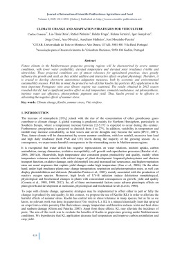
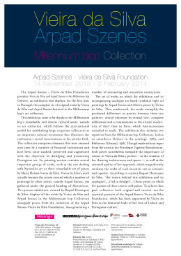
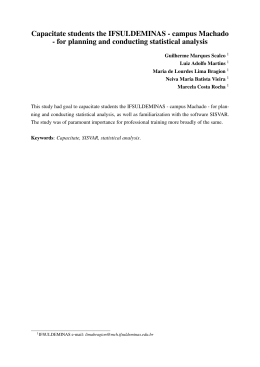
Baixar