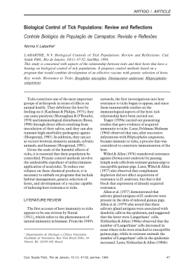
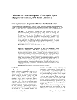
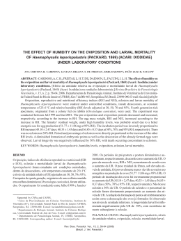
STUDIES ON THE Anaplasma marginale THEILER, 1910 INFECTION IN Boophilus
microplus (CANESTRINI, 1887) USING ‘NESTED’ PCR
ANDERSON B. DE MOURA1*, ODILON VIDOTTO2, MILTON H. YAMAMURA2,
MARILDA C. VIDOTTO3, ADEMIR B. DA LUZ PEREIRA2
ABSTRACT. MOURA A.B. DE, VIDOTTO O., YAMAMURA M.H., VIDOTTO M.C.; PEREIRAA.B. DA L. Studies
on the Anaplasma Marginale Theiler, 1910 infection in Booplhilus microplus (Canestrini, 1887) Using ‘Neste’
PCR. [Estudos da infecção por Anaplasma marginale Theiler, 1910 no Boophilus microplus (Canestrini, 1887)
utilizando a técnica de Nested PCR]. Revista Brasileira de Parasitologia Veterinária, v. 12, n. 1, p. 27-32, 2003.
Universidade Norte do Paraná-UNOPAR, Arapongas, PR 86700-000, Brazil. E-mail: [email protected]
The nested Polymerase Chain Reaction (nPCR) technique was used to detect the presence of Anaplasma
marginale DNA fragments in Boophilus microplus tick progeny (eggs and larvae) and to determine when they
become infected by this rickettsia. Eggs and larvae were obtained by incubation of tick engorged females, harvested
from A. marginale carrier cattle. The nPCR carried out on the tick donor cattle blood and on the engorged females,
eggs and larvae of B. microplus identified DNA bands of A. marginale with 345 bp, in which specificity was
confirmed by cleavage with the Eco R I restriction enzyme. The results showed that animals with a low percent of
infected erythrocytes (0-0.2%) detected by Giemsa staining are capable of infecting B. microplus females. In the
progeny of positive engorged females, fragments of A. marginale DNA were only detected in the eggs of the fourth
day and in the larvae of the first and seventh days of oviposition (three positive samples in 104 analyzed, or 2.88%)
and, in the progeny of negative engorged females, thirty-four of the 152 samples analyzed (22.4%) showed positive
larvae and eggs after the first and second days of oviposition, respectively. Specific fragments of A. marginale
DNA were identified in eggs and larvae from B. microplus engorged females that fed on cattle with low parasitemia
under field conditions, suggesting a possible migration of A. marginale through the general cavity of the tick,
infecting the ovaries and then, eggs and larvae.
KEY WORDS: Anaplasma marginale, Boophilus microplus, migration, ovaries, nested-PCR.
RESUMO
A técnica de nested PCR (nPCR) foi utilizada para detectar a
presença de fragmentos do DNA de Anaplasma marginale na
progênie (ovos e larvas) de Boophilus microplus e determinar quando ela se torna infectada pela rickettsia. Ovos e larvas foram obtidos por incubação de teleóginas colhidas de
bovinos portadores de A. marginale. A nPCR realizada no
1
Universidade Norte Paraná - UNOPAR - Arapongas - PR - Brasil.
Endereço para correspondência: Av Pedro Marques, 271 – Jd. Universitário, Jaboticabal, SP, CEP: 14882-222.
2
CCA/DMVP/ Universidade Estadual de Londrina, Campus Universitário - Cx. Postal 6001, Londrina, PR, Brasil, CEP: 86051-990. Email: [email protected]
3
CCB / MIB / Universidade Estadual de Londrina, Campus Universitário - Cx. Postal 6001, Londrina, PR, Brasil, CEP: 86051-990.
*
sangue dos animais doadores de carrapatos e nas teleóginas,
ovos e larvas identificou bandas de DNA de A. marginale
com 345 pbs, cuja especificidade foi confirmada pela clivagem
com a enzima de restrição Eco R I. Os resultados mostraram
que animais com baixo percentual de eritrócitos parasitados
(0 – 0.2%), detectados na coloração de Giemsa, são capazes
de infectar teleóginas de B. microplus. Na progênie das
teleóginas positivas, fragmentos de DNA de A. marginale
foram detectados somente nos ovos do quarto dia e em larvas
do primeiro e sétimo dias de oviposição (três amostras positivas em 104 analisadas, ou 2,88%) e, na progênie das teleóginas
negativas, 34 das 152 amostras analisadas (22,4%) mostraram
larvas e ovos positivos a partir do primeiro e segundo dias de
oviposição, respectivamente. Fragmentos específicos de DNA
de A. marginale foram identificados em ovos e larvas de
teleóginas de B. microplus que se alimentaram em bovinos
com baixa parasitemia sob condições de campo, sugerindo
Rev. Bras. Parasitol. Vet., 12, 1, 27-32 (2003)
(Brazil. J. Vet. Parasitol.)
28
Moura et al.
uma possível migração de A. marginale através da cavidade
geral do carrapato, infectando os ovários e, então, ovos e
larvas.
PALAVRAS-CHAVE: Anaplasma marginale, Boophilus
microplus, migração, ovários, Nested-PCR.
INTRODUCTION
Ticks and diseases that they transmit to cattle are one of
the major problems of livestock in tropical and subtropical
regions throughout the world. Among these diseases are the
anaplasmosis and babesiosis popularly known in Brazil as
“Tristeza Parasitária Bovina (TPB)”, caused by three distinct
pathogens: the protozoans Babesia bovis (Babès, 1888) and
B. bigemina (Smith; Kilborne, 1893) and the rickettsia
Anaplasma marginale, Theiler (1910).
Anaplasmosis causes meat and milk production losses,
abortion and death during the acute phase of the infection
(ALDERINK; DIETRICK, 1983). It is transmitted mainly by the
tick, Boophilus microplus (THOMPSON; ROA, 1978;
ALONSO et al., 1992; MARTINS; CORRÊA, 1995). It can also
be transmitted by other ticks, as well by blood sucking insects
and surgical materials (ANZIANI, 1979; RICHEY, 1981; EWING
et al., 1997) and through the placenta (ZAUGG, 1985;
ANDRADE, 1998).
The presence of different evolutionary phases of this
rickettsia in B. microplus intestine epithelial cells suggests that
sequential development stages of A. marginale may occur in
the invertebrate host, thus characterizing its replication in this
arthropod (RIBEIRO; LIMA, 1996) which shows its function as
a biological vector.
Although the predominant transmission of rickettsia is by
the tick B. microplus, its biology in this vector is still under
discussion (FARIAS, 1995). Guglielmone (1991), ALONSO et
al. (1992) and Vanzini and Ramirez (1994) quoted several authors
who reported successful transovarial transmission of A.
marginale by B. microplus and other studies indicating the
contrary. Thompson and Roa (1978) and Ribeiro et al. (1996)
working in Colombia and Brazil, respectively, did not confirm A.
marginale transovarial transmission by B. microplus. By the
other hand, Laranja et al. (1975), in Brazil and, Lopes-Valencia
and Vizcaino-Gerdts (1992) in Colômbia, have showed evidences
of transovarian transmission of A. marginale by the B. microplus.
This paper shows the result with A. marginale in engorged B.
microplus females collected from naturally infected cattle
evidencing the rickettsia migration to the eggs and larvae during
the oviposition period.
MATERIALAND METHODS
Farm and carrier animal selection
A farm was selected based on the results obtained in a
serological survey carried out on dairy farms around Londrina city, (ANDRADE, 1998) where 100% of the animals
showed positive serology for A. marginale and there was a
high incidence of clinical anaplasmosis cases. Londrina city is
located in the North of the State of Paraná, Brazil (between
23º 08’47”N and 23º55’46”S), at an altitude of 576 m with an
average annual temperature and rainfall of 22°C and 1876 mm,
respectively.
EDTA blood samples from 12 animals (heifers and cows)
were taken on this farm to select the engorged B. microplus
females donors, by nPCR. Blood smears, stained with
Giemsa, were also made to determine the parasitemia of these
animals (IICA, 1984).
Tick collection and in vitro cultivation
Six to 16 engorged females were collected from four
nPCR identified A. marginale infected animals (parasitemia
ranging from 0 to 0.2% by Giemsa stained blood smears Table 1). Ten engorged females weighing ³200 mg, previously
checked for viability, were selected and incubated in a
chamber at 28°C and 80% relative air humidity for oviposition.
The pre-oviposition, oviposition and egg hatching were
recorded.
The egg mass eliminated every 24 hours was collected
daily from each engorged female, until the end of the
oviposition period. Half of the daily egg production was
kept at –20°C to run the nPCR trying to detect A. marginale
DNA fragments eventually present on different laying days.
The other part of the eggs was incubated under the same
conditions described above until the larvae hatched and
submitted to the nPCR.
Each engorged female was examined by nPCR ten days
after the end of oviposition to see whether or not they were
infected by A. marginale. Based on this information, the
engorged females and their progeny were divided into two
groups: Group 1 had the progeny from five positive females
and the Group 2 had the progeny from five negative engorged
females.
DNA extractions
Blood. EDTA collected blood was washed three times
with PBS buffer to remove the leukocyte layer. The resulting
red blood cell mass was processed to extract the DNA
according to the manufacturer’s recommendations (Purogene,
Gentra Systems). The extracted DNA was kept refrigerated
(4°C) until the nPCR analysis was carried out.
Boophilus microplus engorged females, larvae and eggs
Boophilus microplus engorged females, eggs and larvae
were processed for DNA extraction by the modified silica
technique described by Boom et al. (1990). Engorged females,
eggs and larvae were squashed in TE solution (10 mM TRIS, 1
mM EDTA, pH 8.0) and, 25 ml (eggs and larvae) or 50 ml
(engorged females) of this preparation were added to a 1.5 ml
microtube containing 450 ml of “lise” L6 buffer (120 g guanidine
“isotiocianide” diluted in 100 ml of 0.1M TRIS, pH 6.4 with 22
ml of 0.2M EDTA solution (0.01%). After a short agitation
period, the material was placed at room temperature for 10
minutes and was again agitated and centrifuged at 12,000 x g
for 20 seconds. The supernatant was discarded. The DNA
Rev. Bras. Parasitol. Vet., 12, 1, 27-32 (2003)
(Brazil. J. Vet. Parasitol.)
Studies on the Anaplasma marginale infection in Boophilus microplus using “Nested”PCR
containing pellet was then washed twice in L2 buffer (120 g
guanidine “isotiocianide” diluted in 100 ml 0.1M TRIS, pH 6.4),
twice in 70% ethanol and once in acetone. After the acetone
removal, the microtube was open, incubated at 56°C for 10 minutes
and the DNA was eluted in 100 ml of ultra pure water. The tube
was incubated at 56°C for 10 minutes for elution and the
supernatant was recovered after centrifugation and stored at –
20°C for amplification by nPCR.
“Nested”-PCR
A nPCR was carried out according to TORIONI de
ECHAIDE et al. (1998) using the nPCR master kit (Boehringer
Mannhein) in a MiniCyclerTM thermocycler from MJ Research.
The msp5 sequence from the A. marginale Florida strain
provide the following primers: external forward 5’GCATAGCCTCCCCCTCTTTC-3’; external reverse, 5’TCCTCGCCTTGCCCCTCAGA-3’ and internal forward, 5’TACACGTGCCCTACCGACTTA-3’. The first amplification
was made using 12.5 ml of the master kit solution (2.5 U DNA
Taq polymerase in Brij35 0.005% (v/v), 0.2 mM of each dATP,
dCTP, dGTP, dTTP , 10 mM Tris-HCl, 50 mM KCl and 1.5mM
MgCl2), 1 ml of the external primers (20 mM), 5.5 ml of ultra
pure water and 5ml of DNA template. The volume of 12.5 ml
of the master kit solution, 1 ml of the ‘external reverse’ primer
and 1 ml of the ‘internal forward’ primer (20mm), 8.5 ml of
ultra pure water and 2 ml of the product from the first
amplification were used in the second amplification. A drop
of mineral oil was added to the amplifications to prevent
reagent evaporation. The reactions were processed in the
thermocycler programmed for 5 minutes at 95°C, 35 cycles at
95°C for 1 minute, 65°C for 2 minutes and 72°C for 1 minute
with a final extension at 72 °C for 10 minutes followed by cooling
to 4°C for undetermined period of time in each amplification.
nPCR (10 ml) products were visualized in a 1.5% agarose gel
following electrophoresis staining with 0.015% ethidium
bromide (0,5ml/ml). Negative (cattle blood proven not infected
by A. marginale and water) and positive (A. marginale Florida
29
strain DNA) controls were also subjected to the same
treatment. The 345 bp expected DNA fragment was identified
by comparison with 100 base pairs weight molecular markers
(100 bp Ladder – Gibco BRL).
nPCR reaction specificity
The 345 bp DNA bands were removed from the agarose
gel using the Concert Rapid Gel Extraction System (Gibco
BRL) commercial kit. This DNA was cleaved with the EcoR
I (10 U/ml) (Gibco BRL) restriction enzyme. The cleavage
reaction was performed in a water bath at 37 °C for two
hours and the product was visualized, as previously
described, in SDS polyacrylamide gel (3.5 to 8% gradient
concentration) stained with 0.015% ethidium bromide after
electrophoresis.
RESULTS AND DISCUSSION
In the Group 1, constituted by the progenies of positive
engorged females, fragments of A. marginale DNA was only
detected in the eggs of the fourth day and in the larvae of the
first and seventh days of oviposition (three positive samples
in 104 analyzed, or 2.88%) (Table 1). An opposite situation
was observed for the progenies of the Group 2 (negative
engorged females). Thirty-four of the 152 samples analyzed
(22.4%) showed positive larvae and eggs since the first and
second days of oviposition, respectively (Table 2). Figure 1
shows the result of the nPCR carried out on the tick donor
cattle blood and on the engorged females, eggs and larvae of
B. microplus. DNA bands with 345 bp can be seen in the
positive samples. The nPCR specificity was confirmed by
amplified DNA cleavage with the Eco R I restriction enzyme,
which cleaves the DNA amplified products in two specific
fragments (Figure 2).
These results showed that there was a lower detection of
A. marginale DNA in the positive than in the negative
engorged females. This may be partly explained by the findings
Table 1. Detection of A. marginale on eggs and larvae of positive B. microplus females, 10
days after the oviposition period, by nested PCR.
Engorged
females
1o
2o
3o
4o
5o
1
2
3
4
5
-
-
-
+
-
1
2
3
4
5
+
-
-
-
-
-
Oviposition days
6o
7o
8o
Eggs
Larvae
+
-
9o
10o
11o
12o
13o
-
F
F
F
F
-
F
-
F
F
F
F
-
F
+ positive nPCR; - negative nPCR; F final of oviposition.
Rev. Bras. Parasitol. Vet., 12, 1, 27-32 (2003)
(Brazil. J. Vet. Parasitol.)
30
Moura et al.
Table 2. Detection of A. marginale on eggs and larvae of negative B. microplus females, 10 days after the oviposition period, by
nested PCR.
Engorged
Oviposition days
females
1o
2o
3o
4o
5o
6o
7o
8o
9o
10o
6
7
8
9
10
-
+
+
-
+
+
-
+
+
+
+
+
+
+
-
-
+
+
-
-
-
6
7
8
9
10
+
+
-
-
+
+
+
+
-
-
-
+
+
-
+
-
+
+
-
11o 12o
Eggs
+
Larvae
+
13o
14o
15o
16o
17o
F
-
+
-
+
F
+
F
+
-
+
-
F
-
-
F
-
F
+
+
F
-
F
+
18o
19o
20o
-
-
F
-
-
-
F
+ positive nPCR; - negative nPCR; F final of oviposition.
Figure 1. Nested PCR products of tick donor cattle blood, engorged females, eggs and larvae of Boophilus microplus. Lines 1 and 12, molecular
weight (100bps); line 2, positive control (A. marginale Florida strain); line 3, negative control; line 4, blood from positive cattle; line 5, blood from
negative cattle; line 6, positive engorged female of B. microplus; line 7, negative engorged female of B. microplus; line 8, positive eggs of B.
microplus; line 9, negative eggs of B. microplus; line 10, positive larvae of B. microplus; line 11, negative larvae of B. microplus.
of Ribeiro and Lima (1995), where these authors studied the
influence of temperature on A. marginale development in B.
microplus and reported that ticks kept under ideal temperature
and humidity conditions may begin the oviposition phase
before the rickettsia completes its development cycle in the
gut arthropod epithelial cells. This would result in a lower
number of positive progeny from these infected vectors. But
this does not explain the fact that negative engorged female
progeny were sevenfold more positive.
Our data are not in line with Ribeiro and Lima (1995; 1996)
for either the progeny from the positive or negative engorged
females. They detected the rickettsia presence in engorged B.
microplus females only after 19 days following oviposition,
indicating that transovarial transmission, if occur, would only
be possible after that period. This would only occur in the
winter period under natural conditions, as in other months
more than 90% of the B. microplus oviposition occurs between
the 10th and 13th days after females detachment (VEGA, 1976;
ALVARADO; GONZALES, 1979). Consequently, the
possibility of natural transovarial transmission would be limited
in our environment conditions. The mean oviposition period
was 12 days in the present experiment, with a minimum of nine
and a maximum of 19 days. However, A. marginale DNA
fragments were detected in both eggs and larvae from the 1st
to the 16th oviposition day.
A possible hypothesis to explain the observed results
and even those of Ribeiro and Lima (1995; 1996) may be related
to the moment at which the different tick instars became
infected with A. marginale while feeding on the host blood.
The engorged females whose offspring had a low detection
of rickettsia DNA (Group 1) would have had late infection,
leaving not enough time for A. marginale reaches the ovaries.
On the other hand, the engorged females of the Group 2 may
have been infected earlier, thus enabling contamination of
the ovaries from the beginning of egg production. Connell
(1974), investigating the transovarial transmission in B.
microplus suggested that, the infection did not persist for
sufficient time in the adult ticks to be transmitted to their
Rev. Bras. Parasitol. Vet., 12, 1, 27-32 (2003)
(Brazil. J. Vet. Parasitol.)
Studies on the Anaplasma marginale infection in Boophilus microplus using “Nested”PCR
31
Figure 2. Clivated and intacts Nested PCR products of tick donor cattle blood, engorged females, eggs and larvae of Boophilus microplus. Line
1 and 12, molecular weight (100bps); line 2, positive control (A. marginale Florida strain); line 3, EcoRI cleavage of positive control; line 4, blood
from positive cattle; line 5, EcoRI cleavage of blood from positive cattle; line 6, positive engorged female of B. microplus; line 7, EcoRI cleavage
of positive engorged female of B. microplus; line 8, positive eggs of B. microplus; line 9, EcoRI cleavage of positive eggs of B. microplus; line 10,
positive larvae of B. microplus; line 11, EcoRI cleavage of positive larvae of B. microplus.
progenies. The carrier animal parasitemia level may also
influence the A. marginale transmission by the ticks.
KOCAN et al. (1983) reported that D. andersoni ticks which
were fed on highly infected cattle transmitted the disease to
susceptible animals with a lower pre-patent period than those
which were fed on cattle with a lower percentage of infected
erythrocytes.
Considering that specific fragments of A. marginale DNA
were identified in eggs and larvae from B. microplus engorged
females that fed on cattle with low parasitemia under field
conditions, this would means that A. marginale stages, such
as identified by Ribeiro and Lima (1996) in the intestine of
engorged female, migrated through the general cavity of the
tick, infecting the ovaries and then, eggs and larvae. Whether
and when these infected larvae or other stages of the tick will
infect cattle, still reminder unclear and, more investigations
on A. marginale and B. microplus biology and, on the
potentiality of this vector in the transmission of this rickettsia
to cattle are necessary to better understand the epidemiology
of anaplasmosis in Brazil.
Acknowledgments: We thank Flora Satiko Kano, Marcia K.
Shimada and Richard C. Pacheco for their excellent technical
assistance. This work was supported by grants from the following
agencies: Fundação Coordenação de Aperfeiçoamento de Pessoal de Nível Superior (CAPES), Conselho Nacional de Desenvolvimento Científico e Tecnológico(CNPq) and Coordenadoria
de Pesquisa e Pós-graduação da UEL (CPG/UEL).
REFERENCES
ALDERINK, F.J.; DIETRICH, R.A. Anaplasmosis in Texas:
epidemiologic and economic data from a questionnaire
survey. In: HIDALGO, R.J.; JONES, E. W. (ed), National
Anaplasmosis Conference, 7. 1983, Mississipi. Proceeding.
Mississipi: Mississipi St. Univ. Press, 1983. p. 27-44.
ALONSO, M.; ARRELANO-SOTA, C.; CERESSER, V.H.;
CORDOVES, C.O.; GUCLIELMONE, A.A.; KESSLER, R.;
MANGOLD, A.J.; NARI, A.; PATARROYO, J.H.;SOLARI,
M.A.; VEJA, C.A., VIZCAÍNO, O.; CAMUS, E.
Epidemiology of bovine anaplasmosis and babesiosis in
Latin America and the Caribbean. Rev. Sci. Tech. Off. Int.
Epiz., v. 11, p. 713-733, 1992.
ALVARADO, R.U.; GONZALEZ, J.C. A postura e a viabilidade do Boophilus microplus (Canestrini, 1887) (Acarina,
Ixodidae) em condições de laboratório. Rev. Latinoam.
Microbiol., v. 21, p. 31-36, 1979.
ANDRADE, G.M. Estudos sobre a prevalência e infecção
natural por Anaplasma marginale em bovinos da raça
holandesa na região de Londrina, PR. 1998, Londrina.
Dissertação (Mestrado), 1998.
ANZIANI, O.S. Anaplasmosis em áreas libres de garrapatas.
Reun. An. Inf. Tec. INTA EERA. Rafaela, p. 63-68, 1979.
BOOM, R.; SOL, C.J.A.; SALIMANS, M.M.; NOORDAA,
J.V. Rapide and simple method for purification of nucleic
acids. Journal Clin. Microb., v. 28, p. 495-503, 1990.
CONNELL, M.L. Transmission of Anaplasma marginale by
the cattle tick Boophilus microplus. Queensland J. Agricult.
Ani. Sci., v. 31, 185-195, 1974.
EWING, S.A.; PANCIERA, R.J.; KOCAN, K.M.; GE, N.L.;
WELSH, R.D.; OLSON, R.W.; BARKER, R.W.; RICE, L.E.
A winter outbreak of anaplasmosis in a nonendemic area
of Oklahoma: a possible role for Dermacentor albipticus.
J. Vet. Diagn. Invest., v. 9, p. 206-208, 1997.
FARIAS, N.A.R. Diagnóstico e Controle da Tristeza Parasitária Bovina. Guaíba: Editora Agropecuária. 80 p.,
1995.
GUGLIELMONE, A.A. Epizootiologia de las enfermedades
hemoparasitarias de los vacunos. Santiago: FAO. Oficina Regional para la América Latina y el Caribe. Red de
Cooperación Técnica entre Laboratorios de Investigación
y Diagnostico Veterinario. Serie Rlac-gan. (35) 53 p., 1991.
INSTITUTO INTERAMERICANO DE COOPERACION PARA
LA AGRICULTURA. Técnicas para el diagnóstico de
Rev. Bras. Parasitol. Vet., 12, 1, 27-32 (2003)
(Brazil. J. Vet. Parasitol.)
32
Moura et al.
babesiosis y anaplasmosis bovina. Costa Rica: IICA, 1984.
Serie Salud Animal. Publicacion Científica, n. 8.
KOCAN, K.M.; HOLBERT, D.; EWING, S.A.; HAIR, J. A.;
BARRON, S.J. Influence of parasitemia level at feeding on
development of Anaplasma marginale Theiler in
Dermacentor andersoni Stiles. Am. J. Vet. Res., v. 44, p.
554-557, 1983.
LARANJA, R.J.; ARREGUI, L.A.; ARTECHE, C.C.P. Transmissão dos agentes da Tristeza Parasitária dos Bovinos após
passagem de Boophilus microplus (CANESTRINI, 1887)
em hospedeiros não habituais. Bol. Inst. Pesq. Vet.
Desidério Finamor, v. 03, p. 113-123, 1975.
LOPEZ-VALENCIA, G.; VIZCAINO-GERDTS, O. Trans-mision
transovárica de Anaplasma marginale por la garrapata
Boophilus microplus. Revista ICA., v. 27, p. 437-443.
MARTINS, J.R.; CORRÊA, B.L. (1995). Babesiose e
Anaplasmose bovina: Aspectos destas enfermidades. Pesq.
Agrop. Gaúcha, v. 01, p. 51-58, 1992.
RIBEIRO, M.F.; LIMA, J.D. Influence of temperature on the
development of Anaplasma marginale in Boophilus
microplus. Arq. Bras. Med. Vet. Zoot., v. 47, p. 525-533, 1995.
RIBEIRO, M.F.B.; LIMA, J.D. Morphology and development
of Anaplasma marginale in midgut of engorged female
ticks of Boophilus microplus. Vet. Parasit., v. 61, p. 31-39
1996.
RIBEIRO, M.F.B.; LIMA, J.D.; SALCEDO, J.H.P. Attempted
transmission of Anaplasma marginale by infected
Boophilus microplus. Arq. Bras. Med. Vet. Zootec., v. 48,
p. 397-402, 1996.
RICHEY, E.J. Bovine anaplasmosis. In: HOWARD, R.J. (Ed.)
Current Veterinary Therapy Food Animal Practice.
Philadelphia: The W. B. Saunders Co., 1981. p. 767-772.
THOMPSON, K.C.; ROA, J.C. Transmission (mechanical/
biological) of Anaplasma marginale by the tropical cattle
tick Boophilus microplus. 1978. In: Proceedings of an
International Conference Held In Edinburgh, 1978,
Edinburgh. Proceedings... Edinburgh: University of
Edinburgh, 1978. p. 536-539.
TORIONI DE ECHAIDE, S.; KNOWLES, D.P.; McGUIRE, T.C.;
PALMER, G.H.; SUAREZ, C.E.; McELWAIN, T.F. Detection
of cattle naturally infected with Anaplasma marginale in a
region of endemicity by nested PCR and a competitive
enzyme -linked immunosorbent assay using recombinant
major surface protein 5. J. Clin. Microbiol., v. 36, p. 777782, 1998.
VANZINI, V.R.; RAMIREZ, L.M. Babesiosis y anaplasmosis
bovina. Diagnostico, epidemiologia y control. Rev.
Investigaciones Agropecuarias, v. 25, p. 137-190, 1994.
VEGA, R. Contribuición al estudio de la biologia de Boophilus
microplus (Canestrini, 1887) Acad. Cien. Cuba, Ser. Biol.,
v. 64, p. 1-8, 1976.
ZAUGG, J.L. Bovine anaplasmosis: Transplacental
transmission as it relates to stage of gestation. Am. J. Vet.
Res., v. 46, p. 570-572, 1985.
Received on February 07, 2003.
Accepted for publication on April 17, 2003.
Rev. Bras. Parasitol. Vet., 12, 1, 27-32 (2003)
(Brazil. J. Vet. Parasitol.)
Baixar