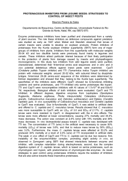
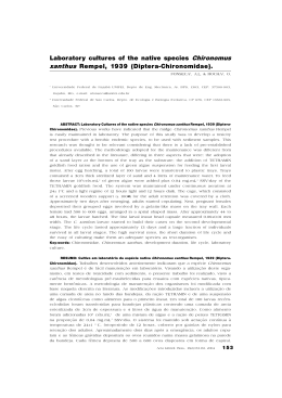
REGULAR PAPER
EFFECT OF TOPICAL APPLICATION OF JUVENILE
HORMONE ON THE OCCURRENCE OF CELL DEATH
IN THE LARVAE OF HONEYBEE (Apis mellifera) *
William Fernando Antonialli Jr¹ and Carminda da Cruz-Landim²
¹State University of Mato Grosso do Sul (UEMS) at Dourados, Dourados, MS,
²Department of Biology, Institute of Biosciences at Rio Claro, Paulista State University (UNESP), Rio Claro, SP, Brazil.
ABSTRACT
Workers and queens of Apis mellifera present marked dimorphism evidenced by the great development
of queen ovaries and atrophy of worker ones as a result of differential diets given by the nurse workers to
the developing larvae. The present paper reports the results of an investigation on how the administration
of juvenile hormone (JH) to worker larvae affects the cell death in their ovaries. Larvae from 2nd and 3rd
instars received topical application of JH III diluted in hexane in the concentration of 1μg/μL. The ovaries
of the treated and non-treated larvae (control) were collected in the 3rd, 4th and 5th instars and prepared
for cell death observation by staining with Hoechst and Propidium Iodide and by labeling with TUNEL
reaction. The results showed that the ovaries of the control larvae present higher rates of cell death, in all
ages, than those found on the JH-treated larvae ovaries and that the effect of the aforementioned treatment
was independent of the larvae stage to which it was applied being thus the same for the 2nd and 3rd instars.
The results suggest that the hormone application partially inhibited the cell death in the workers ovaries
thus affecting the key characteristic essential in the differentiation of larvae fated to be workers and queens.
The exogenous administration of JH to workers larvae reproduces the diet effect responsible for castes
differentiation in Apis mellifera indicating a close relationship between the quality of larval nourishment
and juvenile hormone production, which ultimately evidences the role of the diet in the expression of genes
linked to JH synthesis.
Key words: Apis mellifera, cell death, female castes, juvenile hormone, ovary
INTRODUCTION
Cell death has long been observed during
development and throughout the entire lifetime of
living organisms. The type of cell death is, in general,
classified according to its occurrence, morphology or
biological function. Glucksmann [14] and Hurle [18]
classified the cell death according to its biological
role in: 1. phylogenetic death, when responsible by
the loss of vestigial structures or organs during the
development; 2. metamorphic death, when taking
place during tissues and organs remodeling in
organisms suffering metamorphosis; and 3. histogenic
death, when occurring during differentiation and
_______________
Correspondence to: Dr. William Fernando Antonialli Junior
Universidade Estadual do Mato Grosso do Sul (UEMS), Cidade
Universitária de Dourados, Rodovia Dourados-Itahum, KM 12, CEP
79804-970, Dourados, MS, Brasil. E-mail: [email protected]
*Research performed in the laboratories of the Biology Department of
Biosciences Institute (UNESP)
maturation of the organism. Saunders [33] referring
to these physiological cell death used the expression
“death clock” meaning that each cell type has
an innate determined lifespan and are, therefore
programmed from the beginning to death.
Henceforth, as well established since long ago,
the programmed cell death has an important role in
the organism modulation and functioning during
the embryonic development, morphogenesis and
metamorphosis as it has been observed in some
organisms as the holometabolous insects. Besides,
the programmed cell death is important in regulating
the size of cell population during the growth and
maturity phases, by exerting a force equal and
opposite to mitosis. This kind of programmed cell
death, generally called apoptosis, occurs without
causing inflammatory reactions in the tissues, and
is morphologically and physiologically well defined
[5,17,19,23,28,35,36].
Braz. J. morphol. Sci. (2006) 23(3-4), 377-383
378
W. F. Antonialli Jr and C. Cruz-Landim
Although the cells are programmed to die at
a certain time, several exogenous factors, others
than non-physiological stimulation, such as virus
infections or other causes of illness [2,4,10,21,37],
may unchain the cell death programming such as
hormones, cytokines, interleukins, and so on. The
presence or absence of specific growth factors, the
increase or decrease of hormonal titers may promote
or suppress the activation of the programmed cell
death [8,13]. In addition, the same stimulus may
produce opposite effects in different cell types [1].
During the occurrence of the apoptosis, the
DNA is cleaved by the endonucleases activation,
preferentially at the internucleosomal sites, leaving
free ends that can be identified by TUNEL reaction
(Terminal-transferase dUPT Nick end Labeling).
The DNA fragments under electroforesis in agarose
gel gives a characteristic ladder pattern [37].
In eusocial bees such as Apis mellifera in which
the ovaries present a differential development in the
female castes, these organs constitute a good model
for studying the role of cell death in the modulation
of the castes fertility. Both castes, the queens - the
fertile females, and the workers - the sterile females,
originate from the same kind of egg, undergo the same
embryonic development, only diverging during their
larval development due to the differential type of
nutrition that they receive. The castes differentiation
in A. mellifera is, therefore, determined by their
larval diet [3,22,34].
While 2 to 3-day-old larvae fated to be queens
are fed with royal jelly, the same aged larvae fated
to be workers receive a less rich diet from the nurse
workers, what downregulates the rates of juvenile
hormone (JH) production and, consequently, its titer
in the hemolymph. The hormones titers, mainly
the JH but also the ecdysteroids are, therefore,
directly responsible for the changes that occur in
the post-embryonic determination of castes in bees
[7,9,15,25,27,32]. Hartfelder et al. [16] found that the
titers of this hormone affect the ovarian development
during lifetime. Nevertheless, the morphological
features of the mechanisms causing the atrophy
that takes place in the workers ovaries are poorly
known. The differential nutrition and its eventual
implication on JH titers is the unchaining factor of
the expression of the genes that control the castes
differentiation. How this expression manifests itself
Braz. J. morphol. Sci. (2006) 23(3-4), 377-383
in the ovaries is the scope of this work. Therefore,
this work’s intent is to verify the effect of extra
doses of JH in the death of ovary cells when these
doses are topically applied to workers larvae of Apis
mellifera in the 2nd and 3rd instars or more precisely
between 48 and 72 h after larvae eclosion.
MATERIAL AND METHODS
Brood combs isolated by wire screen were made
available of a queen with a good performance of posture,
for a period of approximately 6 h. The comb containing
the eggs was, afterwards, transferred to a queenless
colony where they were left to develop. The 48 h-old(2nd instar) and the 72 h-old- (3rd instar) ecloded larvae
were separated in two groups. One group received a
topical treatment with 1 μL of JH III (Sigma) diluted
in hexane in the concentration of 1 μg/μL and the other
was the non-assisted control group. The application was
performed directly on the comb by using an automatic
micropipette Gibson P². The development observed in the
assisted larvae was compared to the one observed in the
non-assisted larvae of the control group.
The developmental instar of the larvae was determined
by measuring the width of the cephalic capsule and by
applying the Dyar law [11]. The results showed that each
instar lasts 24 h. Therefore, samples of the JH-treated
and JH-non-treated larvae were collected at each 24 h
after the hormone administration, a procedure which
was maintained up to the 6th day after larvae eclosion
(fifth instar). Afterwards, the larvae were anesthetized by
cold and had their ovaries dissected. Three larvae, or six
ovaries of each larval age were studied.
Ovaries staining
The ovarioles of the dissected ovaries from both the
JH-treated larvae and the control larvae were released by
the extraction of the outer capsule of the organ, and prior
to fixation the ovarioles were incubated in a solution of one
drop of Hoechst at 0.001% in sterile distilled water during
10 min, followed by incubation in Propidium Iodide ( 2.5
μg/μL) during 5 min. Immediately after staining the ovaries
were examined and photographed under a fluorescence
microscope (Leica DMLB) using a UV excitation filter of
365 nm and a barrier filter of 400 nm.
TUNEL labeling
After releasing and dissecting the ovarioles as
described above, the ovaries of both experimental and
control larvae groups were fixed in phosphate buffered
4% paraformaldehyde and afterwards they were examined
using the in situ TUNEL technique to detect cell death
according to the POD-1684-817 kit instructions. The
negative control was performed without the treatment with
T-d-T/labeled-deoxinucleotide.
Cell death on the worker larvae ovaries of honeybee
RESULTS
The staining of the fresh ovaries by the
combination of Hoechst and Propidium Iodide
treatments allows the distinction between health
and damaged cells because the Hoechst, a supravital
stain, stains the nuclei of health cells while the
Propidium Iodide only stains the ones with changed
membrane permeability. The ovaries of 48 h-oldlarvae (2nd instar) treated with JH (Fig. 1A-D)
presented a smaller number of cells stained by the
Propidium Iodide, in all of the subsequent instars,
than the one observed in the control group larvae at
the same development stage (Fig. 1H-L). The same
was observed on the 72 h-old larvae (3rd instar)
which were treated with hormone (Fig. 1E-G), when
comparing them with the control group larvae.
However, no visible differences in the number
of stained cells were observed as a result of the
hormone administration being applied either at the
2nd or at the 3rd instars.
Similarly, the results obtained with the TUNEL
technique showed less labeled nuclei in the ovaries
of the JH-treated larvae (Fig. 2A-G) than in the ones
of the control (Fig. 2H-L). Nevertheless, with this
technique it was observed that the treatment in the 2nd
instar was more effective in preventing cell death in the
subsequent instars (Fig. 2A-D). The negative control
of the test, does not show labeled nuclei proving the
validity of the results (Fig. 2M-O).
DISCUSSION
When a colony of A. mellifera looses the queen
the workers produce new queens by feeding 2 to 3day-old larvae with just royal jelly. Older than 2 to
3-day-old larvae fed in the same way will not result
in a well developed queen. One of the differences
between the queen and the worker is on their ovary
size. While each queen ovary consists of 180 to 200
long ovarioles, the ovary of the worker has only 2 to
12 short ones. Therefore, although the worker ovary
is capable of producing eggs the potential fertility of
this caste is several times much lower than that of the
queen and, in addition, the workers cannot mate. It
becomes clear that the differentiation between both
castes is a post-embryonic event due to a less rich
nutrition received by the larvae fated to be worker
from the 2nd or 3rd day on, after eclosion. It is believed
that the worker inflicts itself a kind of nutritional
castration which generates a drastic reduction in the
number and length of their ovarioles.
379
The JH prevents cell death in several tissues
of insects [8]. Knowing that there are differences
in the endogenous hormones titers between the
two castes and that this was correlated with their
differentiation, the possibility of manipulating their
hemolymph contents by reprogramming the tissues
and organs differentiation at the molecular and
cellular levels exists.
Both Hoechst and Propidium Iodide methods
used in this study showed that cell death takes
place at a great rate in the ovaries of the control
larvae. Although the stains here used do not inform
the type of death affecting the cells they are useful
to differentiate intact, healthy cells stained by the
Hoechst, from the ones with impaired membrane
permeability stained by the Propidium Iodide
[20,31].
In accordance with previous results [29,30], the
present study shows that the cell death was observed
in the control ovaries since the 3rd instar. The high
rates of cell death found in the 3rd, 4th, and 5th instars
of the control ovaries, ought to be due to the lower
titers of JH present in worker larvae hemolymph
[6,7,15,27], which were partially diminished in the
case of the experimental larvae due to the topical
application of JH. The hormone concentration
applied was chosen because previous experiments
done with similar objective showed its effectiveness
in provoking changes in the physiology of some
organs of bees [1,12,24].
At the 5th instar the rates of cell death increased
even in the ovaries of the treated larvae. In this
late instar the titers of ecdysteroids were already
increasing while the JH was almost vanished thus
increasing the general rates of cell death [13,16,25,
26,27], a phenomenon typical of the beginning of the
metamorphosis. In the pre-pupae queen an increase
of the JH titer [25,26] prevents the induction of cells
death in the ovaries guaranteeing the survival of the
ovarioles. Therefore, although the extra dose of JH
applied in the 2nd or 3rd instar is enough to partially
prevent cell death during the larval phase in the
treated workers it does not prevent the death that
takes place in late last larval instars and pupation
because the JH is not produced in worker larvae, as
it occurs in queen.
The labeling with TUNEL evidenced a lower
number of dead cells than what was observed
when the Propidium Iodide procedure was used. A
possible explanation is that the Propidium Iodide
Braz. J. morphol. Sci. (2006) 23(3-4), 377-383
380
W. F. Antonialli Jr and C. Cruz-Landim
labels any kind of damaged cells while the TUNEL
reaction marks exclusively the cells where DNA
fragmentation occurs, which on the other hand, is
not the only cell death type occurring in the ovaries
[30]. In fact, the Propidium Iodide stains nuclei of
every cells with disturbed membrane permeability,
an event that precedes cell death.
In conclusion, the present study showed that the
exogenous application of juvenile hormone prevents
Third day of application
Control
Sixth day of larval life
Fifht day of larval life
Fourth day of larval life
Third day of larval life
Second day of application
cell death in the worker ovaries of Apis mellifera
honeybees during their larval development hence
simulating the functional role of the special enriched
nutrition received by the queen larvae. Actually, the
nutrition acts by regulating the hemolymph titers of JH
in the castes, supposedly by modulating the expression
of JH-synthesis linked genes. The results presented
here also confirm that apoptosis is not the only type of
programmed cell death occurring in bees ovaries [30].
Figure 1. Ovarioles from 2 and 3 days old worker larvae of A. Mellifera treated with topical application of 1μL of JH
(A-G), and control without treatment (H-L). The health cells have the nuclei stained in blue (Hoechst). The nuclei of the
dying are stained in red by the Propidium Iodide. ov-ovarioles, ce-ovary capsule. A, B, H, Bars = 30 μm; C, D, E, G, I,
L, Bars = 50 μm; F, J, Bars = 80 μm. (Panels F and J, after Cruz-Landim et al., 2006, Braz. J. Morphol. Sci. 23:27-42).
Braz. J. morphol. Sci. (2006) 23(3-4), 377-383
Third day of larval life
Fourth day of larval life
Fifht day of larval life
Sixth day of larval life
Third day of application
Control
Negative Control of reaction
Figure 2. Ovarioles of experimental (A-G), control larvae (H-L) and negative control (M-N) of A. mellifera 2 and 3 days old labeled with TUNEL showing positive
reaction to cell death (arrows). A, G, Bars = 20 μm, B, C, H, L, O, Bars = 35 μm; D, E, F, I, J, M, N, Bars = 50 μm.
Second day of application
Cell death on the worker larvae ovaries of honeybee
381
Braz. J. morphol. Sci. (2006) 23(3-4), 377-383
382
W. F. Antonialli Jr and C. Cruz-Landim
ACKNOWLEDGMENTS
Financial support for this study was provided by the
Fundação de Amparo à Pesquisa do Estado de São Paulo
(FAPESP), grant # 98/15057-6. (The State of São Paulo
Research Foundation) and by the Conselho Nacional de
Desenvolvimento Científico e Tecnológico/CNPq (National
Council for Scientific and Technological Development) and
to Prof. Dr. Elza da Costa Cruz Vasconcellos for language
correction.
REFERENCES
1. Abdalla FC, Gracioli LF, Salles HC, Cruz-Landim C
(2001) Effect of topical application of juvenile hormone
(JH) in honeybee worker larvae on the development of
Dufour’s and Koschewnikow’s glands. Sociobiology
37, 185-191.
2. Arends MJ, Wyllie AH (1991) Apoptosis: mechanism
and roles in pathology. Int. Rev. Exp. Pathol. 32,
223-254.
3. Asencot M, Lensky Y (1976) The effect of soluble
sugars and juvenile hormone on the differentiation
of the female honeybee larvae (Apis mellifera L.) to
queens. Life Sci. 18, 693-699.
4. Bowen ID, Bowen SM (1990) Programmed Cell
Death in Tumours and Tissues. Chapman and Hall:
London.
5. Bursch W, Kleine L, Tenniswood M (1990) The
biochemistry of cell death by apoptosis. Biochem.
Cell Biol. 68,1071-1074.
6. Capella ICS, Hartfelder K (1998) Controle hormonal
do desenvolvimento pós-embrionário dos ovários de
Apis mellifera. In: Anais do 3 Encontro sobre Abelhas,
Ribeirão Preto (SP), Brazil. p. 207-211.
7. Copijn GM, Beetsma J, Wirtz P (1979) Queen
differentiation and mortality after application of
different juvenile hormone analogues to worker larvae
of the honeybee (Apis mellifera L.). Proc. K. Ned.
Akad. Wed. Ser- C 82, 29-42.
8. Dai JD, Gilbert LI (1998) Juvenile hormone prevents the
onset of programmed cell death in the glands of Manduca
sexta. Gen. Comp. Endocrinol. 109, 155-165.
9. Dietz A, Herman HR, Blum MS (1979) The role of
exogenous JH-I, JH-II, JH-III and anti-JH (Precocene
II) on the queen induction of 4.5 day-old-worker honey
bee. J. Insect Physiol. 25, 503-512.
10. Doi T, Nishida K, Matsuo M,Yoshida A, Murakami
T, Inoue H (2002) Evidence of oncotic cell death
and DNA fragmentation in human hypertrophic
chondrocytes in chondro-ostehyte. Osteoarthritis
Cartilage 10, 270-276.
11. Dyar HG (1890) The number of molts of lepidopterous
larvae. Psiche 5, 420-422.
12. Farinha EK, Torneiros LM, Silva-de-Moraes RM,
Cruz-Landim C (1988) Influência hormonal sobre
o conteúdo de DNA nas células da glândula salivar
larval de Melipona quadrifasciata anthidioides Lep.
Braz. J. morphol. Sci. (2006) 23(3-4), 377-383
(Apidae, Meliponinae). I. Hormônio juvenil. Naturalia
13, 75-83.
13. Fujiwara H, Ogai S (2001) Ecdiysteroid-induced
programmed cell death and cell proliferation during
pupal wing development of silkworm, Bombyx mori.
Dev. Genes Evol. 211, 118-123.
14. Glücksmann A (1951) Cell deaths in normal vertebrate
ontogeny. Biol. Rev. Cambridge Philosoph. Soc. 26,
59-86.
15. Hartfelder K, Engels W (1998) Social insect
polymorphism: hormonal regulation of plasticity in
development and reproduction in the honeybee. Curr.
Top. Dev. Biol. 40, 45-77.
16. Hartfelder K, Köstlin K, Hepperle C (1995)
Ecdysteroid-dependent protein synthesis in castespecific development of the larval honey bee ovary.
Roux Arch. Dev. Biol. 205, 73-80.
17. Huppertz B, Frank HG, Kaufmann P (1999) The apoptosis
cascade -- morphological and immunohistochemical
methods for its visualization. Anat. Embryol. 200, 1-18.
18. Hurle JM (1988) Cell death in developing systems.
Methods Achiev. Exp. Pathol. 13, 55-86.
19. Kerr JFR, Willie AH, Currie AR (1972) Apoptosis:
a basic biological phenomenon with wider ranging
implications in tissue kinetics. Br. J. Cancer 26,
239-257.
20. Locke SJ, Peng YS, Cross NLA (1990) Supravital
staining technique for honey bee spermatozoa. Physiol.
Entomol. 15, 187-192.
21. Majno G, Joris I. (1995) Apoptosis, oncosis, and
necrosis. An overview of cell death. Am. J. Pathol.
146, 3-19.
22. Michener CD (1974) The Social Behavior of Bees.
Harvard University Press: Cambridge.
23. Osborne BA, Schwartz LM (1994) Essential genes
that regulate apoptosis. Trends Cell Biol. 4, 394-398.
24. Paes-de-Oliveira VT, Cruz-Landim C (2001)
Experimental control of the extra doses of juvenile
hormone on bee development: the case of the wax
glands of Apis mellifera (Hymenoptera: Apidae).
Sociobiology 38, 513-521.
25. Pinto LZ, Hartfelder K, Bitondi MM, Simões ZLP
(2002) Ecdysteroid titers in pupae of highly social
bees relate to distinct modes of caste development. J.
Insect Physiol. 48, 783-790.
26. Rachinsky A, Engels W (1995) Caste development in
honeybees (Apis mellifera): juvenile hormone turns on
ecdysteroids. Naturvissenchaften 82, 378-379.
27. Rachinsky A, Strambi C, Strambi A, Hartfelder K
(1990) Caste and metamorphosis: hemolymph titers
of juvenile hormone and ecdysteroids in last instar
honeybee larvae. Gen. Comp. Endocrinol. 79, 31-38.
28. Raff MC, Barres BA, Burne JF, Coles HS, Tshizaki
Y, Jacobson MD (1993) Programmed cell death and
control of cell survival: lessons from the nervous
system. Science 262, 695-700.
383
Cell death on the worker larvae ovaries of honeybee
29. Reginato RD, Cruz-Landim C (2001) Differentiation of
the worker’s ovary in Apis mellifera L. (Hymenoptera,
Apidae) during life of the larvae. Invert. Reprod.
Develop. 39, 127-134.
30. Reginato RD, Cruz-Landim C (2002) Morphological
characterization of cell death during the ovary
differentiation in worker honey bee. Cell Biol. Intern.
26, 243-251.
31. Reid S, Cross R, Snowe C (1996) Combined Hoechst
33342 and merosyanine 540 staining to examine
murine B cell cycle stage, viability and apoptosis. J.
Immunol. Meth. 192, 43-54.
32. Rembold H, Czoppelt CH, Rao PJ (1974) Effect of
juvenile hormone treatment on caste differentiation
in honeybee Apis mellifera. J. Insect Physiol. 127,
1193-1202.
33. Saunders JW (1996) Death in embryonic system.
Science 154, 604-612.
34. Snodgrass RE (1956) Anatomy of Honeybee. Comstock
Publishers: Ithaca.
35. Steller H (1995) Mechanism and genes of cellular
suicide. Science 267,1445-1449.
36. Wyllie AH, Kerr JFR, Currie AR (1980) Cell death:
the significance of apoptosis. Int. Rev. Cytol, 68, 251306.
37. Wyllie AH, Duvall E, Blow JJ (1984) Intracellular
mechanism in cell death in normal and pathological
tissues. In: Cell Aging and Cell Death (Davies I, Singer
DC, eds). pp. 269-294. Cambridge University Press:
Cambridge.
_________________
Received: March 13, 2006
Accepted: August 14, 2006
Braz. J. morphol. Sci. (2006) 23(3-4), 377-383
Baixar