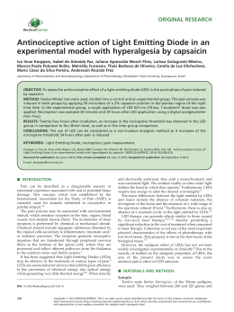
Lasers Med Sci (2009) 24:209–213
DOI 10.1007/s10103-008-0551-5
ORIGINAL ARTICLE
Low-level laser therapy (670 nm) on viability of random skin
flap in rats
Paulo Sérgio Bossini & Renan Fangel &
Rafael Malfará Habenschus & Ana Claudia Renno &
Benedito Benze & José Antônio Zuanon &
Carlos Benatti Neto & Nivaldo Antonio Parizotto
Received: 14 July 2007 / Accepted: 11 February 2008 / Published online: 20 March 2008
# Springer-Verlag London Limited 2008
Abstract This study investigated the effects of 670 nm
laser, at different fluences, on the viability of skin flap in
rats. One hundred male animals were used. The animals
were divided into control group; group treated with 3 J/cm2;
group treated with 6 J/cm2; group treated with 12 J/cm2 and
group treated with 24 J/cm2. The skin flap was made on the
backs of all animals studied, with a plastic sheet interposed
between the flap and the donor site. Laser irradiation was
done immediately after the surgery and on days 1, 2, 3 and
4 after surgery. The percentage of necrosis of the flap was
calculated at the 7th postoperative day. Additionally, a
sample of each flap was collected to enable us to count the
blood vessels. Treated animals showed a statistically
significant smaller area of necrosis than did the control
group. The necrosis in the treated groups was 41.82%
P. S. Bossini : R. Fangel : R. M. Habenschus : N. A. Parizotto
Department of Physiotherapy, Federal University of São Carlos,
Sao Carlos, SP, Brazil
A. C. Renno
Department of Health Science, Federal University of Sao Paulo,
Santos, SP, Brazil
B. Benze
Department of Statistic, Federal University of São Carlos,
Sao Carlos, SP, Brazil
J. A. Zuanon : C. B. Neto
Department of Physiology and Pathology,
Estadual University of the State of Sao Paulo (UNESP),
Araraquara, SP, Brazil
A. C. Renno (*)
Department of Physiotherapy, Federal University of São Paulo,
Av. Ana Costa, 95,
Santos, SP 15011-000, Brazil
e-mail: [email protected]
(group 2), 36.51% (group 3), 29.45% (group 4) and 20.37%
(group 5). We also demonstrated that laser irradiation at
670 nm, at all doses used, had a stimulatory effect on
angiogenesis. Our study showed that the 670 nm laser was
efficient to increase the viability of the skin flap, at all
fluences used, with a tendency of reaching better results at
higher doses.
Keywords Laser therapy . Microcirculation . Necrosis .
Surgical flaps
Introduction
Skin flap is a common surgical procedure used in plastic
and reconstruction surgeries. However, the failure of the
flap, mainly due to inadequate vascularization, is a frequent
problem found in this type of intervention [1]. Ischemia is
responsible for tissue necrosis, causing an undesirable
failure of the proposed treatment. In contrast, if blood flow
is sufficient in the distal portion of the flap, then flap
necrosis becomes much less of a problem [2].
Some authors have studied the effects of low-level laser
therapy (LLLT) on the viability of skin flaps [3–5]. The
action of LLLT is based on the absorption of the light by
tissues, which will generate modifications in cell metabolism [6, 7]. Studies have shown that laser irradiation
increased mitochondrial respiration and ATP synthesis in
isolated cells in culture [8]. Other studies have shown that
laser light affects calcium exchange through the cell
membrane, causing transient changes in the cytoplasmic
calcium levels [6, 7]. These modifications can increase the
synthesis of DNA, RNA and cell-cycle regulatory proteins,
stimulating cell proliferation, which could, therefore, be
beneficial for the re-establishment of connective tissue
210
Lasers Med Sci (2009) 24:209–213
during tissue repair and wound healing, contributing to the
increase of skin flap viability [7].
Pinfildi et al. [3] and Prado et al. [4] demonstrated that
laser irradiation was able to decrease the area of necrosis of
the skin flap. In addition, other authors suggest that the
positive effects of laser irradiation on skin flap viability is
due to positive effects on stimulating the formation of new
blood vessels, improving blood flow in the skin flap area
[8–10].
The use of LLLT on tissue regeneration has increased in
recent years. However, knowledge about the mechanisms by
which laser therapy acts and the lack of protocols of
treatment are still limited and need further investigation
[11, 12].
In order to progress our understanding of the clinical
parameters involved in the field of laser therapy and to
determine the responses of tissue regeneration to different
fluences of LLLT, the aim of this study was to investigate
the dose–response effects of 670 nm laser irradiation on the
viability of skin flaps in rats.
Methods
This study was conducted in accordance with the Guide for
Care and Use of Laboratory Animals and was approved by
the Animal Ethics Committee of the Federal University of
Sao Carlos. At the beginning of the experiment, 100 adult
male rats (12 weeks, 260–320 g) were randomly divided
into five groups, with 20 animals each: group 1 (control
group); group 2 (treated with 3 J/cm2); group 3 (treated
with 6 J/cm2); group 4 (treated with 12 J/cm2) and group 5
(treated with 24 J/cm2).
All animals were anesthetized with ketamine (95 mg/kg)
and xylazine (12 mg/kg) intraperitoneally and they were
also depilated. A random skin flap measuring 10 cm×4 cm
was made with a cranial base on the back of each rat [3]. A
plastic barrier with the same dimensions was placed
between the flap and its donor site. Flaps were closed with
simple nylon 4–0 stitches (Fig. 1).
Laser irradiation was performed immediately after the
surgery and on days 1, 2, 3 and 4 after surgery. A low-
Percentage of necrosis area of the flap: ¼
Fig. 1 Random skin flap (10 cm×4 cm)
energy AlGaInP laser, 670 nm (Ibramed Equipamentos
Médicos Ltda), continuous wave (CW), 0.6 mm beam
diameter, 30 W cm−2 was used. Laser irradiation was at
fluences of 3 J/cm2 for 6 s (total energy 4.32 J), 6 J/cm2 for
12 s (total energy 8.64 J), 12 J/cm2 for 24 s (total energy
17.28 J) and 24 J/cm2 for 48 s (total energy 34.56 J).
Twenty-four points, on the skin flap surface and surrounding it, were irradiated by the punctate contact technique
(Fig. 2). The irradiation was performed with a plastic
template on the skin flap with demarcation points for each
group [3].
Analysis
Skin flap necrosis
The percentage of skin flap necrosis was calculated on the
7th post-operative day through the paper-template method.
The rats were sedated, and the limit between viable tissue
characterized by soft skin, reddish, warm and haired, and
necrotic tissue (stiff, dark, cool, and hairless skin) was
demarcated on the animals [3]. A mold of the entire flap
and the necrotic area was drawn on transparent paper and
cut out, being checked with a precision balance (0.001 g
error). After that, the following equation was used:
Weight of paper template of flap necrosis
X 100
Weight of paper template of total area of flap
After this procedure, a sample of the skin flap was taken
for histological analysis. Then, the rats were humanely
killed by an overdose of general anesthetic so that we could
verify the necrotic areas.
Histological analysis
The specimens were retrieved en bloc. A sample of 2 cm2
in the region of the necrosis line transition for each skin
Lasers Med Sci (2009) 24:209–213
211
Fig. 2 Scheme of laser irradiation on 24 points, using the punctate
technique
flap was taken and processed (Fig. 3). The flap samples
were fixed in 10% buffered formalin (Merck, Darmstadt,
Germany), embedded in paraffin blocks and cut into
transverse sections (5 µm). Five laminae of each part of
the sample were stained with hematoxylin and eosin (HE,
Merck) and analyzed. Histologic evaluation was performed
under a light microscope (Zeiss Axioshopt, Carl Zeiss, Rio
de Janeiro, Brazil), with a ×40 objective. The number of
blood vessels were counted for each lamina by two
experienced pathologists, who were blind to the treatment.
A mean of the number of blood vessels for the laminae of
each skin flap was considered for statistical analysis.
Statistical analysis
The results are given as means and standard deviations. We
used analysis of variance (ANOVA) to compare changes
among the groups and the Tukey test to identify the
differences. Correlation between the areas of necrosis and
the number of blood vessels was assessed with the
Pearson’s correlation coefficients. A P level ≤0.05 was
considered as being statically significant.
Results
Figure 4 shows the means and the standard deviations (SD)
of the necrotic areas found in each group. Non-irradiated
animals (control group) showed statistically significant
Fig. 3 Blood vessel
Fig. 4 Percentage of tissue necrosis. G1 control group; G2 treated
with 3 J/cm2); G3 treated with 6 J/cm2; G4 treated with 12 J/cm2 and
G5 treated with 24 J/cm2. * Statistically significant when compared to
control
higher values of necrotic areas (49.92%) than did the other
groups. Moreover, LLLT produced a statistically significant
increase in skin flap viability, mainly at higher fluences
(24 J/cm2). The animals of group 5 presented a lower mean
percentage of necrotic area (20.37%) than the other groups
(group 2, 41.84%; group 3, 36.51% and group 4, 29.45%).
Moreover, animals irradiated with 3 J/cm2 demonstrated
no difference when compared to animals irradiated with
6 J/cm2 and showed lower values than animals irradiated
with 12 J/cm2 and 24 J/cm2. Mean percentage of necrosis
shown in animals irradiated with 6 J/cm2 was statistically
significant different when compared with that in animals
irradiated with 12 J/cm2 (P<0.05) and was highly statistically significant different when compared with that in
animals irradiated with 24 J/cm2, P<0.01). Animals irradiated with 12 J/cm2 and 24 J/cm2 were highly statistically
significant different (P<0.01).
We also demonstrated that laser irradiation at 670 nm, at
all fluences used, had a stimulatory effect on the increase of
the number of blood vessels at the necrosis transition line
(Figs. 5 and 6). The irradiated animals showed a statistically significant higher number of blood vessels than did
the non-irradiated animals, mainly at the dose of 24 J/cm2.
Fig. 5 Number of blood vessels. G1 control group; G2 treated with
3 J/cm2); G3 treated with 6 J/cm2; G4 treated with 12 J/cm2 and G5
treated with 24 J/cm2. * Statistically significant when compared to
control
212
Fig. 6 Number of blood vessels. a control group; b treated with 24 J/cm2
Figure 6 demonstrates the analysis of blood vessels in the
control group compared to group 5.
Pearson’s correlation coefficient showed a significant
negative correlation between the number of blood vessels
and the area of necrosis (P=−0.972; P=0.0001), suggesting
that the higher number of blood vessels was one of the
factors that could have contributed to the decrease of the
necrotic areas.
Discussion
We investigated the effects of 670 nm laser irradiation at
different laser fluences. Our observations indicated that
laser irradiation produced an increase in skin flap viability,
at all fluences used, especially at 24 J/cm2.
Recent studies have shown that laser stimulated mitochondrial activity, which led to a stimulation of cell
proliferation, promotion of fibrin absorption from injured
tissue and enhancement of the conversion of myofibroblasts
Lasers Med Sci (2009) 24:209–213
from fibroblasts, which could explain the enlarged surviving area of the skin flap after laser irradiation [13].
Our results corroborate those of Pinfildi et al. [3] and
Amir et al. [14], who also found decreases in tissue necrosis
of the skin flap after 632.8 nm laser irradiation at fluences
of 3 J/cm2 and 2.9 J/cm2, respectively.
Furthermore, our study demonstrated a stimulatory
effect of different 670 nm laser doses on blood vessel
growth. Adequate blood perfusion is essential to guarantee
skin flap survival and consequently the success of the
repair procedure [15, 16]. Moreover, Chang et al. [17]
demonstrated that both arterial and venous augmentation
were effective for increasing flap survival. Vascular photomodulation can be associated with the reduction of
inflammatory cells and with the stimulation of macrophages, T-lymphocytes, endothelial cells, fibroblast migration during the healing process, decreasing flap necrosis
[18].
Enwemeka et al. [19] stated that the development of new
blood vessels is an essential part of the healing process and
that the re-establishment of the circulation at the injury site
limits ischemic necrosis and permits repair. Salate et al. [20]
observed that the 660 nm laser was able to stimulate the
formation of new blood vessels in injured tendons of rats
compared to control animals. Kubota [1] found that 830 nm
laser irradiation produced higher vascular perfusion and
larger flap survival areas than control flaps that were not
irradiated.
Moreover, our findings further support the existence of a
dose–response curve, demonstrating that higher fluences
were more likely to produce a response than lower fluences
were. The same results were found in other studies
investigating the effects of laser on hard and soft tissues
[13, 16, 21, 22]. Probably, the fluence of 24 J/cm2 was
more efficient in accelerating the inflammatory response
and in stimulating the migration of cells related to tissue
regeneration, as macrophages and fibroblasts. Moreover, as
discussed above, the higher dose used in this study was
more efficient at stimulating angiogenesis in the skin flap,
which probably contributed to the increased flap viability.
Similarly, Prado et al. [4] and Kubota and Oshiro [23]
found an increase in vascular perfusion and a decrease in
tissue necrosis in skin flaps at high fluences (36 J/cm2).
However, the best fluence to be used in skin flap regeneration is still controversial and need further investigation.
The methodology employed in this study is aligned with
previous reports found in the literature [3, 4, 14]. The
plastic film used between the flap and the donor site
prevents revascularization of the flap through donor site
vessels, assuring homogeneous ischemia [14]. In addition,
the paper-template method is a simple and quick method to
measure necrotic areas and only requires transparent paper
and an accurate scale [3, 4]. Moreover, the punctate contact
Lasers Med Sci (2009) 24:209–213
technique of irradiation used (on the skin flap surface and
around it) was effective in stimulating skin flap viability.
We should point out some limitations of our work.
Although we found an increase in the number of blood
vessels at the region of the necrosis transition line in the
irradiated animals, we did not measure the blood flow or
the proportion of the different types of vessels (arteries or
capillaries). Moreover, as the region of the transition line on
the backs of the animals varied among groups, the region of
the histological analysis varied as well. The inclusion of a
group that had not been operated on would allow us to have
a standard of the behavior of the number of blood vessels
on the backs of the animal. Moreover, the analysis of blood
flux in the skin flap would give more specific information
about the distribution of the blood flow.
Although the effects of LLLT have been demonstrated in
many studies, the regulatory mechanisms of laser on tissues
are poorly understood [23, 24]. Such mechanisms probably
involve increases in cell proliferation through changes in
cell metabolism, affecting RNA synthesis and the expression of various cell regulatory proteins [9, 10, 25, 26].
However, the reasons for the stimulatory effects of laser
remain unclear and need further investigation.
Conclusion
Our study demonstrated the positive effects of 670 nm laser
on skin flap viability, mainly at the higher dose used. These
data highlight the importance of using adequate laser
wavelength and dose to elicit the best tissue response.
Studies investigating the effects of different fluences of
laser on tissue repair are important in determining the
efficacy of laser therapy. Further studies are required to
investigate possible mechanisms of action that may explain
the effects of laser on skin flap viability.
References
1. Kubota J (2002) Effects of diode laser therapy on blood flow in
axial pattern flaps in the rat model. Lasers Med Sci 17:146–153
2. Kerrigan CL (1993) Skin flap failure: pathophysiology. Plast
Reconstr Surg 72:766–777
3. Pinfildi CE, Liebano RE, Hochaman BS, Ferreira LM (2005)
Helium-neon laser in viability of random skin flap in rats. Lasers
Surg Med 37:74–77
4. Prado RP, Pinfildi CE, Liebano RE, Hochaman BS, Ferreira LM
(2005) Diode laser in viability of random skin flap in rats
(abstract). Photomed Laser Surg 23:115
5. Smith RJ, Bindorf M, Gluck G, Hammond D, Moore WD (1992)
The effect of low-energy laser on skin-flap survival in the rat and
porcine animal models. Plast Reconstr Surg 89:306–310
6. Reddy GK (2003) Comparison of the photostimulatory effects of
visible He-Ne and infrared Ga-As lasers on healing impaired
diabetic rat wounds. Lasers Surg Med 33:344–351
213
7. Mester E, Mester AF, Mester A (1985) The biomedical effects of
laser application. Lasers Surg Med 5:31–39
8. Dortbudak O (2000) Biostimulation of bone marrow cells with a
diode soft laser. Clin Oral Implants Res 16:540–545
9. Karu TI, Lubart R (2000) Effects of low-power light on biological
systems V. Proceedings of SPIE, Amsterdam, Netherlands, pp 01–
17
10. Basford JR (1995) Low-intensity laser therapy: still not an
established clinical tool. Lasers Surg Med 16:331–342
11. Beckerman H, de Bie R, Boulter L, De Cuyper H, Oostendorp R
(2002) The efficacy of laser therapy for musculoskeletal and skin
disorders: a criteria based meta-analysis of randomized clinical
trials. Phys Ther 72:483–491
12. Yu SY, Chiu JH, Yang SD, Hsu YC, Lui WY, Wu CW (2006)
Biological effect of far-infrared therapy on increasing skin
microcirculation in rats. Photodermatol Photoimmunol Photomed
22:78–86
13. Coombe AR, Ho C, Philips JR Chapple CC (2006) The effects of
low level laser irradiation on osteoblastic cells. Clin Orthod Res
4:3–14
14. Amir A, Solomon AS, Giler S, Cordoba M, Hauben DJ (2000)
The influence of helium-neon laser irradiation on the viability of
skin flaps in the rat. Br J Plast Surg 53:58–62
15. Sasaki GH, Pang CY (1980) Hemodynamics and viability of acute
neurovascular island skin flaps in rats. Plast Reconstr Surg 198
65:152–158
16. Kaufman T, Angel MF, Eichenlaub EH, Levin M, Hurwitz DJ,
Futrell JW (1985) The salutary effects of the bed on the survival
of experimental flaps. Ann Plast Surg 14:64–73
17. Chang H, Minn KW, Imanishi K, Minabe T, Nakajima T (2007)
Effect of venous superdrainage on a four-territory skin flap
survival in rats. Plast Reconstr Surg 119:2046–2051
18. Corazza AV, Jorge J, Kurachi C, Bagnato V (2007) Photobiomodulation on the angiogenesis of skin wounds in rats using
different light sources. Photomed Laser Surg 25:102–106
19. Enwemeka CS, Conhen-Kornberg E, Duswalt EP, Weber DM,
Rodriguez IM (1994) Biomechanical effects of three different
periods of GaAs laser photostimulation on tenotomized tendons.
Laser Ther 6:181–188
20. Salate ACB, Barbosa G, Gaspar P, Carrinho P, Koeke P,
Parizotto N (2005) Effect of In-Ga-Al-P diode laser irradiation
on angiogenesis in partial ruptures of Achilles tendon in rats.
Photomedicine Laser Surg 23:470–475
21. Wilden L, Karthein R (1998) Import of radiation phenomena of
electrons and therapeutic low-level laser in regard to the
mitochondrial energy transfer. J Clin Laser Med Surg 16:159–165
22. Kipshidze N, Nikolaychik V, Keelan MH, Shankar LR, Khanna L,
Kornowski R, Leon M, Moses J (2001) Low-power helium-neon
laser irradiation enhances production of vascular endothelial
growth factor and promotes growth of endothelial cells in vitro.
Lasers Surg Med 28:355–364
23. Kubota J, Oshiro T (1996) The effects of diode laser LLLT on flap
survival: measurement of flap microcirculation with laser speckle
flowmetry. Laser Ther 8:241–246
24. Byrnes KR, Waynant RW, Ilev IK, Wu X, Barna L, Smith K,
Heckert R, Gerst H, Anders JJ (2005) Light promotes regeneration
and functional recovery and alters the immune response after
spinal cord injury. Lasers Surg Med 36:171–85
25. Moore P, Ridgway TD, Higbee RG, Howard EW, Lucroy MD
(2005) Effect of wavelength on low-intensity laser irradiationstimulated cell proliferation in vitro. Lasers Surg Med 1:8–12
26. Kujawa J, Zavodnik L, Zavodnik I, Buko V, Lapshyna A,
Bryszewska M (2004) Effect of low-intensity (3.75–25 J/cm2)
near-infrared (810 nm) laser radiation on red blood cell ATPase
activities and membrane structure. J Clin Laser Med Surg 22:111–
117
Baixar