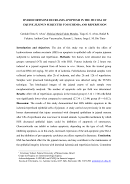
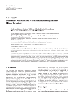
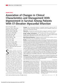
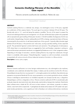
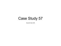
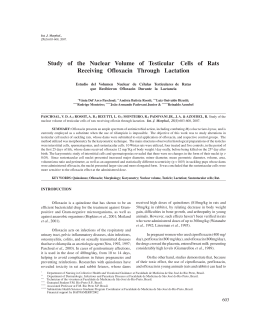
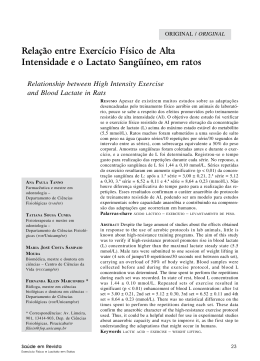
ORIGINAL ARTICLE
Rev Bras Cir Cardiovasc 2009; 24(2): 150-156
Evaluation of ischemic postconditioning effect on
mesenteric ischemia treatment. Experimental study
in rats
Avaliação do pós-condicionamento isquêmico no tratamento da isquemia mesentérica. Estudo
experimental em ratos
Carlos Henrique Marques dos SANTOS1, José Carlos Dorsa Vieira PONTES2, Otoni Moreira GOMES3, Luciana
Nakao Odashiro MIIJI4, Marco Aurélio Feltrin BISPO5
RBCCV 44205-1070
Abstract
Objective: To assess the preconditioning and
postconditioning effect on intestinal mucosal lesions in rats
undergone mesenteric ischemia and reperfusion procedure.
Methods: Thirty Wistar rats were studied and divided
into three groups: Group A, 10 rats undergone mesenteric
ischemia (30 minutes) and reperfusion (60 minutes); Group
B, 10 rats undergone mesenteric ischemia and reperfusion
preceded by ischemic preconditioning for three cycles of
ischemia and reperfusion for two minutes each; Group C,
10 rats undergone mesenteric ischemia and reperfusion
and, preceding the beginning of reperfusion, ischemic
postconditioning was performed for three cycles of
reperfusion and ischemia for two minutes each. Then, a
segment of small intestine was resected for histological
analysis. We assessed the results by Chiu et al. score and
the statistical analysis was performed.
Results: According to Chiu et al. score, the means of lesion
degree were: In the group A, 3.5; Group B, 1.2; Group C, 1.
The difference between group A with the groups B and C
was considered statistically significant (P < 0.05).
Conclusion: Ischemic pre- and postconditioning were
capable of minimizing - in a similar intensity - the tissue
injury on the intestinal mucosa of rats undergone
mesenteric ischemia and reperfusion process.
Resumo
Objetivo: Avaliar o efeito do pré e pós-condicionamento
isquêmico sobre a lesão tecidual na mucosa intestinal de ratos
submetidos ao processo de isquemia e reperfusão mesentérica.
Métodos: Foram estudados 30 ratos Wistar, distribuídos em
três grupos: grupo A, em que se realizou isquemia (30 minutos)
e reperfusão (60 minutos) mesentérica; grupo B, isquemia e
reperfusão mesentérica precedidos pelo pré-condicionamento
isquêmico por três ciclos de isquemia e reperfusão com duração
de dois minutos cada; grupo C, isquemia e reperfusão
mesentérica e, precedendo o início da reperfusão, foi realizado
o pós-condicionamento isquêmico por três ciclos de reperfusão
e isquemia com duração de dois minutos cada. Ao final, ressecouse um segmento do intestino delgado para análise histológica.
Avaliaram-se os resultados pela classificação de Chiu et al. e
procedeu-se ao tratamento estatístico.
Resultados: As médias dos graus de lesão tecidual segundo
a classificação de Chiu et al. foram: no grupo A, 3,5; grupo
B, 1,2; grupo C, 1. A diferença entre o resultado do grupo A
com os resultados dos grupos B e C foi considerada
estatisticamente significativa (P < 0,05).
Conclusão: O pré e pós-condicionamento isquêmico foram
capazes de minimizar, com intensidade semelhante, a lesão
tecidual na mucosa intestinal de ratos submetidos ao
processo de isquemia e reperfusão mesentérica.
Descriptors: Ischemia. Reperfusion injury. Ischemic
preconditioning.
Descritores: Isquemia. Traumatismo por reperfusão.
Precondicionamento isquêmico.
1.
2.
3.
4.
5.
Correspondence address:
Carlos Henrique Marques dos Santos
Rua Aluisio de Azevedo, 606 – São Bento – Campo Grande, MS,
Brasil – CEP 79004-050.
E-mail: [email protected]
PhD, Assistant Professor.
PhD, Adjunct Professor.
Full Professor, Titular Professor.
Master’s Degree, Public Employee.
Specialist, Public Employee.
This study was carried out at the Federal University of Mato Grosso
do Sul, Campo Grande, MS, Brazil.
150
Article received on October 16th, 2008
Article accepted on May 12th, 2009
SANTOS, CHM ET AL - Evaluation of ischemic postconditioning
effect on mesenteric ischemia treatment. Experimental study in rats
INTRODUCTION
In 1986, two publications have brought a great
advancement for the treatment of ischemia and reperfusion
syndrome. The first of them was the study from Parks and
Granger [1], showing that reperfusion is more damaging
than ischemia alone, a fact unknown up to now, promoting
a major change in current concepts and initiating several
studies that came to elucidate the physiopathology of
ischemia and reperfusion. The second publication of great
importance was the study from Murry et al. [2], which
brought the concept of ischemic preconditioning (IPC) as
a way to minimize the lesions of ischemia and reperfusion.
These authors described the short-term beneficial effect of
coronary occlusion also followed by short periods of
reperfusion, before initiating the stage of ischemia itself,
by noting reduction in ischemic myocardial lesion in dogs.
However, there are situations in which ischemia is
identified when the lesions are already installed, with no
opportunity for employment of the preconditioning.
In 2003, Zhao et al. [3] presented the concept of ischemic
postconditioning (IPC), that consisted in performing one
or more short cycles of reperfusion followed by one or
more short cycles of ischemia, immediately after the phase
of ischemia and before establishment of permanent
reperfusion. These authors showed that IPreC was as
effective as the IPostC in the prevention of reperfusion
lesions.
This study led to other studies that similarly have
demonstrated the ability of IPostC to prevent the lesions
of ischemia and reperfusion, as the IPreC [4].
If confirmed the effectiveness of IPostC in humans, this
could be a breakthrough in the treatment of ischemia and
reperfusion, since it is very common to perform the
diagnosis of lesions of this process when the ischemia has
already occurred, then there would be no space for use of
IPreC. In experimental model, there is already evidence of a
probable protective effect of IPostC on the intestinal
mucosa of rats undergoing mesenteric ischemia and
reperfusion [5].
Thus, considering the current evidences of the value of
the IPostC in reducing tissue lesions resulting from ischemia
and reperfusion process, it becomes of fundamental
importance - and it is the purpose of this study - to evaluate
comparatively the ability of the IPreC and IPostC to mitigate
the intestinal tissue lesion in the mesenteric ischemia and
reperfusion process.
METHODS
This study was approved by the Ethics Committee of
the Federal University of Mato Grosso do Sul.
Rev Bras Cir Cardiovasc 2009; 24(2): 150-156
Animals studied
We studied thirty rats (rattus norvegicus albinos,
Rodentia, Mammalia) of the Wistar strain, male, adults, with
weight ranging from 270 to 350 grams, with an average of
305 grams, from the biotery of the Federal University of
Mato Grosso do Sul.
Groups formed
The animals were divided into the following groups:
• Group A - Ischemia and Reperfusion: ten rats
undergone intestinal ischemia for 30 minutes by occlusion
of the cranial mesenteric artery with vascular forceps,
followed by 60-minutes reperfusion;
• Group B - Ischemic Preconditioning: Ten rats
undergone ischemia procedure for 30 minutes by occlusion
of the cranial mesenteric artery with vascular forceps and
reperfusion for 60 minutes. The phase of ischemia was
preceded by three cycles of ischemia (two minutes each)
interleaved with three cycles of reperfusion (two minutes
each);
• Group C - Ischemic Postconditioning: ten rats
undergone ischemic procedure for 30 minutes by occlusion
of the cranial mesenteric artery with vascular forceps and
reperfusion for 60 minutes. Between the ischemia and
reperfusion were performed three cycles of reperfusion (two
minutes each) interleaved with three cycles of ischemia
(two minutes each).
Anesthesia
The animals were weighed on accuracy electronic scales
and anesthetized with intraperitoneal injection of 2:1
solution of ketamine hydrochloride (Cetamin ®), 50 mg/ ml,
and Xylazine hydrochloride (Xilazin ®), 20mg/ml,
respectively, in dose of (0.1 ml/ 100 g).
Surgery procedure
The rats were maintained on spontaneous ventilation
in room air throughout the procedure. They underwent
median longitudinal laparotomy of approximately four
centimeters, exteriorization of the small intestine,
identification and dissection of the cranial mesenteric artery.
In group A, the cranial mesenteric artery was occluded
by atraumatic vascular forceps, which remained for thirty
minutes (ischemic phase). After placing the forceps, the
small intestine was repositioned in the abdominal cavity
and the surgical wound was closed with continuous suture
of the skin using 4-0 monofilament nylon yarn (mononylon
®). When the phase of ischemia has finished, the abdominal
wall was opened again by the removal of the suture yarn
and vascular forceps was removed, initiating the phase of
reperfusion, for 60 minutes. In all three groups, the phase
151
SANTOS, CHM ET AL - Evaluation of ischemic postconditioning
effect on mesenteric ischemia treatment. Experimental study in rats
Rev Bras Cir Cardiovasc 2009; 24(2): 150-156
of reperfusion was initiated, the abdomen was again closed
by continuous suture of the skin using 4-0 monofilament
nylon yarn until the end of the experiment.
In group B, before the stage of ischemia (30 minutes),
the ischemic preconditioning was performed by three cycles
of ischemia, lasting two minutes each (occlusion of the
cranial mesenteric artery by atraumatic vascular forceps),
intervealed by three cycles of reperfusion, also lasting two
minutes each (atraumatic vascular forceps removed from
the cranial mesenteric artery). After the stage of ischemia,
reperfusion was performed for 60 minutes.
In group C, the phase of ischemia (30 minutes) and
reperfusion (60 minutes) was performed. Preceding the
reperfusion phase, the ischemic postconditioning was
performed through three cycles of reperfusion (removal of
atraumatic vascular forceps from the cranial mesenteric
artery) lasting two minutes each, intervealed with three
cycles of ischemia (occlusion of the cranial mesenteric
artery by atraumatic vascular forceps), also lasting two
minutes each.
After finishing of the reperfusion phase in the three
groups, the abdominal wall was opened again by the removal
of the suture yarn and a segment of approximately one cm
of the ileum was resected, five centimeters proximal to the
ileocecal transition, that was opened on its antimesenteric
border, washed with saline solution and placed in a 10%
formaldehyde solution for further histological analysis.
The animals were sacrificed by deepening the anesthetic
plan.
longer being observed any glandular structure, but only
amorphous material deposited on the submucose tissue.
Histopathological study
The resected intestinal segments, after fixation in 10%
formaldehyde solution, underwent histological processing
and the slides were stained with hematoxylin-eosin and
analyzed under optical microscopy by the pathologist,
without his prior knowledge on the group that each rat
belonged, and were classified according to the degree of
tissue lesion according Chiu et al. [6]:
• Degree 0: mucosa without changes;
• Degree 1: well-formed villosity, without cell lysis or
inflammatory process, but with formation of the
Grunhagen’s subepithelial space;
• Degree 2: presence of cell lysis, formation of the
Grunhagen’s subepithelial space and increased spacing
between the villi;
• Degree 3: destruction of the free portion of the villi,
presence of dilated capillaries and inflammatory cells;
• Degree 4: structural destruction of the villi, with only
draft of one of them, formed by inflammatory cells and
necrotic material, with hemorrhage and basal glandular
ulceration;
• Degree 5: destruction of the entire tunica mucosa, no
152
Statistical analysis
The results obtained underwent statistical treatment,
by using the non-parametric Kruskal-Wallis test, with
establishment of a significance level of P <0.05. We used
the Bioestat software 2.0.
RESULTS
After the histological analysis of the degree of lesion of
the intestinal mucosa according Chiu et al. [6], we found
the results summarized in Table 1 and Figure 1.
Table 1. Result of histological analysis of the lesion degree of the
intestinal mucosa of rats in groups A, B and C according
to the classification of Chiu et. al. [6]
Rats
1
2
3
4
5
6
7
8
9
10
Mean
Group A
3
3
3
3
4
4
4
4
4
3
3.5
Mucosa lesion degree
Group C
Group B
0
0
1
1
3
1
1
2
2
3
1
3
0
1
0
0
1
0
1
1
1.0
1.2
Fig. 1 – Comparative graph of the histological analysis of the
lesion degree of the intestinal mucosa of rats in groups A, B and C
according to the classification of Chiu et. al. [6]
In group A, the mean degree of tissue lesion was 3.5.
Half of the animals in this group presented lesion degree 3
and the other half, degree 4.
In group B, the mean tissue lesion degree was 1.2. In
this group, three animals showed no lesion (degree 0), four
presented lesion degree 1, one degree 2, and two, degree 3.
SANTOS, CHM ET AL - Evaluation of ischemic postconditioning
effect on mesenteric ischemia treatment. Experimental study in rats
Rev Bras Cir Cardiovasc 2009; 24(2): 150-156
In group C, the mean tissue lesion degree was 1. Three
animals showed no lesion (degree 0), five presented lesion
degree 1, one presented 2 and one degree 3.
By using the non-parametric Kruskal-Wallis test, it was
observed that the groups B and C were similar statistically
(P=0.7507), however, there is a statistically significant
difference between these groups and group A (between A
and B P=0.0004 and between A and C P=0.0002).
Figures 2 to 6 show the histological aspect of the tissue
lesion degree found.
Fig. 5 – Photomicrography of the 3 degree according to Chiu et. al.
classification [6] (HE, A x 100, B x 400) – rat 4 from group A
Fig. 2 – Photomicrography of the 0 degree according to Chiu. et.
al. classification [6] (HE, A x 100, B x 400) – rat 1 from group B
Fig. 6 – Photomicrography of the 4 degree according to Chiu et. al.
classification [6] (HE, A x 100, B x 400) – rat 8 from group A
DISCUSSION
Fig. 3 – Photomicrography of the 1 degree according to Chiu et. al.
classification [6] (HE, A x 100, B x 400) – rat 2 from group C
Fig. 4 – Photomicrography of the 2 degree according to Chiu et. al.
classification [6] (HE, A x 100, B x 400) – rat 4 from group B
Since the publication of Murry et al. [2], ten years have
passed for the assessment of the IpreC effects in the
intestine. In 1996, Hotter et al. [7] published an article in
which the authors used effectively the IPreC in the
prevention of lesion caused by intestinal ischemia and
reperfusion in rats. The authors compared a group that
underwent only intestinal ischemia and reperfusion with
another IPreC group, and showed lower intensity of the
lesions in the second group.
In the present study the ability of the IPreC to minimize
the harmful effects of the mesenteric ischemia and
reperfusion was shown, since the means of tissue lesion
degree were 3.5 and 1.2 in the control groups and IPreC,
respectively, according to classification of Chiu et al. [6].
Also, Sola et al. [8] proved the benefit of the use of
IPreC in mesenteric ischemia and reperfusion, in a study on
which the authors found that in the group of rats undergone
only ischemia and reperfusion, the mean degree of intestinal
lesion according Chiu et al. [6] was 5, and in the group on
153
SANTOS, CHM ET AL - Evaluation of ischemic postconditioning
effect on mesenteric ischemia treatment. Experimental study in rats
Rev Bras Cir Cardiovasc 2009; 24(2): 150-156
which the IPreC was performed, such degree was 3. The
differences between the group ischemia-reperfusion and
the group IPreC was statistically significant both in this
study as in that from Sola et al. [8], although the values are
quite different. The reason for this difference may be related
to the time of ischemia and reperfusion and IPreC. In the
study by Sola et al. [8], the animals underwent 90 minutes
of ischemia and 30 minutes of reperfusion, in contrast with
this study, on which is used 30 and 60 minutes respectively.
The IPreC was also performed differently. Sola et al. [8]
performed the IPreC with a single cycle of ischemia and
reperfusion lasting ten minutes each, while in this study
three cycles of ischemia and reperfusion were performed
lasting two minutes each.
Moore-Olufemi et al. [9] also used the classification of
Chiu et al. [6] in their study, on which means were 3 and 1.2
in groups ischemia and reperfusion and IPreC, respectively.
Although very similar to the results of this study, the method
used was different. Moore-Olufemi et al. [9] performed the
ischemia and reperfusion lasting 30 minutes and six hours
respectively. The IPreC was performed with three cycles of
ischemia and reperfusion lasting four and ten minutes each,
respectively.
Other studies also showed the ability of IPreC to reduce
intestinal lesions of animals undergone mesenteric ischemia
and reperfusion [10-16], in such a way to become evident
that the IPreC is able to minimize the harmful effects of the
mesenteric ischemia and reperfusion.
The IPostC has also presented similar results to IPreC
in the prevention of lesions resulting from the process of
ischemia and reperfusion. However, currently there is only
one publication [5] that shows that this method may be
similar to IPreC in respect to its effect in the aforementioned
process in mesenteric circulation.
Zhao et al. [3] were the first to report the similarity of the
outcomes of IPreC and IPostC in prevention of lesions from
the process of ischemia and reperfusion. The study was
performed in dogs, by performing occlusion of the left
anterior descending artery for 60 minutes and reperfusion
for three hours. The IPreC was performed by a cycle of
ischemia lasting five minutes and reperfusion for ten
minutes. The IPostC consisted of three cycles of ischemia
and reperfusion lasting 30 seconds each. The area of
infarction was similar in groups IPreC and IPostC and
significantly lower than in the control group.
In the last 20 years, many forms of treatment for ischemia
and reperfusion were tested without great success [17,18].
With the advent of IPreC, there was great progress in the
treatment of Ischemia-Reperfusion Syndrome, however,
specifically in relation to mesenteric ischemia and
reperfusion, its use is limited due the fact that in most cases
the ischemia is already installed at the time of diagnosis
[19]. This is the main reason that values the IPostC because
when its efficacy is also confirmed in mesenteric ischemia
and reperfusion, its clinical applicability could be of great
importance. Nevertheless, up to now there is no studies
assessing its action in mesenteric ischemia and reperfusion.
In this study we observed the mean of the tissue lesion
degree 1.2 and 1 in groups IPreC and IPostC respectively,
according to the classification of Chiu et al. [6], in clear
contrast with the control group, in which the result was 3.5.
This shows that in this study the IPreC and IPostC were
similar in ability to prevent the lesion of ischemia and
reperfusion in the intestine of rats.
Most of the studies that analyzed the effects of IPostC
was performed in myocardial ischemia and reperfusion.
Donato et al. [20] compared the IPreC and IPostC in isolated
rabbit hearts, by performing ischemia and reperfusion of 30
minutes. The IPreC was performed by a cycle of ischemia
and reperfusion of five minutes each, and the IPostC, two
cycles of ischemia and reperfusion of 30 seconds each.
The authors concluded that the IPostC reduces the size of
infarction in the same magnitude as the IPreC without
changing the post-ischemic ventricular dysfunction.
These results were also obtained by Darling et al. [21]
in an experimental study that assessed infarction in rabbit
hearts. The IPostC was able to minimize the area of infarction
in comparison to the control group, by performing four
cycles of ischemia and reperfusion lasting 30 seconds
before initiation of the reperfusion phase.
It is believed that the IPostC may have protective effects
by activation of the adenosine receptors, based on studies
that showed that when administering agonists of such
receptors during reperfusion, the infarction is significantly
reduced [22.23].
Tang et al. [24] showed that the IPostC is as effective as
the IPreC in prevention of lesions from the process of
coronary ischemia and reperfusion in rats, since the time of
ischemia does not exceed 45 minutes. After this period,
according to these authors, the IPreC provides better results
on the reduction of the infarcted area.
In addition to the assessment of the IPostC in animal
hearts under ischemia and reperfusion, there is also a report
of its use in spinal cord. Huang et al. [25] showed that the
IPostC was as effective as the IPreC in the prevention of
tissue lesions in the spinal cord of rabbits undergone
ischemia and reperfusion.
The IPostC was also assessed in humans. Staat et al.
[26] reported its beneficial effect by performing intermittent
reperfusion during angioplasty in patients with acute
myocardial infarction, noting reduction of myocardial lesion.
Loukogeorgakis et al. [27] also performed an experimental
study in humans on which caused the transient upper limb
ischemia followed by reperfusion, observing the protective
effect from IPostC.
The protection mechanism of the IPostC in the
154
SANTOS, CHM ET AL - Evaluation of ischemic postconditioning
effect on mesenteric ischemia treatment. Experimental study in rats
Rev Bras Cir Cardiovasc 2009; 24(2): 150-156
mesenteric ischemia and reperfusion process is still not
entirely clear, but there are evidences that the IPostC may
be related to the significant decrease in levels of
malondialdehyde and products related to lipid peroxidation.
These observations suggest a reduction in the production
of Reactive Oxygen Species (ROS) and less lesion mediated
by oxidants with the IPostC [3].
An abundant production of ROS during the initial phase
of reperfusion has been implicated as the main factor in the
pathogenesis of tissue lesion. The peak production of ROS
occurs between the first and seventh minutes after the
initiation of reperfusion, although these substances are
detectable in later periods [21,28,29]. The IPostC acts at
this stage in such a way not yet fully clarified, possibly
reducing the production of ROS by gradual release of
oxygen to tissues [16.30]. For this reason, in this study the
IpostC was performed soon after ischemia, by performing
short cycles of ischemia and reperfusion, since there is no
one definition of which is the better time to be used in
these cycles, nor the number of cycles that would lead to
better outcomes.
We should consider that this study has limitations
regarding the assessment of tissue lesion. It was used the
histological analysis, a method widely used for this purpose,
however, thus becoming difficult to compare with other
studies on which different methods for assessment of lesion
by ischemia and reperfusion are used, such as the dosage
of myeloperoxidase and plasma or tissue malondialdehyde.
Thus, it is believed that the results found in this study
show that, similar to what is shown in the literature in respect
to the equivalence of the IPostC and IPreC capacity to
minimize the lesions caused by ischemia and reperfusion in
heart and spinal cord in the intestine rats, the IPostC
minimizes such lesions in the same proportion as the IPreC.
2. Murry CE, Jennings RB, Reimer KA. Preconditioning with
ischemia: a delay of lethal cell injury in ischemic myocardium.
Circulation. 1986;74(5):1124-36.
3. Zhao ZQ, Corvera JS, Halkos ME, Kerendi F, Wang NP,
Guyton RA, et al. Inhibition of myocardial injury by ischemic
postconditioning during reperfusion: comparison with
ischemic preconditioning. Am J Physiol Heart Circ Physiol.
2003;285(2):H579-88.
4. Schipke JD, Halkos ME, Kerendi F, Gams E, VintenJohansen J. Postconditioning: a brief review. Arch Med Sci.
2006;2:137-45.
5. Santos CHM, Gomes OM, Pontes JCDV, Miiji LNO,
Bispo MAF. Post-conditioning: preliminary results of
this new option in the treatment of mesenteric ischemia
and reperfusion. Cardiovascular Sciences Forum.
2007;2(2):13-24.
6. Chiu CJ, McArdle AH, Brown R, Scott HJ, Gurd FN. Intestinal
mucosal lesion in low-flow states. I. A morphological,
hemodynamic, and metabolic reappraisal. Arch Surg.
1970;101(4):478-83.
7. Hotter G, Closa D, Prados M, Fernández-Cruz L, Prats N,
Gelpí E, et al. Intestinal preconditioning is mediated by a
transient increase in nitric oxide. Biochem Biophys Res
Commun. 1996;222(1):27-32.
8. Sola A, Hotter G, Prats N, Xaus C, Gelpi E, RosellóCatafau J. Modification of oxidative stress in response to
intestinal
preconditioning.
Transplantation.
2000;69(5):767-72.
9. Moore-Olufemi SD, Kozar RA, Moore FA, Sato N, Hassoun
HT, Cox CS Jr, et al. Ischemic preconditioning protects against
gut dysfunction and mucosal injury after ischemia/reperfusion
injury. Shock. 2005;23(3):258-63.
CONCLUSION
The ischemic pre- and postconditioning were able to
reduce tissue lesion in the intestinal mucosa of rats
undergone process of mesenteric ischemia and reperfusion
in a similar manner.
REFERENCES
1. Parks DA, Granger DN. Contributions of ischemia and
reperfusion to mucosal lesion formation. Am J Physiol.
1986;250(6 Pt 1):G749-53.
10. Mallick IH, Yang W, Winslet MC, Seifalian AM. Ischaemic
preconditioning improves microvascular perfusion and
oxygenation following reperfusion injury of the intestine. Br J
Surg. 2005;92(9):1169-76.
11. Ceylan H, Yüncü M, Gürel A, Armutçu F, Gergerlioglu HS,
Bagci C, et al. Effects of whole-body hypoxic preconditioning
on hypoxia/reoxygenation-induced intestinal injury in newborn
rats. Eur J Pediatr Surg. 2005;15(5):325-32.
12. Wu B, Ootani A, Iwakiri R, Fujise T, Tsunada S, Toda S, et al.
Ischemic preconditioning attenuates ischemia-reperfusioninduced mucosal apoptosis by inhibiting the mitochondriadependent pathway in rat small intestine. Am J Physiol
Gastrointest Liver Physiol. 2003;286(4):G580-7.
13. Sola A, Alfaro V, Hotter G. Intestinal ischemic preconditioning:
less xanthine accumulation relates with less apoptosis.
Apoptosis. 2004;9(3):353-61.
155
SANTOS, CHM ET AL - Evaluation of ischemic postconditioning
effect on mesenteric ischemia treatment. Experimental study in rats
Rev Bras Cir Cardiovasc 2009; 24(2): 150-156
14. Tamion F, Richard V, Lacoume Y, Thuillez C. Intestinal
preconditioning prevents systemic inflammatory response in
hemorrhagic shock. Role of HO-1. Am J Physiol Gastrointest
Liver Physiol. 2002;283(2):G408-14.
23. Budde JM, Morris CD, Velez DA, Muraki S, Wang NP,
Guyton RA, et al. Reduction of infarct size and
preservation of endothelial function by multidose
intravenous adenosine during extended reperfusion. J Surg
Res. 2004;116(1):104-15.
15. Wang SF, Li GW. Early protective effect of ischemic
preconditioning on small intestinal graft in rats. World J
Gastroenterol. 2003;9(8):1866-70.
16. Cinel I, Avlan D, Cinel L, Polat G, Atici S, Mavioglu I, et al.
Ischemic preconditioning reduces intestinal epithelial apoptosis
in rats. Shock. 2003;19(6):588-92.
17. Santos CHM, Gomes OM, Pontes JCDV, Miiji LNO, Higa
EI. Uso do propofol (2,6 diisopropilfenol) como inibidor da
lesão tecidual na isquemia e reperfusão mesentérica. Estudo
experimental em ratos. Acta Cir Brás. 2003;18(4):347-54.
18. Santos CHM, Gomes OM, Pontes JCDV. Terapêutica
medicamentosa na isquemia e reperfusão mesentérica: revisão
da literatura. Rev Bras Coloproctol. 2006;26(1):28-33.
19. Santos CHM. Evolution and challenges in the phisiopatology
of the ischemia and reperfusion. Cardiovasc Sciences Forum.
2006;1(3):6-8.
20. Donato M, D’Annunzio V, Sabán M, Flor L, Gelpi RJ.
Poscondicionamiento: un nuevo mecanismo protector. Su
comparación con el precondicionamiento en el infarto
experimental. Rev Arg Cardiol. 2004;72(4):258-62.
21. Darling CE, Jiang R, Maynard M, Whittaker P, Vinten-Johansen
J, Przyklenk K. Postconditioning via stuttering reperfusion
limits myocardial infarct size in rabbit hearts: role of ERK1/2.
Am J Physiol Heart Circ Physiol. 2005;289(4):618-26.
22. Xu Z, Downey JM, Cohen MV. Timing and duration of
administration are crucial for antiinfarct effect of AMP 579 infused
at reperfusion in rabbit heart. Heart Dis. 2003;5(6):368-71.
156
24. Tang XL, Sato H, Tiwari S, Dawn B, Bi Q, Li Q, et al.
Cardioprotection by postconditioning in conscious rats is
limited to coronary occlusions < 45 min. Am J Physiol Heart
Circ Physiol. 2006;291(5):H2308-17.
25. Huang H, Zhang L, Wang Y, Yao J, Weng H, Wu H, et al. Effect
of ischemic post-conditioning on spinal cord ischemicreperfusion injury in rabbits. Can J Anaesth. 2007;54(1):42-8.
26. Staat P, Rioufol G, Piot C, Cottin Y, Cung TT, L’Huillier I, et
al. Postconditioning the human heart. Circulation.
2005;112(14):2143-8.
27. Loukogeorgakis SP, Panagiotidou AT, Yellon DM, Deanfield
JE, MacAllister RJ. Postconditioning protects against
endothelial ischemia-reperfusion injury in the human forearm.
Circulation. 2006;113(7):1015-9.
28. Bolli R, Patel BS, Jeroudi MO, Lai EK, McCay PB.
Demonstration of free radical generation in "stunned"
myocardium of intact dogs with the use of the spin trap
alpha-phenyl N-tert-butyl nitrone. J Clin Invest.
1988;82(2):476-85.
29. Schwartz LM, Lagranha CJ. Ischemic postconditioning during
reperfusion activates Akt and ERK without protecting against
lethal myocardial ischemia-reperfusion injury in pigs. Am J
Physiol Heart Circ Physiol. 2005;290(3):H1011-8.
30. Kin H, Zhao ZQ, Sun HY, Wang NP, Corvera JS, Halkos ME,
et al. Postconditioning attenuates myocardial ischemiareperfusion injury by inhibiting events in the early minutes of
reperfusion. Cardiovasc Res. 2004;62(1):74-85.
Download