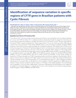
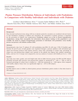
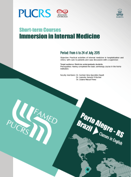
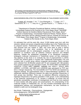
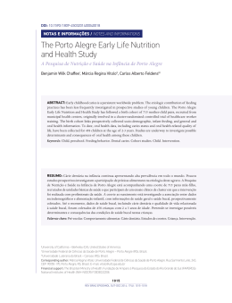
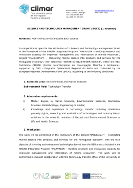
RELATOS DE CASOS
Cicatricial ectropion correction in a patient
with pyoderma gangrenosum: case report
Correção de ectrópio cicatricial em paciente com pioderma gangrenoso:
relato de caso
Fernando Procianoy1
Mariana Tremel Barbato2
Luiz Eduardo Osowski3
Francisco José de Lima Bocaccio4
Lucio Bakos5
ABSTRACT
A 19 year old female with pyoderma gangrenosum was referred to the
oculoplastic clinic for evaluation of severe cicatricial ectropion of the
right lower eyelid. Examination evidenced important scarring of lower
eyelid and malar region with secondary ectropion. A full-thickness skin
graft associated with a lateral tarsal strip procedure and scar tissue
release was performed. The patient’s corticosteroid dose was raised for
the procedure, and after surgery there was no healing problem or ulcer
formation in the eyelid or in the graft donor site. As pyoderma gangrenosum is associated with a pathergy phenomenon in up to 25% of the
cases, the onset of new lesions is a relevant concern when performing
surgery in these patients. In this case, the surgery was safely performed
under corticosteroid immunosuppression.
Keywords: Pyoderma gangrenosum; Ectropion; Eyelid diseases; Skin transplantation;
Immunosuppression/therapeutic use; Case reports [Publication type]
INTRODUCTION
Trabalho realizado no Departamento de Oftalmologia e
Otorrinolaringologia da Faculdade de Medicina da Universidade Federal do Rio Grande do Sul - UFRGS Porto Alegre (RS) - Brasil.
1
2
3
4
5
Setor de Trauma e Plástica Ocular do Serviço de Oftalmologia do Hospital de Clínicas de Porto Alegre da
Universidade Federal do Rio Grande do Sul - UFRGS
- Porto Alegre (RS) - Brasil.
Serviço de Dermatologia do Hospital de Clínicas de
Porto Alegre da UFRGS - Porto Alegre (RS) - Brasil.
Setor de Trauma e Plástica Ocular do Serviço de Oftalmologia do Hospital de Clínicas de Porto Alegre da
UFRGS - Porto Alegre (RS) - Brasil.
Setor de Trauma e Plástica Ocular do Serviço de Oftalmologia do Hospital de Clínicas de Porto Alegre da
UFRGS - Porto Alegre (RS) - Brasil.
Serviço de Dermatologia do Hospital de Clínicas de
Porto Alegre da UFRGS - Porto Alegre (RS) - Brasil.
Address for correspondence: Fernando Procianoy.
Serviço de Oftalmologia - Zona 17 - Hospital de Clínicas de Porto Alegre. Rua Ramiro Barcelos, 2.350 Porto Alegre (RS) CEP 90035-003
E-mail: [email protected]
Recebido para publicação em 11.04.2007
Última versão recebida em 15.11.2008
Aprovação em 08.12.2008
Nota Editorial: Depois de concluída a análise do artigo
sob sigilo editorial e com a anuência da Dra. Midori
Osaki sobre a divulgação de seu nome como revisora,
agradecemos sua participação neste processo.
Pyoderma gangrenosum (PG) is a rare neutrophilic dermatosis frequently associated to other systemic diseases such as ulcerative colitis, Crohn’s
disease, polyarthritis, gammopathy and other conditions. It is characterized
by painful necrotic ulcerations with eritematous and irregular borders. The
initial lesion is usually a nodule or sterile pustule, which rapidly breaks
down to form a progressively enlarging ulcer(1-3).
The extremities are more frequently involved, more commonly the inferior limbs. However, PG can occur in the skin anywhere: head, face, arms,
chest, periocular region and mucocutaneous areas(1). Etiology is still uncertain, but these patients were already described to have alfa-TNF, IL-8(4)
and dermic necrosis factor(5) increase. Pathergy phenomenon is found in
25% of the patients(1).
Diagnosis is based on clinical findings, and histopathology is not
specific. Differential diagnosis with other cutaneous ulcers may be complicated, but the treatment must be readily started since the ulcers cause
important deformities and impair in life quality(5).
The mainstay of PG treatment is immunosuppressive treatment for systemic disease, and therapy focusing on underlying disease when present.
The systemic administration of glucocorticoids is the most effective treatment of PG, however long term therapy produces side effects in up to 50%
of the patients. Sulfa drugs (dapsone, sulfapyridine and sulfasalazine) are
beneficial, but not all patients respond equally well to them. Cyclosporine
Arq Bras Oftalmol. 2009;72(3):384-6
72(3)09.pmd
384
2/7/2009, 14:34
Cicatricial ectropion correction in a patient with pyoderma gangrenosum: case report
385
and tacrolimus are very effective in the treatment of PG, and
other immunosuppressive agents as azathioprine and methotrexate have been beneficial for some patients(6).
Ocular involvement in PG has already been described in
different presentations that may vary from orbital destruction
to sclerokeratitis(7-11). Eyelid necrosis and destruction have
been reported, but not eyelid malpositions due to facial sequelae of ulcers. There is no current data or recommendation in
the literature on the surgical treatment of the eyelid and its
safety under this condition.
CASE REPORT
A 19 year old female with pyoderma gangrenosum was
referred to the oculoplastic clinic for evaluation of severe cicatricial ectropion of the right lower eyelid that developed after
the healing of malar PG ulcers 12 months before. Patient’s
history consisted of chest and face papules that eventually
ulcerated for the last 4 years, being examined by dermatologists, who diagnosed PG based on the clinical aspects of the
skin lesions, after exclusion of other specific ulcerative processes and the prompt clinical response to corticosteroid treatment. Histopathology of the lesions was unspecific (chronic
ulcer) and she had no systemic disease associated. Medical
therapy with prednisone 80 mg/day and dapsone 50 mg/day
was started as she was pregnant at that time. After healing of
the ulcers, prednisone dose was tapered.
Examination evidenced important scarring of the right lower
eyelid region and the midface region, secondary ectropion and
retraction, and extensive lower conjunctival exposure (Figure 1
and Figure 2A). The eyelid skin close to the margin had a normal
aspect. The patient complaints included eye irritation and aesthetic issues.
As the problem was caused essentially by shortage of the
anterior lamella, a lateral tarsal strip procedure and scar tissue
release with full-thickness skin grafting was were performed
under local anesthesia. After an infraciliar incision, canthotomy
and inferior cantholysis were performed. A tarsal strip was
fashioned and the suture was passed through the periosteum of
the lateral orbital rim with 5-0 polyester fiber suture in order to
provide horizontal tightening. The supraclavicular skin graft
was transposed into the lower eyelid defect and sutured to the
adjacent skin with 6-0 nylon sutures. The patient was already in
corticosteroid use for several months, and the disease was
considered quiescent for the last 6 months under treatment. The
prednisone dose was raised for the procedure from 40 mg/day
to 60 mg/day one week before the surgery. This dose was
maintained during the first month post-operative and posteriorly tapered.
There were no signs of pathergy phenomenon, disease activation or new lesions in both eyelids and donor site in an 18month post-operative follow-up. The eyelid remained with minor
retraction and no ectropion recurrence after this follow-up (Figure 2B). The donor site developed a hypertrophic scar (Figure 3).
Figure 1 - Cicatricial ectropion of right lower eyelid
DISCUSSION
Surgical treatment of cicatricial ectropion is well established, with consistent results over time. The need for eyelid
surgery and skin grafting turns into a challenge when the
patient has a disease in which both the operated eyelid and
the graft donor site can turn into a painful necrotic ulcer after
surgery.
PG is associated with a potential pathergy phenomenon in
25% of the patients. This means that skin trauma like intradermal skin testing or injections, pricks, insect bites, biopsies
and operations may induce new lesions. The reactivation of
the disease or the onset of new lesions are important concerns
when performing surgery in these patients(1). This risk must be
in mind when choosing the donor site, as the onset of PG
lesions in an otherwise healthy area may occur. In this case,
supraclavicular area was chosen as the graft donor site because there was previous scarring on both retro auricular spaces,
and a possible lesion induction on the supraclavicular space
would be less significantly disfigurating. Other surgical tech-
Arq Bras Oftalmol. 2009;72(3):384-6
72(3)09.pmd
385
2/7/2009, 14:34
386 Cicatricial ectropion correction in a patient with pyoderma gangrenosum: case report
Although it’s not possible to know in whom and when the
pathergy phenomenon will occur, in the above reported case,
eyelid surgery and skin graft harvesting was safely performed
in a quiescent phase under corticosteroid.
ACKNOWLEDGEMENTS
To Tiago Lansini, MD for the pleasant support.
RESUMO
A
B
Figure 2 - Detail: A) Preoperative; B) 6-month post-operative
Paciente do sexo feminino de 19 anos com pioderma gangrenoso foi encaminhada ao setor de Plástica Ocular para avaliação
de ectrópio cicatricial grave da pálpebra inferior direita. O exame
evidenciou cicatrização importante na pálpebra inferior e região
malar com um ectrópio secundário. Foi realizado liberação do
tecido cicatricial e um enxerto de pele total associado a “tarsal
strip”. A dose de corticóide da paciente foi aumentada para a
realização do procedimento e não houve problemas de cicatrização ou formação de úlceras na pálpebra ou no sítio doador após
a cirurgia. Como o pioderma gangrenoso é associado ao fenômeno de patergia em até 25% dos casos, o surgimento de novas
lesões é uma preocupação relevante ao indicar cirurgia nestes
pacientes. Neste caso, a cirurgia foi realizada com segurança
sob imunossupressão com corticóide.
Descritores: Pioderma gangrenoso; Ectrópio; Doenças palpebrais; Transplante de pele; Imunossupressão/uso terapêutico;
Relatos de casos [Tipo de publicação]
REFERENCES
Figure 3 - Donor site hypertrophic scar
niques for cicatricial ectropion correction such as flap rotation
or sub orbicularis oculi fat (SOOF) lift were not suitable in this
case because of the extensive scarring around the inferior
eyelid. The risks and benefits of the intervention must be
discussed with the patient before surgery.
1. Su WP, Davis MP, Weenig RH, Powell FC, Perry HO. Pyoderma gangrenosum: clinicopathologic correlation and proposed diagnostic criteria. Int J
Dermatol. 2004;43(11):790-800.
2. Vidal D, Puig L, Gilaberte M, Alomar A. Review of 26 cases of classical
pyoderma gangrenosum: clinical and therapeutic features. J Dermatol Treat.
2004;15(3):46-52. Comment in: J Dermatol Treat. 2004;15(3):135.
3. Von Den Driesch P. Pyoderma gangrenosum: a report of 44 cases with follow-up.
Br J Dermatol. 1997;137(6):1000-5.
4. Sapienza MS, Cohen S, Dimarino AJ. Treatment of pyoderma gangrenosum
with infliximab in Crohn’s disease. Dig Dis Sci. 2004;49(9):1454-7.
5. Dantzig PI. Pyoderma Gangrenosum. N Eng J Med. 1975;292(1):47-8
6. Wolff K, Goldsmith LA, Katz SI, Gilchrest BA, Paller A, Leffell DJ.
Fitzpatrick’s Dermatology in General Medicine.7th ed. New York; McGrawHill; 2007.
7. Rose GE, Barnes EA, Uddin JM. Pyoderma gangrenosum of the ocular adnexa:
a rare condition with characteristic clinical appearances. Ophthalmology. 2003;
110(4):801-5.
8. Browning DJ, Proia AD, Sanfilippo FP. Pyoderma gangrenosum involving the
eyelid. Arch Ophthalmol. 1985;103(4):551-2.
9. Newman WD, Frank HJ. Pyoderma gangrenosum of the orbit. Eye. 1993;
7(Pt 1):89-94.
10. Teasley LA, Foster CS, Baltatzis S. Sclerokeratitis and facial skin lesions: a
case report of pyoderma gangrenosum and its response to dapsone therapy.
Cornea. 2007;26(2):215-9.
11. Miserocchi E, Modorati G, Foster CS, Brancato R. Ocular and extracutaneous involvement in pyoderma gangrenosum. Ophthalmology. 2002;109(10):
1941-3.
Arq Bras Oftalmol. 2009;72(3):384-6
72(3)09.pmd
386
2/7/2009, 14:34
Baixar