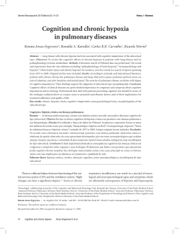
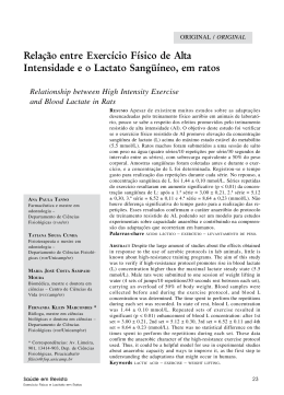
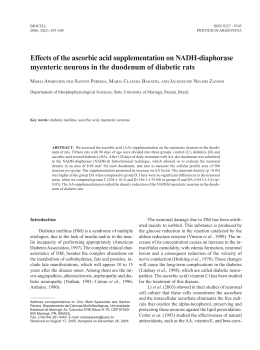
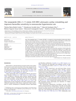
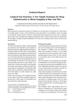
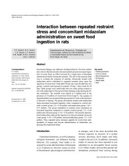
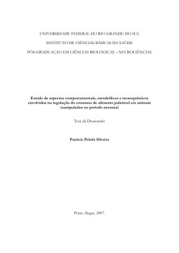
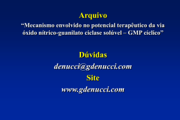
Brazilian Journal of Medical and Biological Research (1999) 32: 1389-1398
NO and hypoxia-induced hypothermia and hyperventilation
ISSN 0100-879X
1389
Role of nitric oxide in hypoxia-induced
hyperventilation and hypothermia:
participation of the locus coeruleus
G. Fabris1,
J.A. Anselmo-Franci2
and L.G.S. Branco2
1Departamento
de Fisiologia, Faculdade de Medicina de Ribeirão Preto and
de Morfologia, Estomatologia e Fisiologia,
Faculdade de Odontologia de Ribeirão Preto, Universidade de São Paulo,
Ribeirão Preto, SP, Brasil
2Departamento
Abstract
Correspondence
L.G.S. Branco
Departamento de Fisiologia
Faculdade de Odontologia de
Ribeirão Preto, USP
14040-904 Ribeirão Preto, SP
Brasil
Fax: +55-16-633-0999
E-mail: [email protected]
Presented at the Meeting
“NO Brazil, Basic and Clinical
Aspects of Nitric Oxide”,
Foz do Iguaçu, PR, Brazil,
March 10-13, 1999.
Research supported by FAPESP and
CNPq. G. Fabris was the recipient
of a postgraduate fellowship from
FAPESP.
Received June 17, 1999
Accepted September 9, 1999
Hypoxia elicits hyperventilation and hypothermia, but the mechanisms involved are not well understood. The nitric oxide (NO) pathway
is involved in hypoxia-induced hypothermia and hyperventilation,
and works as a neuromodulator in the central nervous system, including the locus coeruleus (LC), which is a noradrenergic nucleus in the
pons. The LC plays a role in a number of stress-induced responses, but
its participation in the control of breathing and thermoregulation is
unclear. Thus, in the present study, we tested the hypothesis that LC
plays a role in the hypoxia-induced hypothermia and hyperventilation,
and that NO is involved in these responses. Electrolytic lesions were
performed bilaterally within the LC in awake unrestrained adult male
Wistar rats weighing 250-350 g. Body temperature and pulmonary
ventilation (VE) were measured. The rats were divided into 3 groups:
control (N = 16), sham operated (N = 7) and LC lesioned (N = 19), and
each group received a saline or an NG-nitro-L-arginine methyl ester (LNAME, 250 µg/µl) intracerebroventricular (icv) injection. No significant difference was observed between control and sham-operated rats.
Hypoxia (7% inspired O2) caused hyperventilation and hypothermia in
both control (from 541.62 ± 35.02 to 1816.18 ± 170.7 and 36.3 ± 0.12
to 34.4 ± 0.09, respectively) and LC-lesioned rats (LCLR) (from
694.65 ± 63.17 to 2670.29 ± 471.33 and 36 ± 0.12 to 35.3 ± 0.12,
respectively), but the increase in VE was higher (P<0.05) and hypothermia was reduced (P<0.05) in LCLR. L-NAME caused no significant change in VE or in body temperature under normoxia, but abolished both the hypoxia-induced hyperventilation and hypothermia.
Hypoxia-induced hyperventilation was reduced in LCLR treated with
L-NAME. L-NAME also abolished the hypoxia-induced hypothermia
in LCLR. The present data indicate that hypoxia-induced hyperventilation and hypothermia may be related to the LC, and that NO is
involved in these responses.
Key words
·
·
·
·
·
·
Nitric oxide
Locus coeruleus
Hypoxia
Ventilation
Body temperature
Rat
Braz J Med Biol Res 32(11) 1999
1390
G. Fabris et al.
Introduction
Hypoxic activation of arterial chemoreceptors increases the excitatory synaptic drive
of respiratory neurons in vivo (1). An increase in central respiratory activity also
occurs in in vitro preparations in which
chemoreceptor afferents are deleted (2,3).
Hence, activation of medullary chemosensory neurons (4) or direct stimulation of
respiratory neurons (2) contributes to the
increased respiratory response to hypoxia.
Moreover, oxygen consumption and body
temperature decrease during acute exposure
to hypoxia in a variety of animal species (5).
Although the precise nature for this hypoxic
hypothermia is not fully understood, it is
generally admitted that it results from a reduction in the thermoregulatory set point,
which has been referred to as anapyrexia (6),
rather than from a reduction in body temperature due to an impairment of the thermoregulatory effector mechanisms caused by a limitation in oxygen availability (7). The importance of this response is emphasized by reports that show an increase in survival of the
tested species if they are allowed to become
hypothermic during hypoxia exposure (8).
Sympathetic activation is one of the major components of the adaptive responses to
oxygen reduction, especially in the regulation of cardiovascular events that accompany hyperventilation (9-11). Sympathetic
neurons receive both excitatory and inhibitory influences from noradrenergic cell
groups located in the brainstem (12). The
locus coeruleus (LC), a pair of nuclei located
in the pons, is an assembly of densely packed
noradrenergic neurons; its extensive projections provide noradrenergic innervation to
many brain areas and to the spinal cord (13).
Hypoxia causes a significant increase in cfos in the LC (14). Although little is known
about the central pathways and neuromodulators involved in the neuro-physiological
adjustments occurring during hypoxia, some
candidates have been proposed, including
Braz J Med Biol Res 32(11) 1999
nitric oxide (15).
The diffusible lipophilic gas nitric oxide
(NO) has been recognized as a physiological
molecule because of its role in body temperature regulation (16,17). A previous study
indicated that the hypoxia-induced hypothermia depends on the NO pathway (17).
Also, NO may serve as a neurotransmitter in
the central nervous system (CNS) and inhibit inhibitory synaptic transmission that is
triggered by CNS hypoxia (15).
The purpose of the present study was to
test the hypothesis that the LC participates in
the control of body temperature and pulmonary ventilation (VE) under normoxic and
hypoxic conditions, and that NO is involved
in this process in the CNS.
Material and Methods
Animals
Experiments were performed on adult
male Wistar rats weighing 250-350 g, housed
at controlled temperature (25 ± 2oC) and
exposed to a daily 12:12-h light-dark cycle.
The animals were allowed free access to
water and food. Experiments were performed
between 10:00 a.m. and 3:00 p.m.
Animals were divided into three groups:
control (N = 16), sham-operated (N = 7) and
LC-lesioned rats (LCLR) (N = 19). The experimental group comprised rats submitted
to bilateral lesion within the LC. Each group
was divided into 2 other subgroups: 1) animals which received an intracerebroventricular (icv) injection of saline (1 µl) and 2)
animals which received an icv injection of
NG-nitro-L-arginine methyl ester (L-NAME,
250 µg/µl; Sigma Chemical Co., St. Louis,
MO, USA).
Surgery
Rats used for LC lesion were submitted
to general anesthesia with 2,2,2-tribromoethanol (Aldrich, Milwaukee, WI, USA) and
1391
NO and hypoxia-induced hypothermia and hyperventilation
mounted in a Kopf stereotaxic apparatus.
Bilateral electrolytic lesions (2 mA, 10 s,
positive electrode) were made using a constant current source and coated stainless steel
electrodes in the LC (coordinates: A -3.4
mm from lambda, L ±1.2 mm, D 6.8 mm)
(18). Sham-operated rats were similarly prepared, but the electrode was placed 2 mm
above the dorsal coordinate and no current
was passed through the electrodes. This distinction was made to test the possibility of
alteration in body temperature and VE induced by the surgical intervention. Experiments were initiated 24 h or seven days after
lesion.
At the end of the experiment, the animal
was killed with ether and the brain was
removed and post-fixed in formol saline
(10%) for 1 week. After fixation, the brainstem was embedded in paraffin. Serial 13µm frozen sections were cut and stained by
the Nissl method for histological verification of the location and extension of the
lesions (or their absence in the case of shamoperated rats) and schematically presented
in neuroanatomic maps (19). The LC lesions
comprised a necrotic core surrounded by a
zone of gliosis tissue. The sham-operated
rats had an identifiable electrode tract, but
no discernible lesion. Only the animals with
50 to 100% bilateral LC lesion were used for
analysis of the results.
Rats used for icv injection of L-NAME
were also anesthetized with 2,2,2-tribromoethanol and fixed in a Kopf stereotaxic apparatus. A stainless steel guide cannula (14
mm) was implanted into the right lateral
ventricle of the brain (coordinates: A -1.0
mm from bregma, L ±1.6 mm, D -3.2 to -3.7
mm) (18). The displacement of the meniscus
in a water manometer ensured correct positioning of the cannula in the right lateral
ventricle. The cannula was attached to the
bone with stainless steel screws and acrylic
cement. A tight-fitting stylet was kept inside
the guide cannula to prevent occlusion. The
surgical procedures were performed over a
period of 40 min. Six days after cannula
placement, the animals were submitted to
LC lesion or sham operation.
Experimental protocol
Experiment 1. Determination of the combined effect of lesion and hypoxia on body
temperature and VE. Experiments were performed using animals first exposed to humidified room air and then to a humidified
hypoxic mixture of 7% O2 (AGA, Sertãozinho, SP, Brazil). VE was determined by the
plethysmograph method (20), and body temperature was determined by inserting a thermocouple probe into the colon. Before the
experiment, the animals were habituated to
temperature measurements in order to avoid
stress-induced elevations in body temperature.
Each animal was placed in a 5-liter
plexiglass chamber and allowed to move
about freely while the chamber was flushed
with humidified air. After the animals remained calm (at least 60 min), control body
temperature was measured. Control VE was
measured and the test gas mixture (7% inspired O2) was flushed through the chamber
for 60 min. Ventilation was also measured
after 5, 15, 45 and 60 min of exposure to
hypoxia, and body temperature was measured at the end of the hypoxic period. The
chamber was then flushed with humidified
air for 30 min and during this period VE was
measured at 15-min intervals. At the end of
the experiment, body temperature was measured again.
During the ventilation measurements, the
flow was interrupted and the chamber sealed
for short periods of time, and the oscillations
in air temperature caused by breathing were
measured as pressure oscillations. Calibration for volume was obtained during each
measurement by injecting the chamber with
a known amount of air (0.2 ml) using a
graduated syringe. Signals from a differential air transducer displayed on a paper reBraz J Med Biol Res 32(11) 1999
1392
G. Fabris et al.
corder (HP) allowed the calculation of respiratory frequency (fR) and tidal volume (VT)
by appropriate correction factors (21).
Experiment 2. Determination of the combined effects of NOS blocker and hypoxia on
body temperature and VE. Rats were treated
with an icv injection of L-NAME or saline.
A 705-LT, 50-µl Hamilton syringe and a
Mizzy dental injection needle (200 µm OD)
were used for icv injections. Sterile saline
was the vehicle in which L-NAME was dissolved, in a final volume of 1 µl. The injection was performed over a period of 2 min,
and 1 min was allowed to elapse before the
injection needle was removed from the guide
cannula, to avoid reflux. The same protocol
described above was then used.
Experiment 3. Determination of the combined effects of LC lesion, NOS blocker and
hypoxia on body temperature and VE. Six days
after cannula placement, animals were submitted to LC lesion or sham operation and the
same protocol as described above, with an icv
injection of L-NAME or saline, was used.
A
LC
LC
LC
Figure 1 - Representative
drawings showing the localization of the locus coeruleus (LC) (A) and lesions
(dark areas, B) placed bilaterally in the nucleus.
Sequential sections of
seven different animals.
Braz J Med Biol Res 32(11) 1999
LC
LC
Statistical analysis
Data are reported as means ± SEM.
Changes in body temperature, VT, fR and VE
were evaluated by ordinary analysis of variance (ANOVA) or ANOVA for repeated
measures to analyze the temporal effect of
hypoxia on body temperature and VE. The
differences between means were assessed
by the Tukey-Kramer multiple comparisons
test. Values of P<0.05 were considered to be
significant.
Results
In all experimental protocols, the mean
chamber temperature was 25.41 ± 0.52oC
(SEM), and the room temperature was 24.55
± 0.47oC. Figure 1 shows the lesion sites of
individual rats (N = 7).
During normoxia and the recovery period after hypoxia, body temperature and VE
did not differ between groups. Also, body
temperature and VE during the return to air
B
1393
NO and hypoxia-induced hypothermia and hyperventilation
37
30
LC lesion
Control
+
*
+
20
Body temperature (oC)
VT (ml BTPS/kg)
Hypoxia (7% O2)
+
+
*
+
+
10
+
+
+
+
36
*
35
*
34
0
200
fR (min)
Hypoxia (7% O2)
150
+
100
+
+
+
+
+
+
+
+
+
50
0
4000
VE (ml BTPS min-1 kg-1)
Hypoxia (7% O2)
3000
*
+
2000
+
+
+
*
+
+
+
1000
+
+
+
0
10
+
Figure 3 - Effects of hypoxia on
body temperature of the control
group (N = 16) and locus coeruleus (LC)-lesioned rats (N = 7).
Values are reported as means ±
SEM. *Significant reduction
(P<0.05) of mean body temperature after hypoxia. +Significant
reduction (P<0.05) in the magnitude of hypoxia-induced hypothermia.
30
50
70
90
Time (min)
Figure 2 - Effects of hypoxia on tidal volume (VT), respiratory frequency (fR) and pulmonary ventilation (VE) of
control rats (filled squares) (N = 16) and LC-lesioned
rats (open squares) (N = 7). Values are reported as
means ± SEM. The hypoxia-induced hyperventilation
was higher in LC-lesioned rats. +Significant increase
(P<0.05) in VT, fR and VE after hypoxia compared to
normoxia. *Significant increase in VT and VE of LClesioned rats compared to control group. BTPS, Body
temperature, pressure, saturated with water vapor.
Air
Hypoxia (7% O2)
after hypoxia did not differ from the baseline
value in any group. The sham-operated group
did not differ significantly from control animals in any variable (data not shown). Data
obtained from LCLR 1 week after lesion did
not differ from control or sham-operated rats
(data not shown).
Experiment 1. Combined effect of lesion
and hypoxia on body temperature and VE.
Figure 2 shows the effect of hypoxia on VT,
fR and VE, in control and LCLR. When inspired O2 was reduced from 21 to 7%, a
significant (P<0.05) increase in ventilation
was observed in both groups. The increase in
VE was significantly higher (P<0.05) in LCLR
than in control rats at 5 and 15 min of exposure to hypoxia. The hypoxia-induced hyperventilation of LCLR was primarily the
result of a significant elevation in VT, rather
than a change in fR, which did not differ from
control. The breathing pattern was recovered during return to air, when the experimental and control groups showed similar
recordings.
Figure 3 shows the effect of hypoxia on
body temperature of control and LCLR. After hypoxia, a significant decrease in body
temperature was observed in both control
(P<0.05) and LCLR (P<0.05), but the hypoxia-induced hypothermia was reduced in
LCLR. After hypoxia, the drop in body temperature was significantly lower (P<0.05) in
LCLR compared to control.
Experiment 2. Combined effects of NOS
Braz J Med Biol Res 32(11) 1999
1394
G. Fabris et al.
blocker and hypoxia on body temperature
and VE. Figure 4 shows that the combination
of saline injection and hypoxia caused a
significant increase in VE, similar to that
obtained by application of hypoxia only.
When L-NAME was injected 30 min before
hypoxia, hypoxia-induced hyperventilation
was abolished, a fact primarily due to a
30
Hypoxia (7% O2)
VT (ml BTPS/kg)
Figure 4 - Combined effect of
hypoxia and intracerebroventricular injection of NG-nitro-L-arginine methyl ester (L-NAME;
250 µg/µl) on tidal volume (VT),
respiratory frequency (f R) and
pulmonary ventilation (VE) in LNAME group (open squares) (N
= 7) and saline group (filled
squares) (N = 7). Values are reported as means ± SEM. +Significant increase (P<0.05) in VT,
fR and VE of the saline group after hypoxia compared to normoxia. *Significant difference
(P<0.05), after hypoxia, between
L-NAME-treated rats and salinetreated rats. BTPS, Body temperature, pressure, saturated
with water vapor.
20
+
+
*
*
+
+
+
*
*
10
*
0
200
Hypoxia (7% O2)
150
+
+
+
+
+
significant reduction in VT after hypoxia,
rather than a change in fR.
Hypoxia failed to induce a reduction of
body temperature when L-NAME was given
intracerebroventricularly. These data are plotted in Figure 5.
Experiment 3. Combined effects of LC
lesion, NOS blocker and hypoxia on body
temperature and VE. Figure 6 shows a significant increase (P<0.05) in VE in LCLR +
saline and LCLR + L-NAME after hypoxia,
compared to normoxia, which was significantly higher (P<0.05) in LCLR + saline
than in LCLR + L-NAME, at 5 and 15 min of
exposure to hypoxia. The reduced hypoxiainduced hyperventilation of the LCLR + LNAME group was primarily due to a significant reduction in VT after hypoxia, rather
than a change in fR.
Figure 7 shows that the combination of
LC lesion, saline and hypoxia caused a significant (P<0.05) drop in body temperature,
similar to that obtained by LC lesion and
hypoxia only. When L-NAME was injected
+
100
+
+
+
Control + Saline
50
0
VE (ml BTPS min-1 kg-1)
4000
Hypoxia (7% O2)
3000
+
2000
1000
+
36
35
*
34
Before
injection
1 h after
injection
Hypoxia
(7% O2)
Air
+
+
+
+
*
*
*
*
*
0
10
30
50
Time (min)
Braz J Med Biol Res 32(11) 1999
Control + L-NAME
37
Body temperature (oC)
fR (min)
+
70
90
Figure 5 - Combined effect of hypoxia and intracerebroventricular (icv) injection of NG-nitro-L-arginine methyl
ester (L-NAME; 250 µg/µl) on body temperature of
saline- and L-NAME-treated rats (N = 7 for both groups).
Values are reported as means ± SEM. *Significant
drop (P<0.05) in body temperature after hypoxia of
saline group compared to normoxia. +Significant difference (P<0.05) in the magnitude of hypoxia-induced
hypothermia.
1395
NO and hypoxia-induced hypothermia and hyperventilation
30
37
Body temperature (oC)
VT (ml BTPS/kg)
Hypoxia (7% O2)
20
+
+
+
10
*
+
+
*
+
+
+
*
+
*
+
*
0
*
35
Before
injection
Hypoxia (7% O2)
+
+
fR (min)
36
34
200
150
Figure 7 - Combined effect of
hypoxia and intracerebroventricular injection of NG-nitro-Larginine methyl ester (L-NAME;
250 µg/µl) on body temperature
of locus coeruleus (LC)-lesioned
rats that received an icv injection of saline (N = 5) or L-NAME
(N = 7). Values are reported as
means ± SEM. *Significant drop
(P<0.05) in body temperature
after hypoxia.
LC lesion + Saline
LC lesion + L-NAME
1 h after
injection
Hypoxia
(7% O2)
Air
+
+
+
+
+
+
+
+
Hypoxia
100
50
L-NAME
icv
0
NOCNS
LC
4000
VE (ml BTPS min-1 kg-1)
VE
3000
2000
+
+
+
+
+
+
1000
Tb
Hypoxia (7% O2)
*
+
+
+
30 min before hypoxia in LCLR, hypoxiainduced hypothermia was abolished, as observed by application of L-NAME in control
rats. However, there was no significant difference in body temperature between LCLR
+ saline and LCLR + L-NAME after hypoxia.
+
*
0
10
30
50
Figure 8 - Possible mechanisms
through which the locus coeruleus (LC) and nitric oxide (NO)
could mediate hypoxia-induced
hyperventilation and hypothermia. Hypoxia leads to an increase in NO in the CNS and in
the firing rate of LC neurons,
which cause an increase in pulmonary ventilation (VE) and a reduction in body temperature
(Tb). NG-nitro-L-arginine methyl
ester (L-NAME) prevents hypoxia-induced hyperventilation
and hypothermia by inhibiting
NO synthesis.
70
90
Time (min)
Figure 6 - Combined effect of hypoxia and intracerebroventricular (icv) injection of NG-nitro-L-arginine methyl
ester (L-NAME; 250 µg/µl) on tidal volume (VT), respiratory frequency (fR) and pulmonary ventilation (VE) of LClesioned rats that received an icv injection of saline
(open squares) (N = 5) or L-NAME (filled squares) (N = 7).
Values are reported as means ± SEM. +Significant increase (P<0.05) in VT, fR and VE after hypoxia compared
to normoxia. *Significant difference (P<0.05) in VT and
VE of L-NAME-treated rats compared to saline. BTPS,
Body temperature, pressure, saturated with water vapor.
Discussion
This study provides evidence that the LC
participates in the control of pulmonary ventilation and body temperature during hypoxia challenge, since bilateral electrolytic
lesions of the nucleus caused an increased
ventilatory response to hypoxia and a reduced hypoxia-induced hypothermia. A possible link between the two responses cannot
Braz J Med Biol Res 32(11) 1999
1396
G. Fabris et al.
be excluded since a drop in body temperature
leads to a reduced ventilatory drive. These
data suggest that LC plays an inhibitory role
in the hypoxia-induced hyperventilation and
an excitatory role in the hypoxia-induced
anapyrexia. Moreover, NO seems to play an
important role in these responses to oxygen
deprivation, since icv injection of L-NAME
abolished both hypoxia-induced hyperventilation and hypothermia. Additionally, the
inhibitory role of LC in pulmonary ventilation may depend on the NO pathway, since
LCLR treated with L-NAME had a reduced
ventilatory response to hypoxia.
In a wide variety of animal species, hypoxia elicits a number of compensatory responses, including increased ventilation and
cardiac output (5). Decreases in PaO2 are
monitored by peripheral arterial chemoreceptors that evoke excitation of chemosensory fibers projecting in the brainstem within
the nucleus tractus solitarius (NTS) (12). In
the brainstem, these afferent inputs are processed and integrated together with other
inputs to yield a final command to the respiratory motoneurons resulting in an increase
in respiratory drive (22). However, the increased ventilation is O2 consuming, a fact
that limits its beneficial effect. Hypoxia also
elicits a decrease in body temperature and
oxygen consumption (7), which is considered to be an adaptive response because it
decreases O2 demand according to the Q10
effect, promotes a leftward shift of the oxyhemoglobin dissociation curve, and
blunts the energetically costly response to
hypoxia, e.g., increased cardiac output and
ventilation (5). Previous studies have demonstrated that hypoxia per se causes hypothermia in a variety of organisms ranging
from protozoans to mammals (23), but only
recently did the mechanisms responsible for
the hypoxia-induced anapyrexia start to be
suggested.
The role of CNS in body temperature
control is subjected to numerous modifiers,
such as lactate, adenosine and histamine (for
Braz J Med Biol Res 32(11) 1999
review, see 23), but none of the possible
candidates can trigger a complete hypothermic response. Nitric oxide works as a physiological messenger molecule that may serve
as a neurotransmitter in the CNS (24). NO in
the CNS may have an important role in the
hypoxia-induced hypothermia since during
inhibition of the NO pathway hypoxia failed
to reduce body temperature (17). Besides
mediating hypoxia-induced hypothermia,
oxygen deprivation was recently shown to
lead to activation of the NO-cGMP pathway
in the CNS, contributing to the induction and
maintenance of the hypoxia-induced increased ventilation (15). The importance of
NO can be demonstrated by inhibition of the
effects of NO (25) using L-arginine analogs
such as L-NAME. In the present study we
have chosen L-NAME because it is a nonselective inhibitor of NOS and acts on both the
constitutive and inducible isoforms of the
enzymes.
The present study confirms that hypoxiainduced hypothermia depends on the NO
pathway since L-NAME injection abolished
hypoxia-induced hypothermia in control and
sham-operated rats. Also, NO may be involved in hypoxia-induced hyperventilation
since control animals which received an injection of L-NAME had no ventilatory response to hypoxia (Figure 8). Moreover,
LCLR which received an injection of LNAME showed a significantly lower hypoxia-induced hyperventilation compared to
LCLR treated with saline, indicating that
increased hypothermia-induced hyperventilation of LCLR may depend on the NO
pathway in the CNS. In the present study, LC
lesion caused no significant change in pulmonary ventilation or in body temperature
under normoxic conditions, indicating that
the LC plays no tonic role under control
conditions. Conversely, hypoxia-induced
hyperventilation was greatly increased in the
LCLR, which may be related to the observed
reduction in the hypoxia-induced hypothermia. Accordingly, previous studies have re-
1397
NO and hypoxia-induced hypothermia and hyperventilation
ported that the LC is important in the modulation of sensory processing by the brain and
is activated by a variety of stressful somatic
and autonomic stimuli (26,27), but not during resting conditions (14). Activity in LC
neurons is highest during wakefulness, particularly under conditions requiring increased
alertness (28). The major component of adaptive responses to hypoxia is the stimulation of the sympathoadrenal system (9,11),
and sympathetic neurons are under the control of noradrenergic cell groups located in
the brainstem (12), where the LC is the
major noradrenergic nucleus. Recently, it
has been reported that noradrenergic LC neurons send axons to spinal motoneurons, where
they may participate in the control of respiratory movements (29). Changes in the activity
of LC neurons would then be expected to
elicit widespread effects in the CNS, including those of a respiratory nature. A study
demonstrating the distribution of neuronal
pathways activated by hypoxia reported that
the LC presents an increased c-fos staining
during hypoxia (30). Also, Pérez et al. (31)
observed LC-mediated inhibition of
chemosensory responses in the rat NTS,
which is the zone of termination of afferents
from baroreceptors and chemoreceptors travelling in the carotid sinus (32) and aortic
sinus (33). Furthermore, Moore et al. (34)
observed that LC cooling blocks the fall in
respiratory output during hypoxia in anesthetized neonatal sheep. They concluded that
the fall in the biphasic respiratory response
is mediated by the activation of neurons
inhibitory to respiratory output and involves
either axons of passage or cell bodies lying
in the LC region. In agreement with these
data, the present study suggests that if there
is a CNS inhibitory mechanism of hypoxiainduced hyperventilation, it seems to depend
on neuronal function within the LC.
As to the ventilatory response, LC seems
to be involved in modulation of tidal volume, since the hyperventilation response
during hypoxia of LCLR differed from the
control group due to a significant increase in
tidal volume and not in respiratory frequency.
In summary, the present results suggest
that LC and NO pathway may participate in
the control of body temperature and pulmonary ventilation under hypoxic conditions,
and that the inhibitory role of the LC on
hypoxia-induced hyperventilation and its excitatory role in hypoxia-induced hypothermia may depend on the NO pathway.
Acknowledgments
We thank Mauro Ferreira Silva, Nadir
Martins Fernandes and Rute Aparecida de
Freitas Marcon for excellent technical assistance.
References
1. Lawson EE, Richter DW, Ballantyne D &
Lalley PM (1989). Peripheral chemoreceptor inputs to medullary inspiratory and
postinspiratory neurons of cat. Pflügers
Archive, 414: 523-533.
2. Völker A, Ballanyi K & Richter DW (1995).
Anoxic disturbance of the isolated respiratory network of neonatal rats. Experimental Brain Research, 103: 9-19.
3. Ramirez JM, Quellmalz UJA, Wilken B &
Richter DW (1998). The hypoxic responses of neurons within the in vitro
mammalian respiratory network. Journal
of Physiology, 507: 571-582.
4. Kawai A, Ballantyne D, Mükenhoff K &
Scheid P (1996). Chemosensitive medullary neurons in the brainstem-spinal cord
preparation of the neonatal rat. Journal of
Physiology, 492: 277-292.
5. Wood SC (1995). Oxygen as a modulator
of body temperature. Brazilian Journal of
Medical and Biological Research, 28:
1249-1256.
6. Branco LGS & Malvin GM (1996). Thermoregulatory effects of cyanide and azide
in the toad, Bufo marinus. American Journal of Physiology, 270: R169-R173.
7. Gautier H & Murariu C (1998). Neuro-
modulators and hypoxic hypothermia in
the rat. Respiration Physiology, 112: 315324.
8. Hicks JW & Wood SC (1985). Temperature regulation in lizards: effects of hypoxia. American Journal of Physiology,
248: R595-R600.
9. Dalmaz Y, Pequignot JM, Cotted-Emard
JM, Tavitian E & Peyrin L (1989). Changes
of dopamine, norepinephrine and epinephrine turnover in sympathetic tissues
after long term hypoxia stress. In: Van
Loon GR, Kvetnansky R, McCarty RM &
Axelrod J (Editors), Stress: Neurochemi-
Braz J Med Biol Res 32(11) 1999
1398
10.
11.
12.
13.
14.
15.
16.
17.
18.
G. Fabris et al.
cal and Humoral Mechanisms. Gordon
and Breach, New York, 777-785.
Marshall JM (1987). Analysis of cardiovascular responses evoked following
changes in peripheral chemoreceptor activity in the rat. Journal of Physiology, 394:
385-403.
Richalet JP (1990). The heart and adrenergic system in hypoxia. In: Sutton JR,
Coatesand G & Remmers JE (Editors),
Hypoxia: the Adaptations. Dekker,
Toronto, 231-240.
Soulier V, Cottet-Emard JM, Pequignot J,
Hanshin F, Peyrin L & Pequignot JM
(1992). Differential effects of long-term
hypoxia on norepinephrine turnover in
brain stem cell groups. Journal of Applied
Physiology, 73: 1810-1814.
Yang JJ, Chou YC, Lin MT & Chiu TH
(1997). Hypoxia-induced differential electrophysiological changes in rat locus coeruleus neurons. Life Sciences, 61: 17631773.
Breen S, Rees S & Walker D (1996). Identification of brainstem neurons responding to hypoxia in fetal and newborn sheep.
Brain Research, 748: 107-121.
Haxhiu MA, Chang CH, Dreshaj IA,
Erokwu BNR & Prabhakar Cherniack NS
(1995). Nitric oxide and ventilatory response to hypoxia. Respiration Physiology, 101: 257-266.
Scammell TE, Elmkist JK & Saper CB
(1996). Inhibition of nitric oxide synthase
produces hypothermia and depresses lipopolysaccharide fever. American Journal
of Physiology, 271: R333-R338.
Branco LGS, Cárnio EC & Barros RCH
(1997). Role of nitric oxide pathway in
hypoxia-induced hypothermia of rats.
American Journal of Physiology, 273:
R967-R971.
Paxinos G & Watson C (1986). The Rat
Braz J Med Biol Res 32(11) 1999
19.
20.
21.
22.
23.
24.
25.
26.
27.
Brain in Stereotaxic Coordinates. 2nd edn.
Academic Press, San Diego.
Palkovits M & Jacobowitz DM (1974). Topographic atlas of catecholamine and acetylcholinesterase-containing neurons in
the rat brain. II. Hindbrain (mesencephalon, rhombencephalon). Journal of Comparative Neurology, 157: 29-42.
Bartlett DJ & Tenney SM (1970). Control
of breathing in experimental anemia. Respiration Physiology, 10: 384-395.
Malan A (1973). Ventilation measured by
body plethysmography in hibernating
mammals and in poikilotherms. Respiration Physiology, 17: 32-44.
Teppema LJ, Veening JG, Kranenburg A,
Dahan A, Berkenbosch A & Olievier C
(1997). Expression of c-fos in the rat brainstem after exposure to hypoxia and to
normoxic and hyperoxic hypercapnia.
Journal of Comparative Neurology, 388:
169-190.
Wood SC (1991). Interaction between hypoxia and hypothermia. Annual Review of
Physiology, 53: 71-85.
Moncada S, Palmer RMJ & Higgs EA
(1991). Nitric oxide: physiology, pathophysiology and pharmacology. Pharmacological Reviews, 43: 109-142.
Rees D, Palmer RMJ, Schultz R, Hodson
H & Moncada S (1990). Characterization
of three inhibitors of endothelial nitric oxide synthase in vitro and in vivo. Brazilian
Journal of Pharmacology, 101: 746-752.
Aston-Jones G, Shipley MT, Chouvet G,
Ennis M, Van Bockstaele E, Pieribone V,
Shiekattar R, Akaoka H, Drolet G & Astier
AB (1991). Afferent regulation of locus
coeruleus neurons: Anatomy, physiology
and pharmacology. Progress in Brain Research, 88: 47-75.
Van Bockstaele EJ, Akaoka H & AstonJones G (1995). Integration in the ventral
28.
29.
30.
31.
32.
33.
34.
medulla and coordination of sympathetic,
pain and arousal functions. Clinical and
Experimental Hypertension, 17: 153-165.
Aston-Jones G, Foote SL & Bloom FE
(1984). Anatomy and physiology of locus
coeruleus neurons: Functional implications. In: Ziegler M & Lake CR (Editors),
Norepinephrine (Frontiers of Clinical Neuroscience). Vol. 2. Williams & Wilkins, Baltimore, 92-116.
Fung SJ, Chan JY, Manzoni D, White SR,
Lai YY, Strahlendorf HK, Zhuo H, Liu RH,
Reddy VK & Barnes CD (1994). Cotransmitter mediated locus coeruleus action
on motoneurons. Brain Research Bulletin, 35: 423-432.
Nitsos I & Walker DW (1999). The distribution of FOS-immunoreactive neurons
in the brainstem, midbrain and diencephalon of fetal sheep in response to acute
hypoxia in mid and late gestation. Brain
Research, Developmental Brain Research, 114: 9-26.
Pérez H, Ruiz S, Laurido C & Hernández A
(1998). Locus coeruleus-mediated inhibition of chemosensory responses in the
rat nucleus tractus solitarius is mediated
by alpha2-adrenoreceptors. Neuroscience
Letters, 249: 37-40.
Erickson JT & Milhorn DE (1991). Fos-like
protein is induced in neurons of the medulla oblongata after stimulation of the
carotid sinus nerve in awake and anesthetized rats. Brain Research, 567: 11-24.
Ciriello J (1983). Brainstem projections of
aortic baroreceptor afferent fibers in the
rat. Neuroscience Letters, 36: 37-42.
Moore PJ, Ackland GL & Hanson MA
(1996). Unilateral cooling in the region of
locus coeruleus blocks the fall in respiratory output during hypoxia in anaesthetized neonatal sheep. Experimental Physiology, 81: 983-994.
Baixar