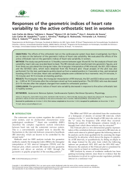
Heart Rate Variability Biofeedback Increases Baroreflex Gain and Peak Expiratory Flow PAUL M. LEHRER, PHD, EVGENY VASCHILLO, PHD, BRONYA VASCHILLO, MD, SHOU-EN LU, PHD, DWAIN L. ECKBERG, MD, ROBERT EDELBERG, PHD, WEICHUNG JOE SHIH, PHD, YONG LIN, PHD, TOM A. KUUSELA, PHD, KARI U. O. TAHVANAINEN, MD, AND ROBERT M. HAMER, PHD Objective: We evaluated heart rate variability biofeedback as a method for increasing vagal baroreflex gain and improving pulmonary function among 54 healthy adults. Methods: We compared 10 sessions of biofeedback training with an uninstructed control. Cognitive and physiological effects were measured in four of the sessions. Results: We found acute increases in low-frequency and total spectrum heart rate variability, and in vagal baroreflex gain, correlated with slow breathing during biofeedback periods. Increased baseline baroreflex gain also occurred across sessions in the biofeedback group, independent of respiratory changes, and peak expiratory flow increased in this group, independently of cardiovascular changes. Biofeedback was accompanied by fewer adverse relaxation side effects than the control condition. Conclusions: Heart rate variability biofeedback had strong long-term influences on resting baroreflex gain and pulmonary function. It should be examined as a method for treating cardiovascular and pulmonary diseases. Also, this study demonstrates neuroplasticity of the baroreflex. Key words: biofeedback, heart rate variability, baroreflex, pulmonary function, neuroplasticity. BP ⫽ blood pressure; HF ⫽ high frequency; HR ⫽ heart rate; HRV ⫽ heart rate variability; LF ⫽ low frequency; RSA ⫽ respiratory sinus arrhythmia. INTRODUCTION iofeedback can enable people to obtain voluntary control over various physiological processes (1), and some biofeedback methods have been used widely as adjuncts to, or substitutes for, medical treatment. Typically, a biofeedback trainee views an instantaneous electronic display of a physiological function and attempts to change it. Most biofeedback methods involve teaching patients to control a level of a physiological function, such as muscle tension, HR, or finger temperature. Recently we have reported using biofeedback to produce increases in heart rate variability (HRV). This method has been used by Russian clinicians to treat autonomic dysfunction with a variety of clinical manifestations, including anxiety and high BP (2), and we recently used it to improve airway function in asthmatic patients (3). These results tentatively suggest that the method can produce long-term changes in multiple organ systems that are affected by autonomic control. This study focuses on arterial baroreflexes and pulmonary function in a healthy population. Arterial baroreflex responses, triggered by stretch receptors in the walls of the aortic arch and carotid artery, modulate vagus nerve traffic to the sinoatrial node, and mediate beatby-beat HR responses to changing arterial pressures (4). Risk B From the Department of Psychiatry Robert Wood Johnson Medical School (P.M.L., R.E., Y.L.), Piscataway, New Jersey; UMDNJ–New Jersey Medical School, Department of Neurosciences, Newark, New Jersey (E.V., B.V.); UMDNJ–School of Public Health, Division of Biometrics (S-E.L., W.J.S.); Medical College of Virginia at Virginia Commonwealth University (D.L.E.), Richmond, Virginia; Department of Physics, University of Turku (T.A.K.), Turku, Finland; Department of Clinical Physiology, Kuopio University Hospital (K.U.O.T.), Kuopio, Finland; and Department of Psychiatry University of North Carolina, School of Medicine, Chapel Hill, North Carolina (R.M.H.). Address reprint requests to: Paul Lehrer, Ph.D., Department of Psychiatry, Robert Wood Johnson Medical School, 671 Hoes Lane, Piscataway, NJ 08854, USA. Email: [email protected] Received for publication June 12, 2002; revision received January 6, 2003. This research was supported by Grant R01HL58805 from the National Heart, Lung, and Blood Institute of the National Institutes of Health. DOI: 10.1097/01.PSY.0000089200.81962.19 796 0033-3174/03/6505-0796 Copyright © 2003 by the American Psychosomatic Society for cardiac events (including sudden death) in patients with heart disease is inversely related to the robustness of their baroreflex responses. La Rovere et al. (5) showed that in patients recovering from myocardial infarction, those with subnormal vagal baroreflex gains have a high risk of fatal cardiac events, especially if the patient also has low HRV. The linkage between vagal baroreflex impairment and mortality may partially reflect patients’ autonomic responses to cardiac rhythm changes. Ventricular tachycardia, a rapid rhythm that commonly precedes sudden death (6), precipitously lowers arterial pressure, and increases muscle-sympathetic (7) and reduces vagal-cardiac (8) nerve activity. During ventricular tachycardia, arterial perfusion pressures recover more rapidly in patients with stronger than weaker vagal and sympathetic baroreflexes (9). In an exercise/ ischemia dog model of sudden cardiac death, ventricular fibrillation occurs when baroreflexes are weak, but does not occur when they are strong (10). Vagal mechanisms also figure importantly in asthma, because the parasympathetic nervous system plays a major role in modulating airway smooth muscle tone (11). Just as increased baroreflex responsiveness may promote successful responses to abrupt rhythm disturbances in cardiac patients, increased vagal activity can cause bronchoconstriction in asthma, and asthma exacerbations can be associated with cholinergic hyperreactivity (12). The therapeutic effects of HRV biofeedback may be through influencing the body’s modulatory processes (eg, the well-known modulation of BP changes by baroreflex activity), through which vagal as well as sympathetic reflexes may be controlled. The levels of baroreflex gain and vagal bronchoconstriction both vary over time, influenced by various neurobehavioral factors. The earliest quantitative analysis of human baroreflex function (13) documented elevated baroreflex gain during sleep. Fritsch et al. (14) reported that changes of arterial pressure lasting only seconds reset the relation between arterial pressure and vagal and sympathetic neural outflows. Systematic changes in pulmonary function also occur during behavioral laboratory tasks (12) and relaxation (15). Psychosomatic Medicine 65:796 – 805 (2003) HEART RATE VARIABILITY BIOFEEDBACK When people try to maximize their respiratory sinus arrhythmia (the vagally mediated HR speeding and slowing that occurs in synchrony with breathing), they spontaneously slow their breathing rates to ~0.1 Hz, about one breath every 10 seconds (16). There are previous reports that slow or deep breathing may acutely increase baroreflex gain in healthy people (17) and can even counteract the bronchoconstrictive effects of inhaled methacholine (18). A controlled trial of slow breathing in the context of yoga documented acute reductions of airway resistance among people with asthma (19). Thus, vagal cardiac and pulmonary mechanisms are linked, and there are reasons to expect that improvements in one vagal limb might spill over into the other. TABLE 1. BIOFEEDBACK AND RESONANT FREQUENCY IN HRV For each individual there is a resonant frequency in HRV at which maximum amplitudes of HRV can be attained by biofeedback. At this frequency, we have found that HR oscillates 180° out of phase with BP and in phase (0° phase relationship) with respiration (20), such that respiratory and baroreflex effects on HRV interact, producing very high amplitudes at a single frequency, accounting for higher total variability. The resonant HRV frequency usually is ~0.1 Hz (6 cycles/min). At this frequency, we previously found that HR and BP oscillate 180° out of phase (20), while HR and Participant characteristics Biofeedback Mean SD N Mean SD N 30.55 144.26 66.38 10.33 27.52 3.66 23 23 23 27.93 144.76 66.56 11.60 29.02 4.14 30 31 31 Sex N % N % Female Male 16 7 69.57 30.43 22 9 70.97 29.03 Age (yr) Weight (lb) Height (in) Fig. 1. Control Recording from one participant before and during biofeedback. In this participant, biofeedback increased systolic pressure and R-R interval oscillations, decreased mean systolic pressure, and increased baroreflex gain. Psychosomatic Medicine 65:796 – 805 (2003) 797 P. M. LEHRER et al. Fig. 2. Heart rate and blood pressure variability during biofeedback from a typical subject. This figure shows the typical 180° phase relationship between heart rate and blood pressure during HRV biofeedback and very high oscillation amplitudes in both measures, all at a single frequency. Fig. 3. Long-term physiological effects across weeks of training. Error bars represent standard deviations. respiration oscillate in phase with each other (0° phase relationship, with inhalation coinciding with HR accelerations and exhalation with decelerations). Thus, when people breathe at their resonant frequency, respiratory effects on HRV stimulate baroreflex effects (ie, as the individual inhales, HR rises, BP falls, and the consequent baroreflex response produces a further increase in HR, with corresponding effects during exhalation). The consequent resonance effects produce very large increases in both HRV and baroreflex gain, which can be 798 obtained only when subjects try to increase HRV at this particular frequency (20). METHOD Participants We recruited participants through media advertisements and personal contacts, and screened out volunteers who smoked, had a history of psychosis, mental deficiency, heart disease or arrhythmias, chronic pulmonary disease (including asthma), serious neurological illness (including epilepsy), or who were taking any medication that affected the autonomic Psychosomatic Medicine 65:796 – 805 (2003) HEART RATE VARIABILITY BIOFEEDBACK Fig. 4. Effects of biofeedback on measures of cardiovascular variability. Error bars represent standard deviations. nervous system. Fifty-four people participated in the study (See Table 1 for participant characteristics), and were assigned to groups using a restricted randomization procedure, balanced for age and sex: 25 to the biofeedback protocol (of whom 2 dropped out before completion), and 32 to the waiting list (of whom 1 dropped out before completion). Participants were paid $100 for each of four testing sessions (see below) and $50 for each of the other training sessions (biofeedback group only). This study was approved by the human research committee of UMDNJ–Robert Wood Johnson Medical School. Physiological data were collected during 4 of the 10 treatment sessions in the biofeedback condition, and in 4 equivalently spaced sessions in the control group. Data were collected during four 5-minute periods: 1) a pretraining rest period (“Task A”) in which subjects were asked to relax as deeply as possible with eyes open, and to try not to move, so as not to disturb the measuring equipment; 2) the first 5 minutes of biofeedback training (“Task B”); 3) the last 5 minutes of an approximately 30-minute biofeedback training period (“Task C”); and 4) a posttraining rest period (“Task D”), with the same Psychosomatic Medicine 65:796 – 805 (2003) instructions as for the pretest rest period. For control subjects, instructions for Tasks B and C were identical to those in Tasks A and D. Instrumentation and Software Physiological data were recorded on a J&J Engineering (Poulsbo, WA) I-330 DSP physiograph unit. EKG data were collected from sensors on the right arm and left leg (Lead II), digitized at the rate of 512 samples/s. Beat-to-beat BP was recorded from a Finapres unit (Ohmeda model 223), and digitized at a rate of 256 samples/s. The sensor was placed on the participant’s left middle finger, and the hand was elevated on a table to approximately the level of the heart. End-tidal CO2 was taken from a Datex 223 capnometer. The intake tube was inserted into a mouthpiece, and subjects wore nose clips and breathed through the mouth during the 5-minute testing periods. A pneumotachometer was used to record respiratory patterns from which measurements of respiratory rate and tidal volume were derived. During biofeedback sessions, strain gauges around the chest and abdomen were also used to display 799 P. M. LEHRER et al. Fig. 5. Effects of biofeedback on measures of tonic physiological activity. Error bars represent standard deviations. respiratory activity for training purposes. Spirometry was done before and after each testing session following procedures recommended by the American Thoracic Society (21) using a Koko pneumotach-based spirometer (PDS Instrumentation, Louisville, CO), calibrated daily with a 3-liter syringe. HR and BP data were edited, and analyzed using the WinCPRS program (Absolute Aliens Oy, Turku, Finland), a program for general analysis of physiological data, including analysis of HR and BP variability and baroreflex gain. Spectral baroreflex gain in the LF range correlates closely with baroreflex gain assessed directly by using phenylephrine injection to alter BP and trigger baroreflex responses BP (22). In cats, baroreceptor denervation abolishes the coherence between systolic pressure and R-R interval oscillations in this frequency range (23). We estimated baroreflex gain over coherent LF (0.04 – 0.15 Hz) segments from the squared coherence between pairs of measurements. In this procedure, the squared cross-spectral densities of systolic pressure and R-R intervals are divided by the product of the individual power densities. The transfer function was calculated as the cross-spectral densities divided by the power spectral densities of the systolic pressure. The modulus of the transfer function was used to estimate baroreflex gain (10, 24). Participants also completed two self-report inventories about their experiences during the testing sessions: 1) the Relaxation Inventory (25), a factor-analytically derived scale that yields a full scale score and three dimensions of the relaxation experience, experience of physical tension, 800 cognitive tension, and subjective assessment of relaxation; and 2) the Side Effects of Relaxation Scale (26), which assesses common adverse experiences of people undergoing various kinds of relaxation training. Procedure for HRV Biofeedback The details of the procedure for HRV biofeedback have been described elsewhere (27). The trainee was first taught to breathe at his/her resonant frequency, ie, the frequency at which maximum amplitudes of HRV could be generated voluntarily for each individual. The resonant frequency was determined in the first session by measuring HR oscillation amplitudes while the individual breathed for intervals of 2 minutes at each of the following frequencies: 4.5, 5, 5.5, 6, and 6.5 breaths/min. We provide a “pacing stimulus” for this purpose: a light display that moved up and down on the computer screen at the target respiratory rate. The trainee was instructed to breathe at the rate of that stimulus. The resonant frequency was determined as the respiratory frequency yielding the highest frequency power peak on a moving Fourier analysis of HR data displayed by the I-330 physiograph. Subjects were instructed to practice breathing at the resonant frequency for 20-minute periods twice daily for the next week. Throughout training, the individual was cautioned to breathe shallowly and naturally, in order to avoid Psychosomatic Medicine 65:796 – 805 (2003) HEART RATE VARIABILITY BIOFEEDBACK TABLE 2. Session 1 4 7 10 Task Pre-Rest Begin Bfk. End Bfk. Post-Rest Pre-Rest Begin Bfk. End Bfk. Post-Rest Pre-Rest Begin Bfk. End Bfk. Post-Rest Pre-Rest Begin Bfk. End Bfk. Post-Rest BR gain (ms/mm) RRI (ms) Medians for cardiovascular variables LF RRI (ms2/Hz) HF RRI (ms2/Hz) RRI tot var (ms2) Syst BP (mm Hg) Tidal vol (ml) Resp Freq (Hz) Bfk Cnt Bfk Cnt Bfk Cnt Bfk Cnt Bfk Cnt Bfk Cnt Bfk Cnt Bfk Cnt 8.2 9.8 13.2 10.4 8.3 9.2 12.3 9.2 8.3 10.7 12.5 8.6 10.7 13 14.8 11.1 10 11.2 11.7 12.4 10 9.9 11.8 9.4 8.1 8.4 9.9 9.8 9.6 9.4 9.4 11.1 830 823 852 899 839 827 880 853 807 790 810 834 871 834 874 899 846 863 864 867 852 849 860 869 852 858 880 885 855 871 879 889 457 3118 4177 502 465 3468 4665 759 659 3178 3936 610 874 4524 5895 897 568 467 610 996 551 495 576 683 545 620 633 552 575 438 413 629 332 232 290 395 546 325 276 513 449 217 194 653 748 459 272 785 1142 879 1225 1121 824 1033 1065 1026 1004 790 1076 956 1086 1165 1210 967 2056 4056 5515 2619 2302 4726 5126 2988 2354 3514 4660 2637 2716 5277 6788 3393 3032 264 284 287 230 281 276 322 245 181 279 270 303 248 233 272 112.7 113.9 117.9 120.1 110.8 115.5 116.7 117.1 104.7 105.7 108.5 111.3 106.9 106.1 111.1 115.6 106.6 108.4 108.8 110.8 101.1 107.4 111.9 109.1 99.9 101.2 105.2 106.1 97.8 101.4 108.2 108.4 510 910 957 557 568 936 955 564 658 928 947 563 607 1030 946 643 602 578 561 586 550 562 517 542 552 549 494 506 575 546 537 513 0.24 0.1 0.1 0.17 0.22 0.1 0.09 0.18 0.2 0.1 0.1 0.2 0.22 0.1 0.09 0.2 0.2 0.21 0.19 0.2 0.21 0.22 0.225 0.21 0.22 0.215 0.235 0.24 0.205 0.215 0.22 0.22 Bfk ⫽ biofeedback; BR ⫽ baroreflex; Cnt ⫽ controls; HF ⫽ high frequency; RRI ⫽ R-R interval. hyperventilation, as can be provoked by this technique (16). Participants also were trained to breathe abdominally and to exhale through pursed lips. At the next session, the participant was given HRV biofeedback in two forms: 1) A beat-to-beat cardiotachometer display superimposed on respiratory activity taken from the strain gauges. The participant was instructed to breathe approximately in phase with HR changes, with the goal of maximally increasing amplitude of RSA; 2) A moving frequency analysis of HR within the band of 0.005– 0.4 Hz, updated approximately every second, reflecting the frequency of HR oscillations within the past 30 seconds. The participant was instructed to increase the spectral power peak that occurred at approximately resonant frequency. In the third session, a stand-alone analog HRV biofeedback device was provided for home practice (Cardiosignalizer KC-3, Biosvyaz Corp., St. Petersburg, Russia), which provided a light-bar display whose height was proportional to amplitude of RSA. The upper and lower limits of the display could be adjusted in order to help shape the participant’s response. Participants’ home practice now was assisted by the machine. RESULTS Figure 1 shows arterial pressure and R-R interval time series (upper panels) and spectral baroreflex gain from a typical participant during 5-minute rest (left) and biofeedback periods (right). Note that biofeedback 1) lowered systolic pressure and oscillation amplitudes, 2) shortened the shortest and lengthened the longest R-R intervals, and 3) increased the average baroreflex gain (from 8.7 to 15.3 ms/mm Hg). Figure 2 shows typical HR and BP variability during biofeedback, with very high amplitudes all at a single frequency, and BP oscillating 180° out of phase with HR. We analyzed data with mixed effects models using SAS Proc Mixed (28), with one between-groups variable (Biofeedback vs. Control) and two repeated measures [Sessions (1, 4, 7, 10) and Tasks (the 5-minute control periods at the beginning and end of each session, ie, Tasks A and D vs. the two 5-minute biofeedback periods in the middle of the sessions, ie, Tasks B and C)]. We fitted each variable with autoregressive (order of one) and compound symmetry models, and identified the better model with Akaike’s Information Criteria (29) [In Psychosomatic Medicine 65:796 – 805 (2003) general, tonic measures such as R-R intervals and arterial pressures, were described better by the autoregressive model (correlations are stronger when measurements are closer in time), and dynamic measures such as baroreflex gain, were better described by compound symmetry (correlations do not depend on their closeness in time).] Baseline age, weight, height and sex were included in the model as appropriate. Our results tended to be skewed by large changes, and therefore, we normalized all data with log transformations. We found a significant pattern of differences in baroreflex gain and HRV between the biofeedback and the control groups across sessions, although mean R-R intervals and systolic pressures were similar (Figs. 3–5; Table 2). During each session, baroreflex gain was significantly (p ⬍ .0001) higher during the two 5-minute biofeedback periods (Table 3) than during the two rest periods. Total R-R interval spectral power also was significantly (p ⬍ .0001) greater during biofeedback than during rest conditions, as was LF spectral power for both BP and R-R interval. Baroreflex gain and R-R interval spectral power did not change in the control group. Because subjects tended to breathe in the LF range, HF variability in RR-interval decreased during biofeedback tasks. The increase in baroreflex gain indicates that the increase in LF R-R variability was greater than that in LF systolic BP variability. Biofeedback also increased baroreflex gain and R-R interval variability cumulatively during sessions. These measures were greater during the postsession rest period (Task D) than during the presession rest (Task A) (Table 2), and they were also greater at the end of the biofeedback training period (Task C) than at the beginning of biofeedback (Task B). Long-term cumulative biofeedback effects were assessed by comparing presession rest measures (Task A) in the first session, before any training had been given, and the last one. By the 10th 801 P. M. LEHRER et al. TABLE 3. Results of mixed models analysis on log values Long-Term (Between-Sessions) Effects Measure and Group Effect Alpha LF baroreflex gain Biofeedback Control Controlled for tidal volume and respiration rate: Biofeedback Control % Expected peak expiratory flow Biofeedback Control Treatment ⫻ Sessions Pre-sess. Rest, Sess. 10 Pre-sess. Rest, Sess. 10 Treatment ⫻ Sessions Pre-sess. Rest, Sess. 10 Pre-sess. Rest, Sess. 10 Treatment ⫻ Sessions Pre-sess. Rest, Sess. 10 Pre-sess. Rest, Sess. 10 Measure and group Alpha LF baroreflex gain Biofeedback Control Biofeedback Control Biofeedback Control Controlled for tidal volume and respiration rate Biofeedback Control Biofeedback Control Biofeedback Control HF RRI variability Biofeedback Control Biofeedback Control Biofeedback Control LF RRI variability Biofeedback Control Biofeedback Control Biofeedback Control Total RRI variability Biofeedback Control Biofeedback Control Biofeedback Control LF systolic pressure Biofeedback Control Biofeedback Control Biofeedback Control 802 Test Statistic vs. 1 vs. 1 vs 1 vs 1 vs. 1 vs. 1 F ⫽ 2.91 t ⫽ 2.47 t ⫽ 0.31 F ⫽ 3.18 F ⫽ 2.25 F ⫽ 0.27 F ⫽ 15.57 t ⫽ 5.01 t ⫽ 1.74 df p 3,149 577 577 3,149 577 577 3,141 188 188 ⬍.04 ⬍.02 NS ⬍.03 ⬍.03 NS ⬍.0001 ⬍.0001 ⬍.09 Short-Term (Within-Sessions) Effects Effect Treatment D vs. A D vs. A B vs. C B vs. C BC vs. AD BC vs. AD Treatment D vs. A D vs. A B vs. C B vs. C BC vs. AD BC vs. AD Treatment D vs. A D vs. A B vs. C B vs. C BC vs. AD BC vs. AD Treatment D vs. A D vs. A B vs. C B vs. C BC vs. AD BC vs. AD Treatment D vs. A D vs. A B vs. C B vs. C BC vs. AD BC vs. AD Treatment D vs. A D vs. A B vs. C B vs. C BC vs. AD BC vs. AD vs. task vs. task vs. task vs. task vs. task vs. task Test Statistic F ⫽ 2.74 t ⫽ 3.03 t ⫽ 1.60 t ⫽ 2.51 t ⫽ 1.65 t ⫽ 6.41 t ⫽ 0.66 F ⫽ 0.3 t ⫽ 2.42 t ⫽ 1.68 t ⫽ 2.15 t ⫽ 1.88 t ⫽ 0.83 t ⫽ 0.67 F ⫽ 7.69 t ⫽ 0.57 t ⫽ 0.75 t ⫽ 1.29 t ⫽ 1.60 t ⫽ 11.26 t ⫽ 0.62 F ⫽ 20.28 t ⫽ 3.28 t ⫽ 2.45 t ⫽ 2.64 t ⫽ 1.39 t ⫽ 19.86 t ⫽ 0.8 F ⫽ 8.96 t ⫽ 3.11 t ⫽ 2.51 t ⫽ 3.94 t ⫽ 3.20 t ⫽ 12.75 t ⫽ 0.70 F ⫽ 14.73 t ⫽ 1.69 t ⫽ 1.20 t ⫽ 1.08 t ⫽ 0.26 t ⫽ 16.85 t ⫽ 0.6 df 12,577 577 577 577 577 577 577 12,577 577 577 577 577 577 577 12,580 580 580 580 580 580 580 12,579 579 579 579 579 579 579 12,580 580 580 580 580 580 580 12,578 578 578 578 578 578 578 p ⬍.01 ⬍.003 NS ⬍.02 NS ⬍.0001 NS NS ⬍.02 NS ⬍.04 ⬍.07 NS NS ⬍.0001 NS NS NS NS ⬍.0001 NS ⬍.0001 ⬍.002 ⬍.02 ⬍.009 NS ⬍.0001 NS ⬍.0001 ⬍.002 ⬍.02 ⬍.0001 ⬍.002 ⬍.0001 NS ⬍.0001 NS NS NS NS ⬍.0001 NS Psychosomatic Medicine 65:796 – 805 (2003) HEART RATE VARIABILITY BIOFEEDBACK TABLE 3.—Continued Long-Term (Between-Sessions) Effects Measure and Group Effect RRI Treatment ⬍.003 D vs. A D vs. A B vs. C B vs. C BC vs. AD BC vs. AD Treatment D vs. A D vs. A B vs. C B vs. C BC vs. AD BC vs. AD Treatment D vs. A D vs. A B vs. C B vs. C BC vs. AD BC vs. AD Treatment D vs. A D vs. A B vs. C B vs. C BC vs. AD BC vs. AD Biofeedback Control Biofeedback Control Biofeedback Control Systolic blood pressure Biofeedback Control Biofeedback Control Biofeedback Control Respiratory frequency Biofeedback Control Biofeedback Control Biofeedback Control Tidal volume Biofeedback Control Biofeedback Control Biofeedback Control Test Statistic df p 12,580 580 580 580 580 580 580 12,578 578 578 578 578 578 578 12,580 580 580 580 580 580 580 12,581 581 581 581 581 581 581 ⬍.0001 ⬍.0008 ⬍.0001 ⬍.006 ⬍.0001 NS NS ⬍.0001 ⬍.0001 ⬍.0001 ⬍.004 ⬍.02 NS ⬍.0001 ⬍.006 NS NS NS ⬍.0001 NS ⬍.0001 NS NS NS NS ⬍.0001 NS vs. task vs. task vs. task vs. task F ⫽ 2.58 t ⫽ 5.13 t ⫽ 3.40 t ⫽ 8.51 t ⫽ 2.80 t ⫽ 3.94 t ⫽ 0.15 F ⫽ 0.98 t ⫽ 6.37 t ⫽ 5.27 t ⫽ 4.05 t ⫽ 2.93 t ⫽ 2.55 t ⫽ 0.6 F ⫽ 48.06 t ⫽ 2.82 t ⫽ 0.12 t ⫽ 1.75 t ⫽ 0.73 t ⫽ 31.58 t ⫽ 0.22 F ⫽ 20.22 t ⫽ 0.42 t ⫽ 1.27 t ⫽ 0.92 t ⫽ 1.74 t ⫽ 19.67 t ⫽ 0.61 Mixed models analyses were done on log-transformed values with Treatment as a between-groups variable and with Task and Session as repeated measures. A, B, C, and D designate the 5-min tasks in each session. A ⫽ first rest period; B ⫽ first biofeedback period; C ⫽ second biofeedback period; D ⫽ second rest period. The two 5-min biofeedback periods were separated by 30 min of biofeedback training in the Biofeedback group, and quiet rest in the Control group. RRI ⫽ R-R interval (msec), LF ⫽ low-frequency (0.05– 0.15 Hz), HF ⫽ high-frequency (0.15 Hz– 0.4 Hz). Alpha low-frequency baroreflex gain (ms/mm Hg) is the cross-spectral baroreflex gain within the LF range, where coherence between heart rate and blood pressure oscillations ⬎ 0.8. TABLE 4. Proportions of having one or more negative side effects commonly associated with relaxation training in each treatment group, by testing sessions Session No. 1 4 7 10 Percent Having Side Effect Biofeed Waiting List 27.78 9.26 5.88 5.66 37.04 33.33 27.75 22.64 2 Test for Two Proportions p value NS .01 .02 .05 weekly training session, presession resting baroreflex gain in the biofeedback group was significantly (p ⬍ .003) greater than during the first session. Baroreflex gain did not change across sessions in the control group. There were no long-term between-group differences in mean HRV. Respiration rate slowed during biofeedback training periods to approximately six breaths/min (median ⫽ 0.1 Hz, mean Psychosomatic Medicine 65:796 – 805 (2003) ⫽ 0.092 Hz), and tidal volume increased. However, there were no significant between session differences in either measure at baseline (Task A). In order to determine whether changes in baroreflex gain were explained by changed respiratory patterns, we included tidal volume and respiration rate as factors in the mixed models analysis of baroreflex gain. The immediate effects of biofeedback (ie, the comparison between rest periods [Tasks A and D] and biofeedback periods [Tasks B and C]) were erased by this procedure, but the long-term baseline effects (Task A in Session 1 vs. Task A in Session 10) were not influenced by respiratory patterns (Table 3). Endtidal CO2 was not affected by the experimental procedures. Although pulmonary function was normal in all participants, significant (p ⬍ .0001) increases in peak expiratory flow occurred between the first and last treatment sessions in the biofeedback group (respectively, 95.3 ⫾ 18.5 and 109.6 ⫾ 16.2%), but no changes in the control group and no correlation between baroreflex and pulmonary effects in either group. We found no significant between-group differences in the Relaxation Inventory, but, across sessions, subjects in the biofeedback condition reported significantly fewer negative 803 P. M. LEHRER et al. side effects of relaxation training than subjects in the waiting list condition in Sessions 4, 7, and 10, but not in Session 1 (Table 4), indicating that regular training and/or practice of biofeedback tended to block some of the negative side effects of relaxation that might occur when people are instructed to relax without special training in how to do it. DISCUSSION Biofeedback acutely increased both HRV and baroreflex gain, and chronically increased baroreflex gain and peak expiratory flow even among healthy individuals, in whom these measures ordinarily are thought to be stable. Other interventions known to increase baroreflex gain, including -adrenergic blockade (30) and exercise training (31), also prevent sudden death in high-risk populations. Further research may show that HRV biofeedback training may have similar salutary effects, without the side effects that medication often causes. The acute baroreflex effects are consistent with our hypothesis that stimulation of HRV at its resonant frequency by respiratory activity involves amplification of the vagal baroreflex response, and that this “exercises” the baroreflex. Evidence for resonance in HRV includes the large and highly significant (p ⬍ .0001) increase in total as well as LF HRV (Tables 2– 4) during biofeedback, all at a single frequency. This frequency was close to 0.1 Hz, which appears to be the modal resonant frequency across individuals (20). The acute effects of biofeedback on baroreflex gain were related to respiratory frequency and tidal volume, and were probably produced by the latter. After we adjusted for changes in respiration rate by entering respiration rate as an independent variable in the mixed models analysis, we found that baroreflex gain during biofeedback periods no longer differed significantly from that during rest periods. However, the cumulative changes in baroreflex gain, both within and, more importantly, across sessions, were not simple effects of slow breathing. The effects of biofeedback on baroreflex gain, both within and between sessions, remained significant, after factoring out the effects of respiration rate. Thus, although breathing at participants’ resonant frequencies produced immediate baroreflex augmentation, over time (both within individual sessions and over weeks of practice) the baroreflex became intrinsically more responsive, an effect that no longer depended on breathing rate and volume. Thus, the intrinsic resting baroreflex increased. We suggest that chronic biofeedback-induced increases in baroreflex gain, which, to our knowledge, has not previously been reported, reflects neuroplasticity. There are many opportunities within the baroreflex arc for such plastic changes to occur. It is known that the neurochemical phenotype of autonomic neurons changes continuously in response to changes of neural traffic, feedback by innervated targets, and changing neurotransmitter and hormone levels (32). Also, it is known that baroreflex gain is modulated by higher centers. Electrical hypothalamic stimulation inhibits baroreflex responses (33). It 804 seems likely that biofeedback alters central modulation of baroreflex gain. At the same time, biofeedback appears to modulate traffic over vagal pathways involved in maintenance of airway tone. However, the lack of correlation between baroreflex and pulmonary effects suggests that the mechanisms for the two effects may be different. Similarly, none of the physiological changes were closely associated with self-reported experiences of relaxation, suggesting also that the cardiorespiratory effects cannot be explained by relaxation. The fewer relaxation side effects reported in the biofeedback condition suggest that the training procedures are less stressful than asking people to relax on demand, without special instruction. Our peak flow results are consistent with data from preliminary studies showing that HRV biofeedback may be helpful in treating asthma (3, 16). Similarly, the baroreflex effects suggest that it may be helpful for various cardiovascular disorders linked to impaired baroreflex control, including orthostatic hypotension and perhaps other forms of BP dysregulation, and perhaps other cardiovascular diseases. The principal limitation of our experiment is its duration. Future research should probe the possibility that the trend we identified continues. REFERENCES 1. Schwartz MS. Biofeedback. A Practitioner’s Guide. New York: Guilford Press; 1995. 2. Chernigovskaya NV, Vaschillo EG, Petrash VV, Rusanovskii VV. Voluntary control of the heart rate as a method of correcting the functional state in neurosis. Hum Physiol 1990;16:58 – 64. 3. Lehrer PM, Smetankin A, Potapova T. Respiratory sinus arrhythmia biofeedback therapy for asthma: A report of 20 unmedicated pediatric cases using Smetankin method. Appl Psychophysiol Biofeed 2000;25:193–200. 4. Eckberg DL, Sleight P. Human Baroreflexes in Health and Disease. Oxford: Clarendon Press; 1992. 5. Rovere MT, Bigger JT Jr, Marcus FI, Mortara A, Schwartz PJ. Baroreflex sensitivity and heart-rate variability in prediction of total cardiac mortality after myocardial infarction. ATRAMI (Autonomic Tone and Reflexes After Myocardial Infarction) Investigators. Lancet 1998;351:478 – 484. 6. Bayes de Luna A, Coumel P, Leclercq JF. Ambulatory sudden cardiac death: mechanisms of production of fatal arrhythmia on the basis of data from 157 cases. Am Heart J 1989;117:151–159. 7. Smith ML, Ellenbogen KA, Beightol LA, Eckberg DL. Sympathetic neural responses to induced ventricular tachycardia. J Am Coll Cardiol 1991;18:1015–1024. 8. Huikuri HV, Zaman L, Castellanos A, et al. Changes in spontaneous sinus node rate as an estimate of cardiac autonomic tone during stable and unstable ventricular tachycardia. J Am Col Cardiol 1989;13:646 – 652. 9. Hamdan MH, Joglar JA, Page RL, et al. Baroreflex gain predicts blood pressure recovery during simulated ventricular tachycardia in humans. Circulation 1999;100:381–386. 10. Billman GE, Schwartz PJ, Stone HL. Baroreceptor reflex control of heart rate: a predictor of sudden cardiac death. Circulation 1982;66:874 – 880. 11. Barnes PJ. Neural control of airway function in asthma. In: Barnes PJ, Rodger IW, Thomson NC, editors. Asthma: Basic Mechanisms and Clinical Management, 3rd edition. New York: Academic Press; 1992. 12. Lehrer PM, Hochron S, Carr R, et al. Behavioral task-induced brochodilation in asthma during active and passive tasks: a possible cholinergic link to psychologically-induced airway changes. Psychosom Med 1996; 58:413– 422. 13. Smyth HS, Sleight P, Pickering GW. Reflex regulation of arterial pressure during sleep in man. A quantitative method of assessing baroreflex sensitivity. Circ Res 1969;24:109 –121. 14. Fritsch JM, Eckberg D, Graves LD, Wallin BG. Arterial pressure ramps provoke linear increases of heart period in humans. Am J Physiol 1986; 251:R1086 –R1090. Psychosomatic Medicine 65:796 – 805 (2003) HEART RATE VARIABILITY BIOFEEDBACK 15. Lehrer PM, Hochron S, Mayne T, et al. Relaxation and music therapies for asthma among patients prestabilized on asthma medication. J Behav Med 1994;17:1–24. 16. Lehrer PM, Carr RE, Smetankine A, et al. Comparison of respiratory sinus arrhythmia and neck/trapezius emg biofeedback for asthma: A pilot study. Appl Psychophysiol Biofeed 1997;22:95–109. 17. Bernardi L, Porta C, Spicuzza L, et al. Slow breathing increases arterial baroreflex sensitivity in patients with chronic heart failure. Circulation 2002;105:143–145. 18. Brown RH, Croisille P, Mudge B, Diemer FB, Permutt S, Togias A. Airway narrowing in healthy humans inhaling methacholine without deep inspirations demonstrated by HRCT. Am J Resp Crit Care Med 2000; 161:1256 –1263. 19. Nagarathna R, Nagendra HR. Yoga for bronchial asthma: a controlled study. Br Med J Clin Res Ed 1985;291:1077–1079. 20. Vaschillo E, Lehrer P, Rishe N, Konstantinov M. Heart rate variability biofeedback as a method for assessing baroreflex function: a preliminary study of resonance in the cardiovascular system. Appl Psychophysiol Biofeed 2002;27:1–27. 21. American Thoracic Society. Standardization of spirometry. Am J Resp Crit Care Med 1995;152:1107–1136. 22. Robbe HW, Mulder LJ, Ruddel H, Langewitz WA, Veldman JB, Mulder G. Assessment of baroreceptor reflex sensitivity by means of spectral analysis. Hypertension 1987;10:538 –543. 23. Cerutti C, Barres C, Paultre C. Baroreflex modulation of blood pressure and heart rate variabilities in rats: assessment by spectral analysis. Am J Physiol 1994;266:H1993–H2000. Psychosomatic Medicine 65:796 – 805 (2003) 24. Badra LJ, Cooke WH, Hoag JB, et al. Respiratory modulation of human autonomic rhythms. Am J Physiol Heart Circ Physiol 2001;280: H2674 –H2688. 25. Crist DA, Rickard HC, Prentice-Dunn S, Barker HR. The relaxation inventory: Self-report scales of relaxation training effects. J Personal Assess 1989;53:716 –726. 26. Kotsen CS, Rosen RC, Kostis JB. Adverse side effects of relaxation training in cardiovascular patients. Ann Behav Med. Proceedings of the Society of Behavioral Medicine’s fifteenth anniversary meeting, April (supplement): Boston, MA, 1994. 27. Lehrer PM, Vaschillo E, Vaschillo B. Resonant frequency biofeedback training to increase cardiac variability: rationale and manual for training. Appl Psychophysiol Biofeed 2000;25:177–191. 28. SAS Institute, Inc. Proc Mixed. In: SAS Institute, Inc. SAS/STAT user’s guide: Version 8 (Vol. 2). Cary, NC: SAS Institute; 1999. 29. Akaike H. A new look at the statistical model identification. IEEE Trans Auto Cont 1974;AC19:716 –723. 30. Yusuf S, Peto R, Lewis J, Collins R, Sleight P. Beta blockade during and after myocardial infarction: an overview of the randomized trials. Progr Cardiovasc Dis 1985;27:335–371. 31. La Rovere MT, Bersano C, Gnemmi M, Specchia G, Schwartz PJ. Exercise-induced increase in baroreflex sensitivity predicts improved prognosis after myocardial infarction. Circulation 2002;106:945–949. 32. Kummer W. Neuronal specificity and plasticity in the autonomic nervous system. Anat Anzeig 1992;174:409 – 417. 33. Hilton SM, Spyer KM. Participation of the anterior hypothalamus in the baroreceptor reflex. J Physiol Lond 1971;218:271–293. 805
Baixar