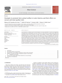
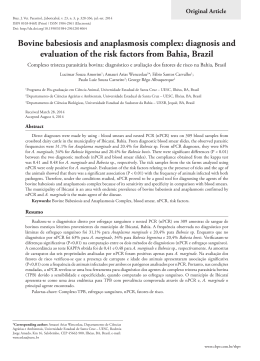
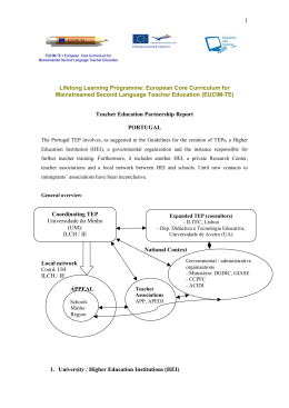
ANDERSON SILVA DIAS
Pochonia chlamydosporia NO CONTROLE BIOLÓGICO DE Fasciola hepatica
EM BOVINOS E COMPARAÇÃO DE TÉCNICAS DE SEDIMENTAÇÃO DE
OVOS
Tese apresentada à Universidade Federal
de Viçosa, como parte das exigências do
Programa de Pós-Graduação em Medicina
Veterinária, para obtenção do título de
Doctor Scientiae.
VIÇOSA
MINAS GERAIS - BRASIL
2014
Ficha catalográfica preparada pela Seção de Catalogação e
Classificação da Biblioteca Central da UFV
T
D541p
2014
Dias, Anderson Silva, 1979Pochonia chlamydosporia no controle biológico de
Fasciola hepatica em bovinos e comparação de técnicas de
sedimentação de ovos / Anderson Silva Dias. – Viçosa, MG,
2014.
xiv, 90f. : il. ; 29 cm.
Inclui anexo.
Orientador: Jackson Victor de Araújo.
Tese (doutorado) - Universidade Federal de Viçosa.
Inclui bibliografia.
1. Controle biológico. 2. Pochonia Chlamydosporia. 3.
Fasciola hepatica. 4. Bovino - Doenças. I. Universidade Federal
de Viçosa. Departamento de Veterinária. Programa de
Pós-graduação em Medicina Veterinária. II. Título.
CDD 22. ed. 632.6
AGRADECIMENTOS
A Deus.
A meus pais: José Marcelino Dias e Maria Lúcia da Silva Dias pelo incentivo.
A meus irmãos pelo estímulo.
A minha esposa, Sandra Helena, pelo apoio e compreensão.
A minha filha Lívia, pela compreensão.
Ao meu orientador, Prof. Jackson, pelas orientações, conselhos, por acreditar e
ajudar sempre.
Ao coorientador, Prof. Fábio, por todo auxílio, orientação e dicas.
Aos senhores José Geraldo, Ademir e Samuel pelo esforço em ajudar.
Aos colegas do curso do Laboratório: Juliana Milani, Fernanda Fernandes e
Sebastião pelo apoio logístico.
Aos colegas da Pós Graduação pelo incentivo.
Ao Vinícius Longo, Odilon e Vinícius Miranda pelo apoio laboral e intelectual.
Aos alunos orientados e estagiários da Facastelo: André Puppin, Wilber, Raul,
Renata, Rafael Fabbri, Diego Serrano, Vitor, Roberto, Gester pelo apoio, auxílio e
execução das tarefas trabalhosas.
Ao senhor José Puppin pela cedição dos animais e espaços e incansável apoio
logístico.
Aos integrantes da banca pelos conselhos, cobranças e observações.
A Rosinéia e Beth pela assistência e apoio.
Ao Departamento de Medicina Veterinária pela sedição de espaço.
Aos colegas pelo incentivo.
A Coordenação de Aperfeiçoamento de Pessoal de Nível Superior (CAPES), ao
Conselho Nacional de Pesquisa (CNPq), a Fundação de Amparo a Pesquisa do
Estado de Minas Gerais – FAPEMIG pelos apoios técnicos e financeiros a essa tese.
ii
BIOGRAFIA
Anderson Silva Dias, nascido em 10 de abril de 1979 em Coronel Fabriciano,
filho de José Marcelino Dias e Maria Lúcia da Silva Dias.
Em 29 de Janeiro de 2004, graduou-se em Medicina Veterinária na
Universidade Federal de Viçosa.
Em março de 2004, ingressou no mestrado no Programa de Pós Graduação da
Universidade Federal de Viçosa. Em 23 de março de 2006, realizou a defesa da tese
de Mestrado.
Em agosto de 2010, ingressou no Doutorado no Programa de Pós Graduação da
Universidade Federal de Viçosa. Em 31 de março de 2014, realizou a defesa da tese
de Doutorado.
iii
SUMÁRIO
LISTA DE TABELAS........................................................................................
viii
LISTA DE FIGURAS.........................................................................................
ix
RESUMO.............................................................................................................
xi
ABSTRACT.........................................................................................................
xiii
1. INTRODUÇÃO...............................................................................................
1
2. REVISÃO DE LITERATURA......................................................................
3
3. OBJETIVOS....................................................................................................
10
3.1. Objetivo Geral..............................................................................................
10
3.1. Objetivos Específicos...................................................................................
10
4. HIPÓTESES...................................................................................................
11
COMISSÃO DE ÉTICA............................................................................
12
REFERÊNCIAS..................................................................................................
13
CAPÍTULO 1 – Biological control of Fasciola hepatica eggs with the
Pochonia chlamydosporia fungus after passing through the cattle
Gastrointestinal…………………………………………………………………... 19
Abstract…………………………………………………………………………..
20
1. Introduction.......................................................................................................
21
2. Materials and methods......................................................................................
22
2.1 Organisms.......................................................................................................
22
2.2 Obtainment of the F. hepatica eggs................................................................
22
2.3 Animals...........................................................................................................
22
2.4. Experimental assay........................................................................................
23
2.5 Statistical analysis..........................................................................................
24
3 Results ..............................................................................................................
24
iv
4 Discussion.........................................................................................................
25
Acknowledgments ..............................................................................................
29
References............................................................................................................
30
CAPÍTULO 2 - Pochonia chlamydosporia in the biological control of
36
Fasciola hepatica in cattle in Southeastern Brazil........................................
Abstract ...............................................................................................................
37
1 Introduction.......................................................................................................
38
2 Materials and methods......................................................................................
39
2.1 Organisms.......................................................................................................
39
2.2 Animals...........................................................................................................
39
2.3 Experimental assay in pasture........................................................................
40
2.4 Collection of faecal samples from the paddock pastures................................
41
2.5 Fungus identification in incubated faecal samples and quantification...........
42
2.6 Statistical analysis...........................................................................................
42
3 Results................................................................................................................
43
3.1 Egg number per gram of faeces.....................................................................
43
3.2 Monthly mean of animal weight gain.............................................................
43
3.3 Faecal samples collected from the paddocks..................................................
44
4 Discussion..........................................................................................................
45
Acknowledgments................................................................................................
48
References............................................................................................................
49
CAPÍTULO
3 – Comparison among three
techniques in the
parasitological diagnosis of Fasciola hepatica in cattle..... ..............................
56
Abstract................................................................................................................
57
1. Introduction......................................................................................................
58
v
2 Materials and methods......................................................................................
59
2. 1 Collection of samples from animals .............................................................
59
2.2. Techniques used in parasitological diagnosis ...............................................
59
2.2.1 Modified technique of Dennis, Swanson & Stone by Belém et al. (1992) .
59
2.2.2 Girão–Ueno filtration and sieving technique ..............................................
60
2.2.3 Modified sedimentation technique ..............................................................
60
2.3 Obtaiment of Fasciola hepatica adults...........................................................
60
2.4 Statistical analysis ..........................................................................................
60
3 Results................................................................................................................
61
4 Discussion .........................................................................................................
62
5. Conclusion.........................................................................................................
64
Acknowledgements .............................................................................................
65
References............................................................................................................
66
CAPÍTULO 4 - A experimental coprological survey of Fasciola hepatica
in cattle in South of Espírito Santo State, Brazil...........................................
72
Abstract.................................................................................................................
73
1 Introduction........................................................................................................
74
2 Materials and methods.......................................................................................
75
2.1 Animals ..........................................................................................................
75
2.2 Coprological examination...............................................................................
76
2.3 Weather conditions.........................................................................................
76
2.4 Statistical analysis .........................................................................................
76
3 Results...............................................................................................................
77
4 Discussion .........................................................................................................
78
vi
Acknowledgments.......................................................................................
80
References.............................................................................................................
81
5 CONCLUSÕES GERAIS................................................................................
86
6 ANEXOS…...…………………………………………………………………
87
6.1 Técnicas de diagnóstico parasitológico em fezes........................................
87
6.1.1 Técnica de sedimentação de Denis, Stone & Swanson modificada
(DSS).............................................................................................................
87
6.1.2 Técnica de filtração e tamisação de Girão–Ueno (QT)............................
88
6.1.3 Técnica de sedimentação modificada de Foreyt......................................
89
6.2 Certificado de Aprovação da Comissão de Ética.......................................
90
vii
LISTA DE TABELAS
CAPÍTULO 1
Página
Table 1 Mean values and standard deviation of the ovicidal activity of the
nematophagous fungus P. chlamydosporia (VC1) on Fasciola hepatica eggs in fecal
samples collected at 12, 18, 24, 48, 72, and 96 h after the pellets' passage containing
25% of the P. chlamydosporia fungus mycelium for the treated and control groups
(without fungal treatment) and recovered on Petri dishes on 2% water–agar with six
replications for each sample, after storage for 35 days.
34
CAPÍTULO 3
Table 1 Number of F. hepatica adults recovered from the bile ducts of cattle
slaughtered after parasitological testing.
70
Table 2 Sensitivity of parasitological examination of Fasciola hepatica eggs
obtained through the modified techniques of Dennis, Stone & Swanson, quatro
tamises, and sedimentation of Foreyt.
71
viii
LISTA DE FIGURAS
Página
CAPÍTULO 2
Fig. 1 Monthly mean number of F. hepatica eggs and standard error per gram of
faeces. Of the egg number of F. hepatica per gram of faeces from the animals in the
control group (A) and treated group (B) with the P. chlamydosporia ovicidal fungus,
collected from July, August, September, October, November and December (2009)
and January, February, March, April, May, June, July, August, September, October,
November and December (2010), in the municipality of Cachoeiro de Itapemirim,
Espírito Santo State, Brazil.
Significant difference (P<0.01) between the treated group (A) and the control (B).
54
Fig. 2 Monthly mean weight and standard error (in kilograms) of the animals in the
control group (A) and the group treated with the P. chlamydosporia ovicidal fungus
(group B), from July, August, September, October, November and December (2009)
and January, February, March, April, May, June, July, August, September, October,
November and December (2010), in Cachoeiro de Itapemirim municipality, Espírito
Santo State, Brazil.
55
ix
Página
CAPÍTULO 3
Fig 1 Means and standard error of number Fasciola hepatica eggs per gram of faeces
obtained by techniques of Dennis, Stone & Swanson (Belém et al. 1992), quatro
tamises (four sieves) (Ueno and Girão, 1982), and sedimentation of Foreyt (2001).
Samples were collected from 12 bovines with 16 repetitions, fortnightly. These
samples were collected from these animals from June to December 2010. The
experimental farm is at the municipality of Cachoeiro de Itapemirim, Espírito Santo
State, southeastern Brazil.
69
CAPÍTULO 4
Fig 1 Mean monthly number of egg per gram of Fasciola hepatica in cattle feces.
Fecal samples were collected from the rectum of 12 bovines, October 2009 to
December 2010, forthnightly, and the sample were submitted at technique modified
of Dennis, Stone and Swanson. The animals were reared at a farm at the municipality
of Cachoeiro de Itapemirim, Espírito Santo State, Brazil.
84
Fig 2 Mean monthly minimum temperature, medium and maximum rainfall and
relative humidity obtained from at a weather station in the region under study, in the
municipality of Cachoeiro de Itapemirim, Espírito Santo State, Brazil. The data
comprehend the period from July 2009 and December 2010 and were collected.
85
x
RESUMO
DIAS, Anderson Silva, D.Sc., Universidade Federal de Viçosa, março de 2014.
Pochonia chlamydosporia no controle biológico de Fasciola hepatica em bovinos
e comparação de técnicas de sedimentação de ovos. Orientador: Jackson Victor de
Araújo. Co-orientador: Fábio Ribeiro Braga.
A presença de helmintos causando prejuízos na pecuária é significativa no Brasil e
no mundo. A Fasciola hepatica figura dentre os principais agentes responsáveis por
essas perdas. Devido ao aumento de ocorrências de problemas de resistência antihelmínticas e falhas no controle desses helmintos, o emprego de agentes de controle
biológico tem sido estudado e tem apresentado resultados promissores. O presente
trabalho tem como objetivos: avaliar a ação do fungo Pochonia chlamydosporia in
vitro e in vivo sobre ovos de F. hepatica, após a passagem através do trato
gastrintestinal de bovinos em formulações peletizadas; avaliar as condições
climáticas sobre a produção de ovos de F. hepatica e avaliar três técnicas de
sedimentação para quantificar os ovos de F. hepatica em fezes de bovinos. Para isso,
amostras de fezes foram coletadas nos bovinos do grupo tratado com péletes
contendo o fungo P. chlamydosporia e grupo controle com péletes se fungo nos
tempos de 12, 18, 24, 48, 72 e 96 h após a administração dos péletes. O efeito
ovicida foi observado após sete dias da interação. O fungo apresentou atividade
ovicida sobre ovos de F. hepatica nas amostras (grupo tratado) em todos os tempos a
partir de 12 h. Diferenças significativas (p<0,01) na destruição de ovos dos animais
do grupo tratado em comparação com o controle foi verificada. Esses resultados
sugerem o emprego desse fungo de forma eficaz para o controle de ovos de F.
hepatica. Trinta dias após a vermifugação, os animais foram separados em dois
piquetes semelhantes e administrados via oral péletes contendo 25% de massa
micelial de P. chlamydosporia (grupo B) e sem fungo (grupo A), a uma dose de 100
xi
gramas, duas vezes por semana, durante 18 meses. A média de ovos de F. hepatica
por grama de fezes foi maior (p<0,01) no grupo A (1,19) em comparação com o B
(0,82). Após 18 meses, os animais do grupo B ganharam a mais 42,33 kg (17,82%)
comparados aos do grupo A (p<0,01). Durante os 18 meses, mensalmente, as
amostras de fezes de bovinos foram recolhidas nas pastagens dos grupos A e B e
foram incubadas para observar estruturas referentes ao fungo P. chlamydosporia e
foram identificados apenas nas amostras coletadas no piquete do grupo B. A média
de ovos por grama de fezes das amostras coletadas entre os períodos de chuva
(outubro a março) e seca (abril a setembro) não foram diferentes (P>0,05) e, dessa
forma, é possível que os animais possam ser infectados por F. hepatica durante todo
o ano. As técnicas de sedimentação modificadas de Dennis, Stone & Swanson (DSS),
Girão e Ueno (quatro tamises) e Foreyt foram comparadas. A técnica de DSS
modificada foi a mais sensível (p<0,01) (48,60%). As três técnicas apresentaram
especificidade de 100%. Não houve correlação na contagem de ovos obtidos pelas
três técnicas e coeficientes significativos não foram observados por análise de
regressão. A técnica DSS modificada foi a mais eficaz para o diagnóstico de F.
hepatica em bovinos (p <0,01). A aplicação de formulação fúngica com P.
chlamydosporia (25%) foi eficaz na redução da disponibilidade de ovos no ambiente
e, por conseguinte, nas reinfecções em bovinos.
xii
ABSTRACT
DIAS, Anderson Silva, D.Sc., Universidade Federal de Viçosa, March, 2014.
Pochonia chlamydosporia in the biological control of Fasciola hepatica and
comparison of techniques of eggs sedimentation. Adviser: Jackson Victor de
Araújo. Co-Adviser: Fábio Ribeiro Braga.
The presence of helminths causing losses in livestock in the Brazil and in the world
is significant. The Fasciola hepatica is among the main agents responsible for these
losses. Due to increased occurrences of problems anthelmintic resistance and failures
in the control of this helminths, the use of agent of biological control have been
studied and they have showed promising results. This study aims to evaluate: the
action of the fungus Pochonia chlamydosporia in vitro and in vivo on eggs of F.
hepatica, after passage through of gastrointestinal tract of cattle in formulations of
pellets; assess the weather conditions on F. hepatica egg production and evaluate
three techniques of sedimentation to quantify F. hepatica eggs in cattle faeces. Thus,
stool samples were collected in cattle of treated group with pellets contained the
fungus P. chlamydosporia and control group with pellets without fungus in times 12,
18, 24, 48, 72 and 96 h after administration of pellets. The ovicidal effect was
observed after seven days of interaction. The fungus showed ovicidal activities on
eggs F. hepatica in the samples (treated group) in all times from 12 h. Significant
differences (p <0.01) in the destruction of the eggs in animals of the treated group
compared to those of the control was verified. This resuts suggest employment this
fungus of effectively to the control of eggs of F. hepatica. Thirty days after
deworming, the animals were divided into two similar paddocks e animals received
per os pellets containing 25% mycelial mass of P. chlamydosporia (group B) and
pellets without fungus (group A), at a dose of 100 g, twice a week over 18 months.
xiii
The mean count of F. hepatica eggs per gram of faeces was higher (p<0.01) in
animals of group A (1.19) compared with those from group B (0.82). After 18
months, the animals from group B gained 42.33 kg above (17.82%) compared to
those of control group (A) (p<0.01). During 18 months, monthly, cattle faecal
samples were collected from the pastures of the paddocks of A and B groups and
they were incubated to observe structures related at P. chlamydosporia fungus e they
was identified only in sample collected in the paddock of B group. Mean number of
eggs per gram of faeces of the samples collected between periods of rain (OctoberMarch) and dry (April-September) were not different (p>0.05) and, thus, it is
possible that the animals can be infected by F. hepatica during all the year. The
modified sedimentation techniques of Dennis, Stone & Swanson (DSS), Girão and
Ueno (quatro tamises) and Foreyt were compared. The modified DSS technique was
the most sensitivity (p<0.01) (48.60%). The three techniques showed a specificity of
100%. There was no correlation among the values of egg obtained by the three
techniques, and significant coefficients were not observed by regression analysis.
The modified DSS technique was the most effective for F. hepatica diagnosis in
cattle (p<0.01). The application of this fungical formulation with P. chlamydosporia
(25 %) mycelial mass was effective in reducing the availability of eggs in the
environment, and consecutive, in the reinfections in calves.
xiv
1. INTRODUÇÃO
A criação de bovinos no Brasil ocupa grande importância socioeconômica
(Honer, 1979) e os helmintos são um dos principais agentes responsáveis por perdas
nessa atividade (Araújo et al., 2004). A Fasciola hepatica, um helminto trematóide,
de distribuição cosmopolita, é responsável por perdas, principalmente na criação de
ovinos e bovinos. Essas perdas se estendem a gastos com tratamentos e condenações
de fígado no abate dos animais (Anderson et al., 1977; Ross et al., 1997; Acha &
Szyfres, 2003), além das casuísticas humanas (Mas-Coma et al., 2005).
Há dificuldades no controle eficaz de F. hepatica, muitas vezes realizado
apenas com o uso de antihelmínticos, que consiste de um método oneroso para a
produção. Além disso, esse helminto tem apresentado resistência cada vez maior a
essas drogas (Winkelhagen et al., 2012; Brockwell et al., 2014). Diante desse quadro,
pesquisas para o controle desses agentes têm sido desenvolvida, dentre elas,
destacam-se a investigação para o desenvolvimento de uma vacina eficaz, o que não
tem obtido êxito, e o estudo do controle biológico, que tem apresentado resultados
animadores in vitro e in vivo. Os fungos helmintófagos tem se destacado como
controladores biológicos, e esses agentes atuam nas formas de vida livre dos
helmintos, onde se alimentam das formas jovens de ovos e larvas dos helmintos e
atuam de forma a prevenir as reinfecções dos animais (Stirling, 1991; Braga &
Araújo, 2014).
1
Três grupos de fungos atuam de diferentes formas sobre helmintos: os
endoparasitos, que se alimentam das porções internas dos helmintos e apresentam a
desvantagem de depender dos helmintos no meio para se propagarem. os fungos
predadores predam as larvas de helmintos no meio e os capturam através de
formação de diversas armadilhas e; os fungos ovicidas ou oportunistas que
apresentam atividade sobre ovos de helmintos parasitos de animais. Os dois últimos
grupos de fungos são constituídos por representantes que produzem formas de
propagação resistentes: os clamidósporos (Stirling, 1991; Braga & Araújo, 2014).
Para o controle da F. hepatica, na qual a fase de vida que está presente no meio
por um período considerável é o ovo, um fungo ovicida seria o mais indicado para o
seu controle. E fungos ovicidas Pochonia chlamydosporia tem se destacado (Braga
et al., 2007, 2008; Araújo et al. 2008). Esse fungo apresenta eficácia satisfatória in
vitro no controle de ovos de F. hepatica, o que não foi verificado ao observar a ação
de fungos predadores como Duddingtonia flagrans e Monacrosporium sinense, que
são eficazes em predar helmintos em sua forma larvária de vida livre e são usados
nesses ensaios como controle negativo (Braga et al., 2008).
Resultados promissores têm sido obtidos em ensaios realizados com o emprego
desses fungos sobre ovos e larvas de diversos helmintos, in vivo e in vitro, no sentido
de se investigar a eficácia da ação desses fungos helmintófagos (Waller et al. 1994;
Araujo et al., 2008; Braga et al., 2007; 2008; 2011; Braga & Araújo, 2014).
2
2. REVISÃO DE LITERATURA
A Fasciola hepatica é um helminto de distribuição mundial pertence ao filo
Platyhelminthes, classe Trematoda, ordem Digenea e possui como hospedeiros
intermediários moluscos dulcícolas da família Lymnaeidae. Morfologicamente ela é
constituída por tegumento, musculatura, sistema nervoso, citoesqueleto, intestino e
sistemas excretor e reprodutivo (Dalton, 1999).
No Brasil, a prevalência desse agente é registrada, principalmente, nas regiões
Sul (regiões de fronteiras do Rio Grande do Sul, Vale do Itajaí, Santa Catarina e
parte do estado do Paraná) e no Sudeste (no Vale do Rio Paraíba do Sul em partes do
sul de Minas Gerais, Nordeste do estado de São Paulo e oeste e noroeste do estado do
Rio de Janeiro e sul do Espírito Santo) (Queiroz et al., 2002). Nessas regiões, esse
agente é responsável por uma parte considerável das perdas produtivas em criações
de bovinos e ovinos. Dentre os prejuízos mais frequentes causados pela F. hepatica
podem ser incluídos: a diminuição na produtividade, gastos com tratamento,
mortalidade, descarte de fígados no abate, além de ser um risco à saúde pública,
causando a mesma sintomatologia no homem (Oliveira, 2008; Lima et al., 2009). A
destruição hepática causada pelas larvas de F. hepatica leva à hepatomegalia, à
diminuição das proteínas plasmáticas e acúmulo de líquido nas cavidades (Honer,
1979; Anderson et al., 1999).
3
O ciclo de Fasciola, no hospedeiro definitivo, inicia-se com a deposição de
ovos pelos adultos no ducto biliar. Os ovos, uma vez liberados no meio e sob
condições climáticas favoráveis (temperatura e umidade altas), liberam o miracídio
que procuram e penetram nos hospedeiros intermediários – moluscos aquáticos
Lymnaeidae. No Brasil, os limnídeos mais importantes na transmissão de fasciolose
são: Lymnaea columela, L. viatrix, L. cubensis (Oliveira, 2008). Nesses moluscos,
ocorre a multiplicação e desenvolvimento do miracídio, que resulta na formação do
esporocisto, um saco alongado formado por células germinativas, que dão origem às
rédias e que se diferenciam até a fase de cercária. As cercárias saem do molusco e
encistam em vegetações, passando a metacercária. Geralmente, ruminantes e outros
animais silvestres ingerem as metacercárias que penetram no trato digestório e se
alojam nos hepatócitos, onde se desenvolvem e, após isso, maturam e atingem os
ductos biliares (Dalton, 1999).
A F. hepatica possui grande capacidade de dispersão, pois é capaz de se
adaptar a novos hospedeiros e ao meio. Além disso, cada espécime presente no ducto
biliar é capaz de ovipor até 20 mil ovos por dia (Mas-Coma et al., 2005). Embora a
F. hepatica possa ser capaz de infectar os mamíferos terrestres em geral, ovinos e
bovinos são as espécies mais susceptíveis à infecção causada por esse agente, onde
as taxas de morbidade e mortalidade nessas espécies variam de região a região, e em
áreas endêmicas não é raro encontrar taxas de infecção superiores a 50% (Ross et al.,
1997). A helmintose causada por esse agente apresenta duas fases, a primeira, aguda,
caracteriza-se por destruição dos hepatócitos quando as formas jovens do parasito
invadem e migram pelo parênquima causando hemorragias, hematomas, rupturas e
inflamação com posterior necrose e destruição do tecido hepático. Nessa fase, podem
ocorrer debilidades repentinas, inapetência, dor à palpação na região hepática, perda
4
de peso, ascite e mortes e hipoproteinemia, essa fase é mais marcante entre ovinos, e
branda entre bovinos (Anderson et al., 1977). E fase crônica, quando os helmintos
estão nos ductos biliares e se caracteriza por perda de peso, emaciação, edema
submandibular, anemia, debilidade, diarréia e ascite (Dalton, 1999; Acha & Szyfres,
2003).
A F. hepatica penetra em caramujos do gênero Lymnaea spp., e os tem como
seus hospedeiros intermediários (Honer, 1979) e a sua permanência numa região está
determinada pela presença desses caramujos nesse local (Pile et al., 2001), além de
ser importante também à presença de hospedeiros definitivos apropriados, em geral,
bons propagadores de ovos para o meio, como os ruminantes e alguns herbívoros
silvestres (Honer, 1979). De acordo com Coelho & Lima (2003), esses caramujos
dulcícolas são influenciados por duas fases climáticas: uma estação seca e menos
úmida, em que esses se encontram em estarvação; e a outra, chuvosa, que se forem
intensas, e houver a presença de áreas não drenadas com ocorrência de inundações,
contribuem para a rápida multiplicação e rápida dispersão desses agentes. Ainda,
segundo os mesmos autores, o aumento da densidade populacional contribui para o
aumento da taxa de infecção dos moluscos pelos miracídios.
Considerando que a F. hepatica é responsável por perdas significativas na
pecuária e que as perdas estão associadas à carga parasitária, a comparação e o
aperfeiçoamento de técnicas de diagnóstico de fasciolose devem ser realizado com a
finalidade de se indicar a técnica mais sensível é essencial, uma vez que, essa técnica
poderia representar de forma mais confiável a carga parasitária nos animais e indicar
se o manejo está ou deverá ser realizado de forma eficaz. Dentre aquelas mais
empregadas destacam-se a de sedimentação simples, a de flukefinder, a de Hofmann
et al., a de quatro tamises e a de Dennis et al. (Ueno & Gonçalves, 1998; Anderson et
5
al., 1999; Kleimann et al., 2005). As técnicas sorológicas apresentam a vantagem de
detectar prévias infecções nos animais (Duménigo et al., 2000). Porém, de acordo
com Kleimann et al. (2005), os testes imunológicos não são capazes de indicar a
carga parasitária, nem o número de ovos que saem nas fezes que são os responsáveis
pela dispersão do agente.
É imprescindível que se conheça as interações que determinam o parasitismo
por helmintos para se estabelecer um sistema de controle. Por isso, conhecer as
condições climáticas e as possibilidades associadas à transmissão de F. hepatica
representa uma ferramenta auxiliar para aperfeiçoar o controle e profilaxia desse
helminto (Araújo et al., 2004). Acha & Szyfres, (2003), por exemplo, relatam que as
condições climáticas são essenciais no mecanismo de contaminação dos caramujos
por larvas e de animais por metacercárias. Malek (1980) observou que em algumas
regiões ocorrem surtos graves de fasciolose depois que as chuvas de fins de verão
ocorrem.
Alguns meios de controles parasitários alternativos têm sido apresentados pela
comunidade científica e dentre elas destacam-se o controle biológico e fitoterápicos
(Waller & Faedo, 1996). As medidas que adotam apenas o uso de vermífugos são
responsáveis pela rápida instalação de resistência pelos parasitos, e isto tem
contribuído para que as pastagens estejam na maioria das vezes contaminadas por
esses agentes (Waller & Faedo 1996; Sangster, 1999). Em relação à F. hepatica, tem
sido registrado, em algumas regiões do mundo, resistência contra anti-helmínticos
mais eficazes no mercado como triclabendanzole, tanto em bovinos e ovinos
(Brockwell et al., 2014) quanto em humanos (Winkelhagen et al., 2012). Além disso,
ocorrem problemas com o alto custo de aquisição desses fármacos e a possibilidade
de ocorrência de resíduos químicos no ambiente e nos produtos de origem animal,
6
além das dificuldades técnicas e econômicas para o desenvolvimento de novas
moléculas (Waller et al., 1994).
Os fungos nematófagos, atualmente chamados de helmintófagos, uma vez que
os mesmos são também eficazes em predar formas de vida livre de outros grupos de
helmintos além dos nematóides, compreendem diferentes tipos de fungos, que podem
ser caracterizados como predadores, endoparasitas de nematóides e oportunistas ou
parasitos de ovos (Braga & Araújo, 2014). Eles são cosmopolitas, ocorrendo em
solos naturais, solos agricultáveis e em todos os tipos de matéria orgânica em
decomposição (Kerry et al., 2008).
O fungo Pochonia chlamydosporia é oportunista, ovicida, classificado como
pertencente ao reino Fungi, filo Ascomycota, subdivisão Pezizomycotina, classe
Sordariomycetes, ordem Hypocreales e família Clavicipitaceae. Possui como
características morfológicas: as hifas que constituem seu micélio de forma septada e
produzem esporos em um saco fechado, denominado ascósporo (Gams & Zare,
2001). Por ser um fungo ovicida e saprófito, não depende da presença de ovos de
helmintos no meio constantemente para se manter (Kerry et al., 2008). Esse fungo é
capaz de parasitar ovos de uma grande variedade de espécies de helmintos e de
moluscos, sua variabilidade genética é considerada ampla. E esse fato poderia estar
ligado à interação com seus diversos hospedeiros e estar associada à sua eficácia no
controle biológico desses agentes (Kerry et al., 2008). Stirling (1991) e Braga et al.
(2011) relatam que esse fungo é capaz de produzir uma grande quantidade de
clamidósporos (esporos de resistência) e possui ampla capacidade de colonizar o
ambiente onde está presente. A produção dessas estruturas foi verificada por Ferreira
et al. (2011) in vitro, após a passagem desse fungo através do trato gastrintestinal de
suínos. Braga et al. (2011) conduziram um ensaio experimental in vitro e observaram
7
que a partir do emprego de concentrações maiores de clamidósporos desse fungo
sobre ovos de Taenia taeniformis houve maior eficácia.
O fungo P. chlamydosporia é adaptado a diversas condições climáticas
(Santiago et al., 2006). Ele cresce bem em meios de cultura e em temperaturas
médias de 24 e 32°C (Lysek et al., 1982). Assim, estes poderiam ser utilizados em
regiões de diferentes condições climáticas sem comprometimento para seu
crescimento (Santiago et al., 2006). De acordo com Lysek et al. (1982), esses fungos
crescem bem em condições climáticas diversas e em condições tropicais e as
atividades ovicidas deles são elevadas.
P. chlamydosporia produz uma estrutura de predação denominada apressório, o
qual é capaz de penetrar em ovos de helmintos por pressão mecânica e ação
enzimática de proteases (Segers et al., 1996). Fungos ovicidas podem apresentar três
formas de interação com o ovo. A saber, o efeito tipo 1, na qual o fungo não penetra
o ovo, mas pode parar o desenvolvimento do embrião ou permitir a produção de uma
larva defectiva; o efeito tipo 2, na qual o fungo não penetra o ovo, mas a casca e o
embrião são enzimaticamente danificados; e o efeito tipo 3, na qual o fungo (as hifas)
penetram a casca, colonizam, crescem (ramificam) e destroem o ovo (Lysek et al.,
1982). Além disso, Lysek & Sterba (1991) e Irving & Kerry (1986) relatam que esse
fungo é capaz de produzir efeito ovicida satisfatório em todas as fases de
desenvolvimento embrionário de ovos de helmintos.
O fungo P. chlamydosporia é promissor in vitro para o controle de helmintos
com ovos como forma de vida livre predominante (Lysek et al., 1982; Lysek &
Sterba, 1991; Braga et al., 2008). esse fungo apresenta atividade ovicida sobre os
ovos de F. hepatica (Braga et al., 2008), mas, testes in vivo com esse fungo sobre
ovos desse trematóide ainda não haviam sido testados até o presente momento.
8
A forma mais prática de se fornecer fungos para o controle biológico é pela
administração oral aos animais (Araújo et al., 2004). Após a passagem pelo trato
gastrintestinal e ser eliminado junto às fezes no meio ambiente, o fungo coloniza o
bolo fecal, estabelece contato com os ovos e alimenta-se dos mesmos (Stirling,
1991). A avaliação desse evento é realizada por execução de testes com alguns
isolados fúngicos, o que tem sido o foco de diversos estudos (Araújo et al., 2004;
Braga et al., 2007; 2008; 2011).
Medidas alternativas como o emprego de agentes de controle biológico tem se
destacado como uma ferramenta auxiliar que visa dar suporte para que se faça o
controle de parasitos, quando o mesmo está presente no ambiente como forma de
ovos, larvas ou no hospedeiro intermediário (Stirling, 1991). Os agentes de controle
biológico atuam sobre os ovos e larvas, atenuando as reinfecções de forma a
minimizar as perdas econômicas, sem impactos ambientais (Garcia et al., 2008).
9
2. OBJETIVOS
2.1 Objetivo Geral
Testar o fungo Pochonia chlamydosporia no controle de Fasciola hepatica in
vitro e in vivo, comparar a sensibilidade de técnicas de sedimentação e avaliar a
produção de ovos de F. hepatica em bovinos no sul do Espírito Santo.
2.2 Objetivos Específicos
Avaliar a viabilidade do fungo P. chlamydosporia em predar ovos de F.
hepatica após a passagem pelo trato gastrintestinal de bovinos.
Testar a eficácia do fungo P. chlamydosporia em bovinos a campo no sul do
Espírito Santo.
Comparar a sensibilidade de três técnicas de sedimentação para o diagnóstico
de ovos de F. hepatica em amostras fecais de bovinos.
Avaliar a produção de ovos de F. hepatica em fezes de bovinos sob condições
tropicais no sul do Espírito Santo em época de chuvas e de estio.
10
4. HIPÓTESES
O fungo ovicida Pochonia chlamydosporia preda ovos de Fasciola hepatica
após a passagem pelo trato intestinal de bovinos.
O fungo ovicida P. chlamydosporia, administrado em formulação peletizada
para bovinos, reduz a infestação nas pastagens das fases de vida livre de F. hepatica.
As técnicas modificadas de sedimentação de Dennis, Stone e Swanson,
Foreyt e Quatro tamises de Girão e Ueno apresentaram diferentes sensibilidades nas
pesquisas por ovos de F. hepatica em fezes de bovinos.
Fatores climáticos interferem na produção de ovos de F. hepatica em bovinos
no sul do Espírito Santo comparando as estações seca e chuvosa.
11
COMISSÃO DE ÉTICA
Esse trabalho foi submetido e aprovado pela Comissão de Ética da
Universidade Federal de Viçosa (processo n.º52/2011 em anexo).
12
REFERÊNCIAS
Acha, P.N.; Szyfres, B. 2003. Zoonosis y enfermedades transmissibles comunes al
hombre y a los animales. 3ed. vol. III. Organización Panamericana de La Salud,
Washington, DC, EUA. 989pp.
Anderson, P.H.; Berrett, S.; Brush, P.J.; Hebert, C.N.; Parfitt, J.W.; Patterson, D.S.
1977. Biochemical indicators of liver injury in calves with experimental fascioliasis.
Veterinary Record, 100, 43–45.
Anderson, N.; Luong, T.T.; Vo N.G.; Bui K.L.; Smooker, P.M.; Spithill, T.W. 1999.
The sensitivity and specificity of two methods for detecting Fasciola infections in
cattle. Veterinary Parasitology, 83,15-24.
Araújo, J.V.; Mota, M.A.; Campos, A.K. 2004. Controle biológico de helmintos
parasitos de animais por fungos nematófagos. In: Congresso de Parasitologia
Veterinária & Simpósio Latino-Americano de Ricketisioses, 13, Ouro Preto, 2004.
Revista Brasileira de Parasitologia Veterinária. Ouro Preto: CBPV. 13, 165-170.
Araújo, J.V.; Braga, F.R.; A.R. Silva; J.M. Araújo; Tavela A.O. 2008. In vitro
evaluation of the effect of the nematophagous fungi Duddingtonia flagrans,
Monacrosporium sinense, and Pochonia chlamydosporia on Ascaris suum eggs.
Parasitology Research, 102, 787–790.
13
Braga, F.R.; Araújo, J.V.; Campos, A.K.; Carvalho, R.O.; Silva, A.R.; Tavela, A.O.;
Maciel, A.S. 2007. Observação in vitro da ação dos isolados fúngicos Duddingtonia
flagrans, Monacrosporium thaumasium e Verticillium chlamydosporium sobre ovos
de Ascaris lumbricoides (Lineu, 1758). Revista da Sociedade Brasileira de Medicina
Tropical, 40,356–358.
Braga, F.R.; Araújo, J.V.; Campos, A.K.; Araújo, J.M.; Silva, A.R.; Carvalho, R.O.;
Tavela, A.O. 2008. In vitro evaluation of the action of the nematophagous fungi
Duddingtonia flagrans, Monacrosporium sinense and Pochonia chlamydosporia on
Fasciola hepatica eggs. World Journal of Microbiology and Biotechnology, 24,
1559–1564.
Braga, F.R.; Silva, A.R.; Carvalho, R.O.; Araújo, J.V.; Pinto, P.S.A. 2011. Ovicidal
activity of different concentrations of Pochonia chlamydosporia chlamydospores on
Taenia taeniaeformis eggs Journal of Helminthology, 85, 7–11.
Braga, F.R.; Araújo, J.V. 2014. Nematophagous fungi for biological control of
gastrointestinal nematodes in domestic animals. Applied Microbiology and
Biotechnology, 98, 71–82.
Brockwell, Y.M.; Elliott, T.P.; Anderson, G.R.; Stanton, R.; Spithill, T.W.; Sangster,
N.C. 2014. Confirmation of Fasciola hepatica resistant to triclabendazole in
naturally infected Australian beef and dairy cattle. International Journal for
Parasitology: Drugs and Drug Resistance, 4, 48–54.
14
Coelho, L.H.L.; Lima, W.S. 2003. Population dynamics of Lymnaea columella and
its natural infection by Fasciola hepatica in the State of Minas Gerais, Brazil.
Journal of Helminthology, 77, 7-10.
Dalton, J.P. 1999. Fasciolosis. 1ed. Cambrige, UK: University Press, pp. 113-149.
Duménigo, B.E.; Espino, A.M.; Finlay, C.M.; Mezo, M. 2000. Kinetics of antibodybased antigen detection in serum and faeces of sheep experimentally infected with
Fasciola hepatica. Veterinary Parasitology, 89,153-161.
Ferreira, S. R.; Araújo, J. V.; Braga, F. R.; Araújo, J. M.; Frassy, L. N.; Ferreira, A.
S. 2011. Biological control of Ascaris suum eggs by Pochonia chlamydosporia
fungus. Veterinary Research Communications, 35, 553–558.
Gams, W.; Zare, R. 2001. A revision of Verticillium sect. Prostrata. III. Generic
classification. Nova Hedwigia, 73, 329–337.
García, L.; Melchor, G.; Domínguez, Y.; Rodríguez, H.; Pino, O.; Hidalgo, L. 2008.
Ecotoxicological evaluation of Pochonia chlamydosporia var. catenulata in
terrestrial invertebrates. Revista de Toxicologia en Línea, 14, 39-50.
Honer, M.R. 1979. Aspectos da epidemiologia da fasciolose. In: Seminário Nacional
sobre parasitoses de bovinos, 1, 1979, Campo Grande. Anais... Brasília:
Embrapa/CNPGC, pp.151-165.
15
Irving, F.; Kerry, B.R. 1986. Variation between strains of the nematophagous
fungus, Verticillium chlamydosporium Goddard. II. Factors affecting parasitism of
cyst nematode eggs. Nematologica, 32, 474-485.
Kerry, B.R.; Morton, C.O.; Mauchline, T.H.; Hirsch, P.R. 2008. Isolates of the
generalist parasite Pochonia chlamydosporia have specific interactions with their
nematode hosts. In: Nematode Interactions Unit, IACR - Rothamsted, Harpenden,
Hert, AL5 2JQ, UK, Posters Sections. 467 pp.
Kleiman, F.; Pietrokovsky, S.; Gil, S., Wisnivesky-Colli, C. 2005. Comparison of
two coprological methods for the veterinary diagnosis of fasciolosis. Arquivo
Brasileiro de Medicina Veterinária e Zootecnia, 57, 181-185.
Lima, W.S.; Soares, L.R.; Barçante, T.A.; Guimaraes, M.P.; Barçante, J.M. 2009.
Occurrence of Fasciola hepatica (Linnaeus, 1758) infection in Brazilian cattle of
Minas Gerais, Brazil. Revista Brasileira de Parasitologia Veterinária, 8, 27-30.
Lysek, H.; Fassatiová, O.; Pineda, N.C.; Hernández, N.L. 1982 Ovicidal fungi in
soils of Cuba. Folia Parasitológica, 29, 265–270.
Lysek, H.; Sterba, J. 1991. Colonization of Ascaris lumbricoides eggs by the fungus
Verticillium chlamydosporium Goddard. Folia Parasitologica, 38, 255-259.
Malek, E. A. 1980. Snail-transmitted parasitic diseases. v. 2. Boca Ratón: CRC
Press, Florida. 334pp.
16
Mas-Coma, S., Bargues, M.D., Valero, M.A. 2005 Fascioliasis and other plant-borne
trematode zoonoses. International Journal for Parasitology 35, 1255-1278.
Oliveira, E.L. de. 2008. Prevalência e fatores associados à distribuição da Fasciola
hepatica Linnaeus, 1758 em bovinos dos municípios de Careaçú e Itajubá, Região da
Bacia do Rio Sapucaí - Minas Gerais. (Tese de Doutorado) Instituto de Ciências
Biológicas, Universidade Federal de Minas Gerais, Belo Horizonte.
Pile, E.; Santos, J.A.A. dos; Pastorello, T.; Vasconcellos, M. 2001. Fasciola hepatica
em búfalos (Bubalus bubalis) no município de Maricá, Rio de Janeiro, Brasil.
Brazilian Journal of Veterinary Research and Animal Science, 38, 42- 43.
Queiroz, V.S.; Luz, E.; Leite, L.C.; Círio, S.M. 2002. Fasciola hepatica (Trematoda,
Fasciolidae): estudo epidemiológico nos municípios de Bocaiúva do Sul e Tunas do
Paraná (Brasil). Acta Biologica Paranaense, 31, 99-111.
Ross, J.G.; Dow, C.; Todd, J.R. 1997. A study of Fasciola hepatica infections in
sheep. Veterinary Record, 80, 543-6.
Sangster, N.C. 1999. Anthelmintic resistance: past, present and future. International
Journal for Parasitology, 29, 115-124.
Santiago, D.C.; Homechin, M.; Silva, J.F.V.; Ribeiro, E.R.; Gomes, B.C.; Santoro,
P.H. 2006. Seleção de isolados de Paecilomyces lilacinus (Thom.) Samson para
controle de Meloidogyne paranaensis em tomateiro. Ciência Rural, 36, 1055-1064.
17
Segers, R.; Butt, T.M.; Keen, J.N.; Kerry, B.R.; Beckett, A.; Peberdy, J.F. 1996. The
role of the proteinase VcP1 produced by the nematophagous fungus Verticillium
chlamydosporium in the infection process of nematode eggs. Mycological Research
100, 421–428.
Stirling, G.R. 1991. Biological control of plant parasitic nematodes. Wallingford:
CAB International, 282pp.
Ueno, W.; Gonçalves, P.C. 1998. Manual para diagnóstico das helmintoses de
ruminantes. 4 ed. Japan International Cooperation Agency, Tokio, Japan, 143pp.
Waller, P.J.; Larsen, M.; Faedo, M.; Hennessy, D.R. 1994. The potencial of
nematophagous fungi to control the free-living stages of nematodes parasites of
sheep: in vitro and in vivo studies. Veterinary Parasitology, 51, 289-299.
Waller, P.J.; Faedo, M. 1996. The prospects of the free-living stages of nematode
parasites of livestock. International Journal for Parasitology, 26, 915-925.
Winkelhagen, A.J.S.; Mank, T.; de Vries, P.J.; Soetekouw, R. 2012. Apparent
triclabendazole-resistant human Fasciola hepatica infection, the Netherlands.
Emerging Infectious Diseases. 18, 1028-1029.
18
CAPÍTULO 1
Biological control of Fasciola hepatica eggs with the Pochonia chlamydosporia
fungus after passing through the cattle gastrointestinal tract
Parasitology Research (2012) 110:663–667, DOI 10.1007/s00436-011-2538-6
19
Abstract
Fasciolosis is a disease caused by Fasciola hepatica responsible for causing
significative losses in livestock. This study aimed to evaluate the Pochonia
chlamydosporia fungus (isolate VC1) on F. hepatica eggs after passing through the
cattle gastrointestinal tract. For this evaluation, 1 g pellet was given in sodium
alginate matrix per kilogram live weight containing 25% of fungal mycelium from
isolate VC1 per animal. Twelve animals were used, six treated and six untreated
(control). Some stool samples were collected from the groups of treated and control
animals, at the times of 12, 18, 24, 48, 72, and 96 h after the pellets' administration.
Then, from each stool sample of treated and control groups, 2 g was placed in a Petri
dish of 9 cm in diameter, containing 2% water–agar and 1,000 eggs of F. hepatica.
The fungus effectively preyed the eggs present in the samples starting at 12 h.
Furthermore, differences were observed (p<0.01) in the destruction of eggs in the
Petri dishes in the treated group compared to the control group. The ovicidal effect
was observed after 7 days of interaction. The ovicidal P. chlamydosporia fungus was
effective in destroying F. hepatica eggs; therefore, this fungus could be used in the
biological control of the helminthes eggs.
20
1. Introduction
Fasciola hepatica has a cosmopolitan distribution, infecting all land mammals,
especially domestic ruminants (Lima et al. 2009). Thus, fascioliasis is the major
cause of losses mainly in some Brazilian coming weight gain redution,
hypoproteinemia, anemia, and liver condemnation at slaughter (Ross et al. 1997;
Dalton 1999; Echevarria 2004). One F. hepatica adult produces ten to 20,000 eggs
per day expelled with the stool. A right humidity, temperature, and light conditions
in the pasture, the miracidia hatch, as the initial larval forms that will infect the
Lymnaea spp. freshwater mollusk (Dalton 1999). Humans and domestic ruminants
maybe contaminate by water consumption. But, most infections in ruminants occur
during the dry season, grazing in these areas, usually not anymore flooded, and
become infected orally by ingestion of metacercaria - the infective form (Pile et al.
2001).
To decrease the Fasciola incidence, chemical control is one of the most-used
measures; however, resistance and the cost of animal treatment, especially small
ruminants, show the necessary some control alternative, especially of the present
eggs in the environment (Echevarria 2004; Braga et al. 2008). On the other hand,
mentioned that biological control appears as a viable tool in an economic and
environmental form, presenting itself as an additional tool for the control of losses by
the responsible agents, among them F. hepatica (Larsen 1999; Singh et al. 2010).
Biological control with ovicidal nematophagous fungi especially Pochonia
chlamydosporia fungus has shown promising results in the control of eggs of various
helminth genera in laboratory and natural conditions (Stirling 1991; Dias et al. 2007;
De et al. 2008; Braga et al. 2010). P. chlamydosporia destroys helminth eggs through
structures known as appressoria (Kerry and Hidalgo 2004). However, there are no
21
reports that mention this fungus's predatory ability on F. hepatica eggs after passing
through the cattle gastrointestinal tract.
This study aimed to evaluate the P. chlamydosporia fungus effectiveness in
preying on F. hepatica eggs after passing through the cattle gastrointestinal tract.
2. Materials and methods
2.1. Organisms
An isolate of P. chlamydosporia fungus (isolate CV1) from the mycology
collection of the Laboratory of Parasitology, from the Veterinary Hospital of Federal
University of Viçosa, was used. This isolate was maintained in test tubes at 4°C, the
culture medium containing corn meal agar (2%).
2.2. Obtainment of F. hepatica eggs
F. hepatica eggs were obtained by dissecting of ten adult samples coming
from slaughtered cattle, and their morphology was analyzed by light microscopy at
×10 objective, according to Dalton (1999). Then, the eggs were washed ten times in
distilled water and centrifuged at 1,000×g for 5 min.
2.3. Animals
Twelve crossbred cattle with a mean age of seven months, weighing
110±7.63 kg, lairaged and previously dewormed orally with the abamectin drug 200
μg/kg and triclabendazole 10 mg/kg was used.
22
2.4. Experimental assay
Fifteen days after deworming, the animals were divided into two groups, one
group treated with the fungus and one control group. Each group was composed of
six animals. These animals received water ad libitum and commercial feed for cattle,
consisting of 18% soybean meal, 71% corn bran, 8% sodium chloride, and 3%
mineral salt. Hereafter, each animal in the treated group received orally, mixed in
commercial feed, 1 g of pellets per kilogram of weight body containing 25%
mycelial mass of the P. chlamydosporia fungus (VC1). The control group animals
received the pellets without the fungus with the commercial feed.
Fecal samples (about 20 g) were collected during the periods of 12, 18, 24,
48, 72, and 96 h after the oral administration of the fungus in the animals from
treated with the fungus and control (without the fungus) groups. After then, two
grams of feces from each sample was removed, homogenized, and placed in a Petri
dish 9 cm in diameter, containing 2% water–agar. One thousand F. hepatica eggs
were poured on Petri dishes of the treated and control groups and were incubated at
26°C for a period of 35 days, according to the methodology by Braga et al. (2010).
Six replications were performed in the treated and control groups for each studied
period.
Everyday, the Petri dishes of both groups were analyzed and it was evaluated
for identify structures research (typical conidia and chlamydospores) for the P.
chlamydosporia fungus in accordance with the ID key Gams and Zare (2001). At the
end of 35 days, approximately 100 eggs were removed from each Petri dish of
treated and control groups (Araújo et al. 1995) and were later analyzed for the
ovicidal activity in accordance with the guidelines established by Lysek et al. (1982),
as described below: type 1 effect, in which one can observe the fungus adherence to
23
the eggshell, but without morphological damage; type 2 effect, in which
morphological damage occurs in the eggshell and to the embryo without hyphal
penetration; and type 3 effect, in which lytic effect occurs on the egg and to the
embryo with hyphal penetration into the egg.
2.5. Statistical analysis
The data obtained from the experimental assay were subjected to the
Friedman nonparametric test, at 1% probability level. For the study, it was used the
Biostat 5.0 software (Ayres et al. 2007).
3. Results
The P. chlamydosporia fungus passed through the cattle gastrointestinal tract,
with viability and ovicidal activity on the F. hepatica eggs. The results for the
ovicidal activity (type 3 effect) (Table 1) showed the following percentages for
ovicidal activity (type 3 effect): “12 (47.2%), 18 (31.6%), 24 (25.1%), 48 (36%), 72
(23.4%), and 96 h (23.2%).” P. chlamydosporia fungus (VC1) showed activity on F.
hepatica eggs, after incubation for 35 days in the collected faecal samples from cattle
at 12 h interval after administration of the pellets. Furthermore, typical structures of
this fungus were observed (conidia and chlamydospores) in the Petri dishes from the
treated group. Moreover, the nematophagous fungi growth was not observed in the
Petri dishes from the control group. The eggs' destruction showed the highest
percentage for the type 3 effect was found on the plates containing the animal feces
samples from the treated at 12 h interval (47.2%). However, fecal pellets were
recovered from the treated animals until 96 h of.
24
A difference in destructed eggs (p<0.01) was observed at the end of 35 days
on Petri dishes in the group treated with the fungus (VC1) compared with the dishes
in the control group. Colonized eggs were found and later destroyed by the P.
chlamydosporia fungus (VC1) as showed by light microscopy, x40 objective.
A comparison between the times of 12 until 96 h was also performed for the
type 1, 2 and 3 effect. It was found that the samples collected during the period of 12
h showed difference in the type 3 effect compared to samples collected at 24, 72 and
96 h (P<0.01). Comparisons between type 1 and 2 effects in the times 12 h until 96
hours was not showed difference (P> 0.01).
4. Discussion
Infective forms (eggs and/or larvae) of gastrointestinal parasites, helminths,
are present in the contaminated environment, causing recurrence infection in
domestic animals, especially in domestic ruminants (Waller et al. 1994; Dias et al.
2007; Tavela et al. 2011). After passing through the cattle gastrointestinal tract, the
P. chlamydosporia fungus grew, germinated, and destroyed the eggs of F. hepatica
in vitro. To authors suggest the applicability of the P. chlamydosporia fungus as an
alternative control to the F. hepatica eggs in the infected cattle feces under natural
conditions. This population control would the helminth cycle, reducing reinfection of
the snails and animals.
The ovicidal activity (type 3 effect) was registered and the P. chlamydosporia
maintained its viability until the end (35 days). The formulation efficacy after an
interval of 96 h suggests that this fungus, can be applied at intervals of twice a week
to the animals. However, there is a lack of studies that aim to identify what would be
25
the best interval for the ovicidal fungi to make use in biological control of
gastrointestinal helminthiasis, and this is the first report.
P. chlamydosporia fungus can be used in biological pest control (García et al.
2004). Moreover, chlamydospore production by fungi is essential to its spread after
stressful conditions (Terrill et al. 2004). This fungus was effective in going through
horses' gastrointestinal tract, keeping its ovicidal activity (Braga et al. 2010).
Comparing our work with the records of the above work, it is noted that the results
were similar, allowing to evaluate some points: “(1) in the two studies, P.
chlamydosporia structures (chlamydospores) were observed present in stool samples
on Petri dishes for the treated group animals; (2) both results mentioned the ovicidal
activity from the interval of 12 h; (3) pellets were recovered in stool samples of the
treated group animals until the last studied interval.” These comparisons are justified
because the transit time physiologies among cattle and horses are different,
suggesting that the P. chlamydosporia fungus can be a potential biological control of
gastrointestinal helminthiasis of domestic animals.
It can be observed that the samples collected at 12 h showed major percentual
of the type 3 effect than those collected at 72 and 96 h, and this tendency can to be
confirmed by fact that samples collected at 24h also have a lower percentage of type
3 effect on the eggs of F. hepatica compared to those samples collected at 12 h. It is
noteworthy that the type 3 effect is what is considered by Lysek et al. (1982) as the
true ovicidal effect.
Also, work, it was observed that at the end of 72 h, 28.4% of Oxyuris equi
eggs (equine nematodes) were destroyed (Braga et al. 2010). Moreover, in the same
interval (72 h), the P. chlamydosporia fungus preyed 23.4% of the F. hepatica eggs.
This information is relevant, since there are morphological differences between the
26
helminthes eggs of the domestic animals that justify the difference in the action of
this fungus's ovicidal activity (Braga et al. 2008, 2011; Araujo et al. 2009).
The period required for egg hatching and subsequent miracidium release from
F. hepatica is around 2 weeks with favorable weather conditions (Dalton1999). In
this study, the P. chlamydosporia ovicidal activity on eggs occurred after 12 h, so it
is suggested that the fungus can be an effective biological control, contributing to the
reduction of environmental contamination by this trematode eggs, since it is
supposed that some of these eggs will be in the stool.
Moreover, it is suggested the stress from the passage through the animals'
gastrointestinal tract could be suggested as a stimulating factor for the
chlamydospore production by the fungus. This event did increase the required time
for the fungal growth (Braga et al. 2008).
The fungus's presence in the environment would reduce the freshwater
mollusk infection, what is an additional of helminth control. Furthermore, the
treatment with benzimidazoles, which has a micostatic effect on fungi, did not inhibit
the proliferation of the P. chlamydosporia fungus after interaction with the
anthelmintic (Singh et al. 2010). These facts are favorable when considering the P.
chlamydosporia fungus introduction in a program of helminth strategic control.
Effective ovicidal action of the P. chlamydosporia fungus on trematode eggs,
namely F. gigantica and Gigantocotyle explanatum under in vitro conditions (De et
al. 2008). ovicidal action on F. hepatica eggs in vitro also obtained by Braga et al.
(2008). The aforementioned authors did not realize the fungus's passage through the
gastrointestinal tract of the domestic animals. However, the fungus made the passage
through the cattle intestinal tract and presented some ovicidal effect.
27
P. chlamydosporia did not show any adverse effect on animals and, compared
to some anthelmintics, also has the advantage of not having any residual effect on the
animals' fauna and flora. The possibility that these agents act only on the parasites is
an attractive and clean alternative.
This was the first report about the feasibility and ability of the
nematophagous P. chlamydosporia fungus after passage through the cattle intestinal
tract without loss of the ovicidal activity on the F. hepatica eggs. The percentage of
the fungus ovicidal effect on the eggs in feces suggests for us to use this agent as a
supplement form for the control of F. hepatica eggs.
28
Acknowledgments
The team authors want to thank CNPq, Fapemig, and Capes by financial support.
29
References
Araujo JM, Araújo JV, Braga FR, Carvalho RO, Ferreira SR (2009) Activity of the
nematophagous fungi Pochonia chlamydosporia, Duddingtonia flagrans and
Monacrosporium thaumasium on egg capsules of Dipylidium caninum. Veterinary
Parasitology 166:6–89
Araújo JV, Santos MA, Ferraz S (1995) Efeito ovicida de fungos nematófagos sobre
ovos embrionados de Toxocara canis. Arquivo Brasileiro de Medicina Veterinária e
Zootecnia 47:37–42
Ayres M, Ayres JRM, Ayres DL, Santos AS (2007) Aplicações estatísticas nas áreas
de ciências biomédicas, 5th edn. Belém Pará, Brasil, 280 pp
Braga FR, Araújo JV, Campos AK, Araújo JM, Carvalho RO, Silva AR, Tavela AO
(2008) In vitro evaluation of the action of the nematophagous fungi Duddingtonia
flagrans, Monacrosporium sinense and Pochonia chlamydosporia on Fasciola
hepatica eggs. World Journal of Microbiology and Biotechnology 24:1559–1564
Braga FR, Araújo JV, Silva AR, Carvalho RO, Araújo JM, Ferreira SR, Carvalho GR
(2010) Viability of the nematophagous fungus Pochonia chlamydosporia after
passage through the gastrointestinal tract of horses. Veterinary Parasitology
168:264–268
30
Braga FR, Silva AR, Carvalho RO, Araújo JV, Pinto PSA (2011) Ovicidal activity of
different concentrations of Pochonia chlamydosporia chlamydospores on Taenia
taeniaeformis eggs. Journal of Helmintology 85:7–11
Dalton JP (1999) Fasciolosis, 1st edn. University Press, Cambridge
De S, Sanyal PK, Sarkar AK, Patel NK, Pal S, Mandal SC (2008) Screening for
Indian isolates of egg-parasitic fungi for use in biological control of fascioliasis and
amphistomiasis in ruminant livestock. Journal of Helminthology 82:271–277
Dias AS, Araújo JV, Campos AK, Braga FR, Fonseca TA (2007) Application of a
formulation of the nematophagous fungus Duddingtonia flagrans in the control of
cattle gastrointestinal nematodioses. World Journal of Microbiology and Biotechnol
38:1245–1252
Echevarria F (2004) Fasciolose. Revista Brasileira de Parasitolologia Veterinária,
13:100–103
Gams W, Zare R (2001) A revision of Verticillium sect. Prostrata. III. Generic
classification. Nova Hedwigia 72:329–337
García U, Bulnes C, Melchor G, Veja E, Ileana M, De Oca NM, Hidalgo L, Marrero
E (2004) Safety of Pochonia chlamydosporia var catenulata in acute oral and dermal
toxicity/pathogenicity evaluations in rats and rabbits. Veterinary and Human
Toxicology 46:248–250
31
Kerry BR, Hidalgo L (2004) Application of Pochonia chlamydosporia in the
integrated control of root-knot nematode on organically grown vegetable crops in
Cuba. IOBC/WPRS Bulletin 27:123–126
Larsen M (1999) Biological control of helminths. International Journal for
Parasitology 29:139–146
Lima WS, Soares LRM, Barçante TA, Guimaraes MP, Barçante JMP (2009)
Occurrence of Fasciola hepatica (Linnaeus, 1758) infection in Brazilian cattle of
Minas Gerais, Brazil. Revista Brasileira de Parasitologia Veterinária18:27–30
Lysek H, Fassatiová O, Pineda NC, Hernández NL (1982) Ovicidal fungi in soils of
Cuba. Folia Parasitologica 29:265–270
Pile E, Santos JAA, Pastorello T, Vasconcellos M (2001) Fasciola hepatica em
búfalos (Bubalus bubalis) no município de Maricá, Rio de Janeiro, Brasil. Brazilian
Journal of Veterinary Research and Animal Science 38:42–43
Ross JG, Dow C, Todd JR (1997) A study of Fasciola hepatica infections in sheep.
Veterinary Record 80:543–6
32
Singh RK, Sanyal PK, Patel NK, Sarkar AK, Santra AK, Pal S, Mandal SC (2010)
Fungus–benzimidazole interactions: a prerequisite to deploying egg-parasitic fungi
Paecilomyces lilacinus and Verticillium chlamydosporium as biocontrol agents
against fascioliasis and amphistomiasis in ruminant livestock. Journal of
Helmintology 84:123–131
Stirling GR (1991) Biological control of plant parasitic nematodes. CAB
International, Wallingford, 282p
Tavela AO, Araújo JV, Braga FR, Silva AR, Carvalho RO, Araujo JM, Ferreira SR,
Carvalho
GR
(2011)
Biological
control
of
cyathostomin
(Nematoda:
Cyathostominae) with nematophagous fungus Monacrosporium thaumasium in
tropical southeastern Brazil. Veterinary Parasitology 175:92–96
Terrill TH, Larsen M, Samples O, Husted S, Miller JE, Kaplan RM, Gelaye S (2004)
Capability of the nematode-trapping fungus Duddingtonia flagrans to reduce
infective larvae of gastrointestinal nematodes in goat feces in the southeastern United
States: dose titration and dose time interval studies. Veterinary Parasitology
120:285–296
Waller PJ, Larsen M, Faedo M, Henessy DR (1994) The potential of nematophagous
fungi to control the free-living stages of nematodes parasites of sheep: in vitro and in
vivo studies. Veterinary Parasitology 51:289–299
33
Table 1 Mean values and standard deviation of the ovicidal activity of the
nematophagous P. chlamydosporia fungus (VC1) on the F. hepatica eggs in fecal
samples collected at 12, 18, 24, 48, 72, and 96 h after the pellets' passage containing
25% of the P. chlamydosporia fungus mycelium for the treated and control groups
(without fungal treatment) and recovered on Petri dishes on 2% water–agar with six
replications for each sample, after storage for 35 days.
GROUPS
Effect at 12 h
Effect type 1*
Effect type 2**
Effect type 3***
VC1
15.6 A ± 7,5
37.2A ± 5.7
47.2A,C ± 8.8
control
0B ± 0
0B ± 0
0B ± 0
VC1
31.5A ± 11.8
36.9A ± 8.2
31.6A,C ± 7.9
control
0B ± 0
0B ± 0
0B ± 0
VC1
33. 6A ± 4.5
41A ± 6.5
25.1A,D ± 6.3
control
0B ± 0
0B ± 0
0B ± 0
VC1
29.6A ± 6.6
34.5 ± 5.8
36A,C ± 6.9
control
0B ± 0
0B ± 0
0B ± 0
VC1
42.1A ± 9.6
34.4A ± 6.5
23.4A,D ± 7.4
control
0B ± 0
0B ± 0
0B ± 0
VC1
42.2A ± 9.4
34.6 A ± 6.6
23.2A,D ± 7.4
control
0B ± 0
0B ± 0
0B ± 0
Effect at 18 h
Effect at 24 h
Effect at 48 h
Effect at 72 h
Effect at 96 h
34
Percentages followed by the same letter (A, B) in the same column are not different
(p<0.01) by the Friedman test. The ovicidal fungus activity on the eggs was
evaluated on Petri dishes on 2% water–agar, after cattle fecal samples plating, when
the cattle were fed with the P. chlamydosporia fungus
a
Lytic effect without morphological damage to the eggshell with the hyphal
adherence in the shell
b
Lytic effect with morphological alteration of the embryo and the eggshell without
the hyphal penetration through the eggshell
c
Lytic effect with morphological alteration of the embryo and the eggshell and the
hyphal penetration in the internal colonization
35
CAPÍTULO 2
Pochonia chlamydosporia in the biological control of Fasciola hepatica in cattle
in Southeastern Brazil
Parasitology Research (2013) 112:2131-2136, DOI 10.1007/s00436-013-3372-9
ISSN 0932-0113
36
Abstract
Biological control with nematophagous fungi has been described as a complementary
control method, for free-living forms of helminths. The efficacy of the fungus
Pochonia chlamydosporia against Fasciola hepatica eggs in faeces was evaluated in
the field. Two bovine groups (six animals each) were used: A (control) and B
(treated with fungus). Thirty days after deworming, the animals were separated into
two similar paddocks with flooded areas and were used pellets with 25% mycelial
mass (group B) or no fungus (group A) at a dose of 1 g/10 kg body weight, twice a
week, during 18 months. Faecal samples were collected fortnightly from the animals
of groups A and B and they were submitted at examination of quantitative
sedimentation. The mean count of F. hepatica eggs per gram of faeces was higher in
group A (1.19) compared with those from group B (0.82) (P<0.01). After 18 months,
animals from group B had gained 42.33 kg above (17.82% more by weight)
(P<0.01), than those of the control group (A). Every month, faecal samples from
paddocks A and B were collected and they were incubated. P. chlamydosporia was
identified only in sample source of the paddock B. The application of this fungical
formulation with P. chlamydosporia at 25% mycelial mass reduced the availability
of eggs in the environment and reinfections in calves in natural conditions.
37
1. Introduction
The overuse of chemotherapy in helminthic control has resulted in the rapid
development of resistance (Brennan et al. 2007). There are several reports of
Fasciola hepatica isolate resistance in the field, including active ingredients
considered among the most efficient for the control of this agent, such as
triclabendazole (Fairweather 2005; Oliveira et al. 2008; McKinstry et al. 2009). On
the other hand, many attempts have been made to control the presence of this agent,
especially among ruminants. Also, some preventive measures have been taken that
may include reuse of pastures by other cultures in flooded areas, monitoring
slaughterhouses
for
determining
dissemination
focus
and
treatment
with
chemotherapy, attempts to introduce competitor mollusk or predators in wetland
environments and attempts to introduce plants that secrete harmful substances to
mollusks (Araújo et al. 2002). An attempt to introduce a vaccine for the control of
this agent has also been the subject of study (Smith and Zarlenga 2006).
Due to the increased incidence of F. hepatica in some Brazilian regions, the
first reports in areas in which previously it had not been found in ruminants
(Echevarria 2004), and the zoonotic potential, concern about this agent has increased.
According to the World Health Organization, this zoonosis is considered emergent,
and, according to Mas-Coma et al. (2005), Latin America is the region where the
largest number of human cases in the world is observed.
Thus, alternative control measures have been the subject of study by several
research groups. Biological control with the use of nematophagous fungi has been
described very successfully by several authors (Waller et al. 2004; Araújo et al.
2006; Braga et al. 2008) and presents itself as a complementary control method,
acting on the free-living forms of helminths. The use of ovicidal fungi, i.e. those able
38
to prey on helminth eggs, for example, F. hepatica, have shown satisfactory results
in vitro (Braga et al. 2008; Dias et al. 2012), and this fungus is a good candidate for
controlling Ascaris suum eggs in vitro (Araújo et al. 2008); however, there are no
reports of the action of this agent under natural conditions. Thus, the fungus
Pochonia chlamydosporia, that is capable of passing through the gastrointestinal
tract and has ovicidal effect, was tested by this study in the field aiming to assess its
ability to reduce the reinfection level of cattle raised in an environment with natural
infection by F. hepatica.
2. Materials and methods
2.1. Organisms
A VC1 strain of P. chlamydosporia from the mycology collection of the
Laboratory of Parasitology, Department of Veterinary Medicine, Federal University
of Viçosa, Minas Gerais State, Brazil. This fungus was isolated from the soil of
pasture in the municipality of Viçosa. This strain has been maintained through
continuous transfer in test tubes at 4 °C, containing the medium culture 2% corn
meal agar.
2.2. Animals
Twelve crossbred calves with a mean age of 7 months and an average weight
of 110.10±7.63 kg were used. They were dewormed orally with a formulation based
on abamectin (200 μg/kg body weight) and triclabendazole (10 mg/kg body weight)
(Avotan Fasciola, Intervet- Schering Plough®), thirty days before the experimental
assay was to be carried out.
39
The project was reviewed and approved by the Commission on Animal
Experimentation of the Federal University of Viçosa (no. 052). About the number of
calves involved, according to the guidelines proposed by the World Association for
the Advancement of Veterinary Parasitology (Wood et al. 1995), as well as other
requirements, the minimum number is six animals per group to test a drug's effect
and its effectiveness on parasites of domestic animals.
2.3. Experimental assay in pasture
At 30 days after deworming, the animals were separated and isolated into two
groups (the criteria of division was body weight), each one with six animals, a
control group (group A) and a group treated with fungus (group B). The paddocks of
the animals in groups A and B were similar in size (47,500 m2) and topographic
character and shaped by Brachiaria decumbens pasture. The paddocks were partially
flooded (about 10 % of area, 7 % of area during rainfall season and 15–20%, during
dry season). In these paddocks before the trial period, cattle had been kept and they
had been infected by F. hepatica, as confirmed by the presence of adults of F.
hepatica in animals slaughtered with a parasite burden similar to animals from
paddocks A and B. In both paddocks, there was a presence of lymnaeids in the
pastures. These animals were kept in extensive farming system, received water ad
libitum and were supplemented with commercial diets. Groups A and B were kept
isolated from each other throughout the experimental period. The animals consumed
commercial feed pellets for cattle, consisting of 18% of soybean meal, 71 % of corn
bran, 8 % of sodium chloride and 3% of mineral salt formed by calcium, phosphorus
and other micro- and macrominerals (Guabiphos 80 Corte, Guabi®). The animals in
group B received a 1 g dose of pellets containing 25% of P. chlamydosporia
40
mycelium (VC1 strain)/10 kg body weight two times per week for a period of 18
months, according to procedures carried out by Dias et al. (2007). Pellets were made
according to Walker and Connick (1983), as modified by Lackey et al. (1993). The
same procedure was performed on animals of group A, but the pellets did not contain
fungal mycelia. Pellets were previously tested to verify the growth of P.
chlamydosporia. The treatment was offered for 18 months starting from July 2009.
Faecal samples were collected directly from the rectum of these animals,
every 2 weeks, in amounts sufficient to perform stool examinations using the
modified qualitative–quantitative sedimentation technique of Dennis, Stone and
Swanson described by Ueno and Gonçalves (1998). Samples were analysed to
quantify the eggs of F. hepatica.
The animals were weighed monthly and their pellet dosage containing fungus
(group B) or no fungus (group A) was corrected according to their weight gain.
Monthly mean, minimum and maximum temperatures, rainfall and relative
humidity were collected at a specialized weather station in the municipality of the
Cachoeiro de Itapemirim, Espírito Santo State, Southeastern Brazil (20°40 60.00 S,
41°12 0.00
W) and were obtained according to the Food and Agriculture
Organization-recommended method of Penman-Monteith (Allen et al. 1998).
2.4 Collection of faecal samples from the paddock pastures
Eight faecal samples were collected from paddocks where animals from
groups A and B remained. These samples were about 20 g and they were collected
monthly and randomly, for 18 months. They were sent to the laboratory, they were
homogenized and they were lightly moistened. From them, 2 g was removed and
plated on 2% water–agar and incubated at 26°C for 15 days. These procedures were
41
conducted according to Larsen et al. (1994). Pellets were also sought in these faecal
samples and, when they were found, they were processed in a Petri dish containing
2% water–agar and were also incubated. Faeces were collected from pastures and
incubated with the aim only to observe whether there was the presence of structures
of P. chlamydosporia identified in the dishes and to count the number of dishes in
which it was possible to observe the recovery of the structures of this fungus.
2.5. Fungus identification in incubated faecal samples and quantification
After the incubation of faecal samples from the animals of groups A and B,
fungus identification was performed according to the classification of Gams and Zare
(2001), using a light microscope (×10 objective). The number of the dishes with
structures of P. chlamydosporia recovered after incubation and the amount of these
structures present in the dishes were considered and analysed.
2.6 Statistical analysis
Egg count per gram of faeces and the weight gain of animals from groups B
and A were statistically analysed using a completely randomized design. Results
were interpreted by variance analysis (F test) at P<0.01. Averages of egg count per
grams of faeces and weight gain of animals were compared by Tukey's test at
P<0.01. To carry out statistical analysis on the data, Biostat 5.0 software was used
(Ayres et al. 2007).
42
3. Results
The average monthly temperatures ranged from 18 in July 2010 to 30°C IN
January and February 2010, with a mean of 25.05±3.46°C. The minimum
temperature was 11°C in June 2010 and the maximum, 37°C in February 2010. The
mean monthly relative humidity was 67% in June 2010 to 82% in December 2010
with a mean of 71.11±4.31%. The monthly rainfall ranged from 12 mm3 in June
2010 to 580 mm3 in December 2010, with a mean of 174.78±156.21 mm3.
3.1. Egg count per gram of faeces
Mean number of F. hepatica eggs per gram of faeces showed after 18 months
was in the Figure 1. Faecal sample collections from animals were every 15 days and
eggs in the faecal sample was not observed in the first 2 months. The egg number per
gram of faeces of group A (1.19±0.94) was higher (P<0.01) than that of group B
(0.82±0.87). In the final of experimental assay (last 4 months), mean count of F.
hepatica eggs in treated group (B) was 67% lower than those of control group (A).
There were difference among the mean count of eggs per gram of faeces in the
treated group (B) compared to the control group (A) with a reduction In July, 47.26;
August, 50.19; September, 75.41; October, 100.00; November, 43.07; and December
(2010), 61.35%.
3.2. Monthly mean of animal weight gain
The monthly mean weight of the animals (Fig. 2) is showed in both groups (A and
B), of animals with weight gain after 18 months of the experimental period. Animals
from group A had a mean weight gain of 194.87±63.49 kg (154.60% in weight gain)
and animals from group B had 237.17±82.62 kg (190.50% in weight gain). The
43
major differences of weight gain were in July (7.32%), August (7.61%), September
(8.02%), October (9.10%), November (10.71%) and December in 2010 (12.60%);
with differences between months. Animal weight gain was 42.33 kg (17.32% weight
gain) major in animals from group B compared to ones from group A (P<0.01).
3.3 Faecal samples collected from the paddocks
Fungus was observed in faecal samples collected from paddock B at 15 days
after the first application. The mean number of Petri dishes with P. chlamydosporia
was 6.50 of eight dishes from the treated group (B). The presence of P.
chlamydosporia was not observed in any samples from group A.
The fungus was recovered in all months from the paddock B. Some pellets
administered to animals were recovered from groups A and B. After 15 days of
incubating in an oven at 26°C, pellets recovered from faecal samples collected from
paddocks of the group B showed a positive result for P. chlamydosporia. However,
this fungus did not grown in Petri dishes from the pellets recovered in samples
collected from the paddocks of group A.
Larger amounts of fungal structures of P. chlamydosporia were recovered in
Petri dishes incubated at 26°C containing 2% water agar and 2 g of stool and they
were observed in microscopy (objective 10x). From the 7th to 15th day, the
structures of P. chlamydosporia were identified in the dishes of the samples
cultivated and derived from the paddock of the group B.
The mean number of Petri dishes in group B with the fungus increased
throughout the period. At first, the mean was two dishes positive (33.33% of
samples) to the specific presence of structures of P. chlamydosporia, and at the end
of the test, P. chlamydosporia was recovered from all eight dishes (100%).
44
4. Discussion
In the present work, the efficacy of P. chlamydosporia on F. hepatica eggs
after passaging through the gastrointestinal tract of cattle under field conditions was
evaluated in faecal dung. There was difference of mean number of eggs per gram of
faeces in the treated group (B) compared to the control group (A) (P<0.01). It was
more pronounced at the end of experimental period. The nematophagous fungus
reduced F. hepatica eggs in the dung, and consequently, decreased eggs survived.
The fungus can reduce number of F. hepatica eggs in faecal sample of cattle after
close to one year, i.e. in the last 6 months of experimental period.
P. chlamydosporia parasite helminth eggs of domestic animals, with effective
control. This fungus showed satisfactory percentage of F. hepatica egg destruction
in Petri dishes with 2% water–agar (Braga et al. 2008). P. chlamydosporia,
Duddingtonia flagrans and Monacrosporium thaumasium fungi were tested on
Dipylidium caninum eggs and only the first fungus showed good ovicidal effect
(Araujo et al. 2009). As obtained with P. chlamydosporia employed on A. suum eggs
(Araújo et al. 2008). Then results are promising because a decrease in the mean
number of F. hepatica eggs per gram of faeces was observed (P<0.01). The
evaluation of ovicidal activity of P. chlamydosporia fungus after passage through the
gastrointestinal tract of cattle on helminth eggs was observed on F. hepatica eggs
(Dias et al. 2012). this fungus was effective after passaging through the horse
gastrointestinal tract, keeping their ovicidal activity against eggs of Oxyuris equi
(Braga et al. 2010).
In addition, the animals showed difference in weight gain (P<0.01) and, in the
last 6 months, showed high values for those treated with the fungus (group B). In the
same period, a difference among number of F. hepatica eggs per grams of faeces
45
compared the treated group (B) and control group (A). Dias et al. (2007) obtained
differences in weight gain, with formulations of D. flagrans on nematode larvae
under field conditions. Similar results were obtained by Fontenot et al. (2003) and
Waller et al. (2004). However, no work using P. chlamydosporia ovicidal fungus had
been made under field conditions. Thus, the present results are summarized as
unprecedented and show the ability to effectively use this agent under field
conditions.
The occurrence of animal's infection by F. hepatica is higher during winter in
southeastern Brazil, when flooded pastures from the rainy season (summer) dry and
become of good quality, although leaving the pasture contaminated by metacercariae
(Amato et al. 1986). The experimental period consisted of two drought periods and a
prolonged rainy period, what say made possible to evaluate the effect of seasonal
variation on this agent. Thus, the monthly rainfall index is a major differential to
infection occurrence (Dalton, 1999). And that minimal rainfall per month to maintain
the F. hepatica life cycle (Fox et al. 2011). The monthly rainfall ranged from 12 mm3
in June 2010 to 580 mm3 in December 2010, wich flooded some of the pastures,
allowing the natural infection by F. hepatica. This climatic factor favoured pasture
contamination by F. hepatica larvae (Dalton 1999; Fox et al. 2011). Amato et al.
(1986) mention that regular rainfall and an average temperature around 25°C are
required in order for F. hepatica eggs to develop feasibly in faeces.
In the initial months of this trial, the absence of F. hepatica eggs is justified
by the following: first, the animals introduced in the area were free from this
trematode; second, the lifetime required for it to become a parasitic form of helminth
is longer than 45 days; and third, this experiment began at the time in which the
animals usually become infected in pastures (dry season).
46
It was verified that from the 7th until the 15th day, P. chlamydosporia were
identified in the dishes of the samples cultivated and derived from the dung in
pasture with animals of group B. This condition indicates that the same animals
decreased the release of F. hepatica eggs in their faeces, and this fungus remained in
organic matter.
After incubation of stool samples, there were a greater number of Petri dishes
with P. chlamydosporia on plates containing fungical structures after the first months
of the application. In the beginning, the mean was only two dishes positive to the
specific presence of structures of P. chlamydosporia, and at the end of the test, it was
possible to recover this fungus on all dishes. Moreover, it was verified an increased
amount of number of fungal structures on the dishes. This observation supports the
premise that the presence of helminth eggs is not necessary for the fungus to develop
in the environment.
This was first report of P. chlamydosporia fungus application in vivo
conditions and with effectively reducing cattle infection. Thus, the pellet
formulations applied to cattle twice a week at a dose of 1 g/10 kg body weight can
effectively reduce the bioavailability of F. hepatica eggs in the environment.
47
Acknowledgments
The authors would like to thank FAPEMIG, CNPq, CAPES and
CAPES/FINEP for financial support and grant. We would also like to thank the
farmer José Puppin for the supply of animals and the use of his property.
48
References
Allen RG, Pereira LS, Raes D, Smith M. (1998) Crop evapotranspiration—
guidelines for computing crop water requirements, FAO Irrigation and drainage
paper 56. Water Resources, Development and Management Service, FAO Rome,
Italy http://www.fao.org]/docrepp/X 0490E/X0490E00.htm. Accessed 23 June 2009
Amato SB, de Rezende HE, Gomes DC, da Serra Freire NM (1986) Epidemiology of
Fasciola hepatica infection in the Paraíba River Valley, São Paulo, Brasil.
Veterinary Parasitology 22:275-284
Araujo JM, Araújo JV, Braga FR, Carvalho RO, Ferreira SR (2009) Activity of the
nematophagous fungi Pochonia chlamydosporia, Duddingtonia flagrans and
Monacrosporium thaumasium on egg capsules of Dipylidium caninum. Veterinary
Parasitology 166:86-89
Araújo JV, Braga FR, Araújo JM, Silva AR, Tavela AO (2008) In vitro evaluation of
the effect of the nematophagous fungi Duddingtonia flagrans, Monacrosporium
sinense and Pochonia chlamydosporia on Ascaris suum eggs. Parasitology Research
102:787-790
Araújo JV, Freitas BW, Vieira TC, Campos AK (2006) Avaliação do fungo predador
de nematóides Duddingtonia flagrans sobre larvas infectantes de Haemonchus
contortus e Strongyloides papillosus de caprinos. Revista Brasileira de Parasitologia
Veterinária 15:76-79
49
Araújo SM de, Pile EAM, Barros JSL, Santos JAA, Vasconcellos MC (2002)
Alterações histológicas em Lymnaea columella provocadas pelo látex de Euphorbia
splendens var. hislopii. Brazilian Journal of Veterinary Research and Animal Science
39:157-159
Ayres M, Ayres JRM, Ayres DL, Santos AS (2007) Aplicações estatísticas nas áreas
de ciências biológicas, 5th edn. Belém, Pará, 380 pp
Braga FR, Araújo JV, Campos AK, Araújo JM, Carvalho RO, Silva AR, Tavela AO
(2008) In vitro evaluation of the action of the nematophagous fungi Duddingtonia
flagrans, Monacrosporium sinense and Pochonia chlamydosporia on Fasciola
hepatica eggs. World Journal of Microbiology and Biotechnology 24:1559-1564
Braga FR, Araújo JV, Silva AR, Carvalho RO, Araújo JM, Ferreira SR, Carvalho GR
(2010) Viability of the nematophagous fungus Pochonia chlamydosporia after
passage through the gastrointestinal tract of horses. Veterinary Parasitology 168:264268
Brennan GP, Fairweather I, Trudgett A, Hoey E, Mccoy M, Mcconville M, Meaney
M, Robinson M, Mcferran N, Ryan L, Lanusse C, Mottier L, Alvarez L, Solana H,
Virkel G, Brophy PM (2007) Understanding triclabendazole resistance. Experimental
Molecular Pathology 82:104-109
Dalton JP (1999) Fasciolosis. 1 ed. CABI, Oxon, 562 pp
50
Dias AS, Araújo JV, Braga FR, Araujo JM, Puppin AC, Fernandes FM, Ramos RF,
Bertonceli RM, da Silva RG, Perboni WR (2012) Biological control of Fasciola
hepatica eggs with the Pochonia chlamydosporia fungus after passing through the
cattle gastrointestinal tract. Parasitology Research 110:663-667
Dias AS, Araújo JV, Campos AK, Braga FR, Fonseca TA (2007) Application of a
formulation of the nematophagous fungus Duddingtonia flagrans in the control of
cattle
gastrointestinal
nematodiosis.
World
Journal
of
Microbiology
and
Biotechnology 38:1245-1252
Echevarria F (2004) Fasciolose. Revista Brasileira de Parasitologia Veterinária
13:100-103
Fairweather I (2005) Triclabendazole: new skills to unravel an old(ish) enigma.
Journal of Helminthology 79:227-234
Fontenot ME, Miller JE, Peña MT, Larsen M, Gillespie A (2003) Efficiency of
feeding Duddingtonia flagrans chlamydospores to grazing ewes on reducing
availability of parasitic nematode larvae on pasture. Veterinary Parasitology
118:203-213
Fox NJ, White PCL, McClean CJ, Marion G, Evans A, Hutching MR (2011)
Predicting impacts of climate change on Fasciola hepatica risk. PLoS One 6:e16126
51
Gams W, Zare R (2001) A revision of Verticillium sect. Prostrata. III. Generic
classification. Nova Hedwigia 73:329-337
Lackey BA, Muldoon AE, Jaffe BA (1993) Alginate pellet formulation of Hirsutella
rossiliensis for biological control of plant-parasitic nematodes. Biological Control
3:155-160
Larsen M, Faedo M, Waller PJ (1994) The potential of nematophagous fungi to
control the free-living stages of nematode parasites of sheep: survey for the presence
of fungi in fresh faeces of grazing livestock in Australia. Veterinary Parasitology
53:275-281
Mas-Coma S, Bargues MD, Valero MA (2005) Fascioliasis and other plant-borne
trematode zoonoses. International Journal for Parasitology 35:1255-1278
McKinstry B, Halferty L, Brennan GP, Fairweather I (2009) Morphological response
of triclabendazole-susceptible and triclabendazole-resistant isolates of Fasciola
hepatica to treatment in vitro with nitroxynil (Trodax). Parasitology Research
104:645-655
Oliveira DR, Ferreira DM, Stival CC, Romero F, Cavagnolli F, Kloss A, Araújo FB,
Molento MB (2008) Resistência da Fasciola hepatica ao triclabendazole em ovinos e
caprinos durante um surto ocorrido em Almirante Tamandaré, Paraná. Revista
Brasileira de Parasitologia Veterinária 17:149-153
52
Smith WD, Zarlenga DS (2006) Developments and hurdles in generating vaccines
for controlling helminth parasites of grazing ruminants. Veterinary Parasitology
139:347-359
Ueno W, Gonçalves PC (1998) Manual para diagnóstico das helmintoses de
ruminantes, 4th edn. Japan International Cooperation Agency, Tokyo, 143 pp
Walker HL, Connick WJ (1983) Sodium alginate for production and formulation of
mycoherbicides. Weed Science 31:333-338
Waller PJ, Schwan O, Ljungstrøm B-L, Rydzik A, Yeates GW (2004) Evaluation of
biological control of sheep parasites using Duddingtonia flagrans under commercial
farming conditions on the island of Gotland, Sweden. Veterinary Parasitology
126:299-315
Wood IB, Amaral NK, Bairden K, Duncan JL, Kassai T, Malone JB Jr, Pankavich
JA, Reinecke RK, Slocombe O, Taylor SM, Vercruysse J (1995) World Association
for the Advancement of Veterinary Parasitology (W.A.A.V.P.) second edition of
guidelines for evaluating the efficacy of anthelmintics in ruminants (bovine, ovine,
caprine). Veterinary Parasitology 58:181-213
53
Fig. 1. Monthly mean number of F. hepatica eggs and standard error per gram of
faeces. Of the egg number of F. hepatica per gram of faeces from the animals in the
control group (A) and treated group (B) with the P. chlamydosporia ovicidal fungus,
collected from July, August, September, October, November and December (2009)
and January, February, March, April, May, June, July, August, September, October,
November and December (2010), in the municipality of Cachoeiro de Itapemirim,
Espírito Santo State, Brazil.
Significant difference (P<0.01) between the treated group (A) and the control group
(B).
54
Fig. 2. Monthly mean weight and standard error (in kilograms) of the animals in the
control group (A) and the group treated with the P. chlamydosporia ovicidal fungus
(group B), from July, August, September, October, November and December (2009)
and January, February, March, April, May, June, July, August, September, October,
November and December (2010), in Cachoeiro de Itapemirim municipality, Espírito
Santo State, Brazil.
Significant difference (P<0.01) between the treated (A) and the control groups (B).
55
CAPÍTULO 3
Comparison among three techniques in the parasitological diagnosis of Fasciola
hepatica in cattle
Archives of Veterinary Science (2014) ISSN 1517-784X
56
Abstract
Helminthiases are responsible by substantial economic losses. Many techniques have
been developed to facilitate parasitological diagnosis and comparisons among these
techniques are essential. The fluke worm Fasciola hepatica causes losses around the
world in rearing of ruminants and it is zoonosis. The parasitological examinations
through faecal samples showed low sensibility, for this, it is necessary to develop
more sensitivities technique to diagnostic F. hepatica. The present work aimed to
compare three techniques of quantitative sedimentation for parasitological diagnosis
of F. hepatica in cattle. Faecal sample was collected from the rectum of 12 cattle
fortnightly, during June to December, 2010. Faecal sample were submitted to three
technique of parasitological sedimentation diagnostic of F. hepatica eggs: the
modified sedimentation techniques of Dennis, Stone and Swanson (DSS), Girão and
Ueno (quatro tamises - QT; four sieves) and Foreyt were compared using analysis of
variance, linear regression and correlation tests. Evaluations of sensitivity, specificity
and agreement were performed using the kappa test. The modified DSS technique
had a high mean egg count and high sensitivity (p<0.01) (48.60%). The three
techniques showed a specificity of 100%. There was no correlation among the values
of egg count obtained by the three techniques, and significant coefficients were not
observed by regression analysis. The egg number of F. hepatica with the techniques
of quatro tamises and Foreyt showed excellent concordance by the kappa test. The
modified DSS technique appeared to be the most effective for F. hepatica diagnosis
in cattle (p<0.01).
Keywords: sedimentation quantitative techniques, diagnostic, eggs, trematode,
ruminants.
57
1. Introduction
Fasciolosis caused by the trematode F. hepatica is a common zoonotic
disease that affects cattle and sheep in endemic areas (Echevarria 2004, Faria et al.
2005) and is considered by the World Health Organization as an emerging disease
(Mas-Coma et al. 1999). The economic losses by F. hepatica in cattle and sheep a
are meaningful (Cringoli et al. 2002; Faria et al. 2005). Previous studies of
prevalence indicate an increase in dispersion and incidence of this agent in endemic
areas (Mas-Coma et al, 1999, Echevarria 2004, Dorchies 2007, Lima et al. 2009).
These events probably are due to the dispersion of the intermediate hosts (lymnaeids
aquatic mollusks) of this agent in some regions (Coelho, Lima 2003).
Due to chemical treatment to reduce the frequency of these agents among
animals, the identification of this agent’s presence in herds by parasitological
examination of the animals’ faeces is often masked, and early diagnosis is essential
so that production losses are reduced. Despite, diagnosis often occurs the animal’s
slaughter; however, parasitological techniques of counting eggs in faeces are the
most widely used, because it is practical and inexpensive. However, the sensitivity of
the most commonly used parasitological techniques, such as the Dennis, Stone and
Swanson (DSS) method (Dennis et al. 1954) quatro tamises Girão and Ueno (1985)
(QT) (four sieves in portuguese), is around 30% (Reichel 2002; Dorchies (2007).
Therefore, attempts to improve existing techniques or their implementation and
studies to review them would be very valuable.
Many efforts have been made to increase the sensitivity of these
parasitological diagnostic techniques, since they would be easier to perform and they
would be low cost, are carried out effectively and do not require expensive reagents,
58
that increasing production costs. The present study aimed to compare the sensitivity
of three sedimentation techniques for F. hepatica diagnosis in cattle faecal samples.
2. Materials and Methods (in annex)
2.1 Collection of samples from animals
A total of 12 animals crossbred with an average age of 2 year 3 months were
reared in a paddock. These animals had been initially free of F. hepatica, they were
previous tested through parasitological technique to detect F. hepatica eggs and, they
were purchased of a region free of F. hepatica. For 16 replications, fortnightly,
during June to December 2010, faecal samples were collected directly from the
animals’ rectum with a plastic bag, chilled and then sent to the laboratory of
Parasitology, Federal University of Viçosa, totalling 192 samples e 576
examinations. The samples were collected from cattle reared in a farm in Cachoeiro
de Itapemirim municipality (Espírito Santo State), 20° 40' 60.00''S, 41° 12' 0.00''W.
The samples were subjected to three diagnostic techniques for F. hepatica, as
described the following. To each technique were used 2 grams of faeces.
2.2. Techniques used in parasitological diagnosis
2.2.1 Modified technique of Dennis, Swanson & Stone by Belém et al. (1992)
Samples were subjected to a sedimentation technique modified by Belém et
al. (1992) that was first described by Dennis, Stone and Swanson (Dennis et al.
1954), which aims to determine and to quantify eggs of F. hepatica through
observation, of the entire content filtered, in 10x objective (100x increase). (in
annex)
59
2.2.2 Girão–Ueno filtration and sieving technique
The samples were also evaluated by the QT modified technique as described
by Girão and Ueno (1985), in which methylene blue is added to the pellet in a Petri
dish, and then the reading is performed in a stereomicroscope increased 50 times (in
annex).
2.2.3. Modified sedimentation technique
This technique was described by Foreyt (2001), in which the sediment is
poured into a Petri dish and examined under a light microscope with 5x objective (in
annex).
2.3. Obtaiment of Fasciola hepatica adults
After period experimental, cattle were slaughtered and the liver were
separated specifically, the trematodes in biliar duct were collected and identified
according to Foreyt (2001) (in annex).
2.4. Statistical analysis
Data were compared for egg counts of F. hepatica per grams of faeces
obtained from the modified techniques of DSS, QT and Foreyt. Statistical analysis
was performed by variance analysis (F test) at the 1% and 5% levels of probability.
Mean of quantitative factors were compared by the Tukey test at the levels of 1% and
5%. Regression analysis was performed to identify the correlation among the results
obtained by the three techniques. A test of sensitivity and specificity of the three
techniques was performed. The kappa index was calculated to assess the degree of
agreement of the presence of F. hepatica eggs in faecal samples through the
60
modified techniques of DSS, QT, and the modified sedimentation of Foreyt. All
above statistical analyses were performed in the BioEstat 5.0 program (Ayres et al.
2007).
3. Results
Mean and standard errors of egg count per grams of faeces through three
techniques are showed in Figure 1. In the table 1 is showed the number of adults of
F. hepatica recovered in the bile duct of the animals after slaughter. In the table 2
displays the results of sensitivity testing of the three techniques for parasitological
diagnosis. The three techniques showed 100% of specificity.
In the 16 different collections (192 samples) and examinations (576 through
three techniques) of faecal samples performed, fortnightly, all animals showed to be
positive count for F. hepatica. The modified technique of DSS had a mean of 0.82 F.
hepatica eggs per grams of faeces and it ranged of 1 to 14; QT gave a mean of 0.04
and it ranged of 1 at 12, and the modified sedimentation method of Foreyt showed a
mean of 0.028 and it ranged of 1 at 16. All techniques showed mode and median
with value 0. A different number of eggs (p<0.01) was observed in the egg count
obtained by the modified technique of DSS compared with counts obtained by the
other two techniques. There were no differences between the number eggs verified
through QT and modified Foreyt techniques (p>0.05).
The mean number of F. hepatica adults verified in biliar duct of slaughtered
cattle was 1.75. In three animals, Fasciola adult in biliar duct was absence, and in
positive animals, the presence of this agent ranged of 1 to 4 adults in biliar duct of
the animals.
61
The correlation coefficients obtained in the linear regression showed no
significance (p>0.05); thus the count eggs obtained by the three techniques of
quantitative sedimentation did not follow a competitor standard of distribution. A
weak linear correlation was observed.
A higher sensitivity of the DSS technique (48.60%) was verified, whereas the
sensitivity obtained by the technique of QT was 15.30% and the modified
sedimentation method of Foreyt was 20.80% (Table 2).
In the analysis of agreement, there was a proportion value of 0.7865 among
the modified sedimentation techniques of DSS and QT, a value of 0.7448 among the
modified sedimentation techniques of DSS and Foreyt, and a value of 0.9583 among
QT and Foreyt. The expected concordance values were 0.6009, 0.6215 and 0.7656.
The kappa index obtained to DSS and QT was 0.4549 (good replicability), to DSS
and Foreyt was 0.3257 (weak) and to QT and Foreyt was 0.8222 (excellent).
4. Discussion
When using the three techniques for counting F. hepatica eggs, low values of
eggs count per grams of faeces were found, and the technique of DSS gave the
highest average (0.82). In this work, a low number of adult parasites in the bile duct
of animals and low egg count in faecal samples were verified, i.e. the results were as
expected. After the slaughter of these animals, an average of 1.75 adults of F.
hepatica per animal was observed. The low parasite load in the animals in this
experimental assay may account for the low sensibility of the techniques to detect F.
hepatica eggs by parasitological examination of faeces (Acha, Szyfres 2003).
The technique of DSS possibly allowed the permanence of much debris at the
bottom of the pellet, because the solution of aluminium potassium sulfate
62
[KAl(SO4)2] allows the sedimentation of less dense particles settling to the bottom of
the cup. Moreover, this method has the advantage of reduced losses of egg losses per
faecal sample.
Technique of Dennis et al. (1954) with the (unmodified) technique of QT
(Girão and Ueno 1985) was compared in an assay by Mattos et al. (2009). They
observed that the (unmodified) QT technique showed higher sensitivity than the
(unmodified) DSS technique, but using faecal samples from sheep. Fasciolosis in
cattle presents a profile of egg release different from that found in sheep. Cattle
infected by F. hepatica had lower egg release than sheep and rodents (Cunha et al.
2007). Thus, under the conditions of the present study (for low parasite load),
possibly the modified technique of DSS showed more reliable results.
Confirming the proposed by authors above cited, Reichel (2002) states that in
regions where the incidence of F. hepatica is low, the sensitivity of diagnostic faecal
tests is low (around 30%) in animals with low egg counts per grams of faeces.
Anderson et al. (1999) reported a sensitivity of 66.70% for an egg-counting method,
while the specificity was 100%. However, this assay was carried out in a region
where the animals were found to be infested with about 100 Fasciola adults in the
bile ducts after slaughter, whereas in the present study the average adult parasite
number was 1.75, which probably contributed to the higher sensitivity than those
obtained through the current work.
Therefore, although modified QT and Foreyt techniques showed a close
mean, the regression between the two mean presented a very small correlation
coefficient, which actually demonstrates that these two techniques showed a
concordance that was not significant.
63
The kappa index between DSS and QT is classified as good replicability; the
kappa value of 0.8222 was considered excellent, when studying the comparison
between the QT and Foreyt techniques. On the other hand, between DSS and Foreyt
the concordance is weak.
At least two replicates are carried out, as more reliable results can then be
reported. The modified technique of DSS showed much greater sensitivity than the
other two studied here, although it is low if compared with diagnosis by necropsy or
even by slaughter; however, it allows early results and the indication of use of
interventional measures.
Finally, when it is possible to compare the diagnostic techniques of F.
hepatica in a quantitative manner in work in which QT and flukefinder techniques
are compared, no difference in the results is obtained quantitatively (Faria et al.
2005; Kleiman et al. 2005). Furthermore, Duménigo et al. (2000) report that the
immunological techniques present more sensitivity; however, they fail to detect
active infections and do not report the number of eggs or predict the number of adult
parasites that produce them.
5. Conclusion
The modified technique of DSS show greater sensitivity to identify cattle
with F. hepatica infection than QT and Foreyt modified techniques.
64
Acknowledgements
The authors would like to thank FAPEMIG, CNPq, CAPES and
CAPES/FINEP for financial support and grant concession. We would also like to
express gratitude to the farmer José Puppin for the supply of animals and for the use
of his property.
65
References
Acha, PN, Szyfres, B. 2003. Zoonoses and enfermidades transmissible common at
human and animals. Pan American Health Organization. 3 ed. Washington.989pp.
Anderson, N, Luong, TT, Voa, NG, Bui, KL, Smooker PM, Spithill TW. 1999. The
sensitivity and specificity of two methods for detecting Fasciola infections in cattle.
Veterinary Parasitology, 83:15–24.
Ayres, M, Ayres, JRM, Ayres, DL, Santos AS. 2007. Aplicações estatísticas nas
áreas de ciências biológicas. 5ed. Brasília, CNPq, 380 pp.
Belém, PAD, Oliveira, MR de, Padovani CR. 1992. Adaptation of the Dennis, Stone
& Swanson's technique for parasitologic diagnostic of Eurytrema sp infection in
cattle. Brazilian Journal of Veterinary Research and Animal Science, 29:303–307.
Coelho, LHL, Lima, WS. 2003. Population dynamics of Lymnaea columella and its
natural infection by Fasciola hepatica in the State of Minas Gerais, Brazil. Journal of
Helminthology, 77:7–10.
Cringoli, G, Rinaldi, L, Veneziano, V, Capelli, G, Malone, JB. 2002. A crosssectional coprological survey of liver flukes in cattle and sheep from an area of the
southern Italian Apennines. Veterinary Parasitology, 108:137–143.
66
Cunha, FOV da, Marques, SMT, Mattos, MJT de. 2007. Prevalence of slaughter and
liver condemnation due to Fasciola hepatica among sheep in the state of Rio Grande
do Sul, Brazil 2000 and 2005. Parasitologia Latinoamericana, 62:188–191.
Dennis, WR, Stone, WM, Swanson, LE 1954. A new laboratory and field diagnostic
test for fluke ova in feces. Journal American of the Veterinary Medical Association,
124:47–50.
Dorchies, P. 2007. Comparison of methods for the veterinary diagnosis of liver
flukes (Fasciola hepatica) in cattle. Bulletin USAMV-CN, 64:14-19,
Duménigo, BE, Espino, AM, Finlay, CM, Mezo, M. 2000. Kinetics of antibodybased antigen detection in serum and faeces of sheep experimentally infected with
Fasciola hepatica. Veterinary Parasitology, 89:153–161.
Echevarria, F. 2004. Fasciolose. Revista Brasileira de Parasitologia Veterinária
13:100–103.
Faria, RN, Cury, MC, Lima, WS. 2005. Prevalence and dynamics of natural infection
with Fasciola hepatica (Linnaeus, 1758) in Brazilian cattle. Revue Médicine
Véterinary, 156:85-86.
Foreyt, WJ. 2001. Veterinary parasitology: Reference manual. 5ed. Blackwell
Publishing Professional, Iowa, USA.
67
Girão, ES, Ueno, H. 1985. Técnica de Quatro tamises para o diagnóstico coprológico
quantitativo de fasciolose em ruminantes. Pesquisa Agropecuária Brasileira, 8:905912.
Kleiman, F, Pietrokovsky, S, Gil S. Wisnivesky-Colli, C. 2005. Comparison of two
coprological methods for the veterinary diagnosis of fasciolosis. Arquivo Brasileiro
de Medicina Veterinária e Zootecnia, 57:181-185.
Lima, W dos S, Soares, LRM, Barçante, TA, Guimaraes, MP, Barçante, JM. 2009.
Occurrence of Fasciola hepatica (Linnaeus, 1758) infection in Brazilian cattle of
Minas Gerais, Brazil. Revista Brasileira de Parasitologia Veterinária, 18:27-30.
Mas-Coma, MS, Esteban, JGF, Bargues, MD. 1999. Epidemiología de la fascioliasis
humana: revisión y propuesta de nueva classificación. Bulletin of the World Health
Organization, 77:340–346.
Mattos, MJT de, Cunha, FOV da, Marques, SMT 2009. Comparação de duas
técnicas parasitológicas na identificação de ovos de Fasciola hepatica. Revista da
Faculdade de Zootecnia, Veterinária e Agronomia, 16:105-112.
Reichel, MP. 2002. Performance characteristics of an enzyme-linked immunosorbent
assay for the detection of liver fluke (Fasciola hepatica) infection in sheep and
cattle. Veterinary Parasitology, 107:.65-72.
68
Figure 1 - Means and standard error of number Fasciola hepatica eggs per gram of
faeces obtained by techniques of Dennis, Stone & Swanson modified (Belém et al.
1992), quatro tamises (four sieves) (Ueno and Girão, 1982), and sedimentation of
Foreyt (2001). Samples were collected from 12 bovines with 16 repetitions,
fortnightly. These samples were collected from these animals from June to December
2010. The experimental farm is at the municipality of Cachoeiro de Itapemirim,
Espírito Santo State, southeastern Brazil.
69
Table 1 Number of Fasciola hepatica adults recovered from the bile ducts of cattle
slaughtered after parasitological testing.
Animal
01
02
03
04
05
06
07
08
09
10
11
12
adult
2
1
2
4
0
3
2
0
3
2
2
0
70
Table 2 Sensitivity of parasitological examination of Fasciola hepatica eggs
obtained through the modified techniques of Dennis, Stone & Swanson, quatro
tamises, and sedimentation of Foreyt
Samples
positive
negative
total
DSS
70 (48,6%)
74
144
4 tamises
22 (15,3%)
122
144
Foreyt
30 (20,8%)
114
144
71
CAPÍTULO 4
A experimental coprological survey of Fasciola hepatica in cattle in
South of Espírito Santo State, Brazil
Abstract
This coprological survey aimed to evaluate the presence of F. hepatica eggs in faeces
72
of cattle under tropical climatic in the south of Espírito Santo State. Experimental
coprological survey of Fasciola hepatica was carried out with cattle in a farm, during
12 months. Twelve cattle were stored in an area with natural transmission of F.
hepatica. Coprological surveys in faecal sample of animals were carried out during
October 2009 to September 2010. Fortnightly, faecal samples of these animals were
collected and subjected to modified parasitological technique of Dennis et al. to
count of F. hepatica eggs per grams of faeces. Climatic data were collected in a
weather station in the region of work. No difference of mean number of eggs per
gram of faeces of the samples collected between periods of rain (October-March) and
dry (April-September). The weather conditions found and the experimental
coprological surveys throughout the year made possible that the animals to be
infected by F. hepatica.
Keywords: fluke, count eggs, climatic conditions, parasitological diagnosis,
ruminants.
1. Introduction
73
Fascioliasis is a disease caused by the liver fluke Fasciola hepatica that
parasite mainly cattle and sheep, possesses a cosmopolitan distribution and is
zoonotic (Cringoli et al. 2002; Lima et al. 2009). The epidemiology of this disease is
closely associated with environmental factors, such as temperature, rainfall and
relative humidity (Garg et al. 2009; Dutra et al. 2010), and with ecology of
freshwater lymnaeids (the intermediate host of F. hepatica). (Acha, Szyfres, 2003).
There are several cross-sectional studies in Brazil are based mainly on parasitological
diagnostic of eggs in faeces of animals and recovery of helminths from slaughtered
animals. On the other land, efforts to understanding the epidemiological pattern and
distribution of fascioliasis for a prolonged time needs to be better studied.
In Brazil, in endemic areas, the contamination of pastures by metacercariae
occur almost all the year (Amato et al. 1986). However, temperate regions with there
a variation of the temperature, humidity and rainfall without a rainy season, in wich
occurs in the contamination of pasture and freshwater lymnaeids by eggs of F.
hepatica and, a dry season, in which freshwater lymnaeids do not multiply, but the
animals, when still are young, they can become infected after grazing in areas
previously flooded (Acha, Szyfres, 2003). Epidemiological dynamics of F. hepatica
in temperate climates was evaluated in southern Brazil by Dutra et al. (2010) and
Silva et al. (2011). Knowledge of the seasonal distribution of the agent is essential
for prophylaxis (Cruz-Mendoza et al. 2005). However, few studies have been
conducted in tropical regions in Brazil for the knowledge of epidemiological
dynamics of this agent.
The objective of this work was to evaluate the production of F. hepatica eggs
in faeces of cattle under tropical weather conditions in the south of Espírito Santo
State.
74
2. Materials and methods
2.1. Animals
Twelve animals were stabled on a farm located in rural area in the
municipality of the Cachoeiro de Itapemirim, Espírito Santo state, Southeastern
Brazil (S 20º 40’ 60.00’’S, W 41º 12’ 0.00’’W). The paddock where animals
remained had 95,000 m2. This area is characterized by predominance of plains
surrounded by small mountains and the elevation is 50 meters. The predominant
vegetation is pastures surrounded by a few of degraded areas in mountains and in
smaller areas with vegetation typical like Atlantic forest. The climatic classification
in the local is Aw, i.e., tropical climate with a dry season according to KoppenGeiger. The period comprehended to these observations occurred during October
2009 to September 2010.
Animals were maintained under grazing conditions. Cattle were crossbred
Holstein x Zebu, and at the beginning of the period showed about 10 months and
weighted 163.17±10.53 kg. They were previously (3 months before) dewormed
orally with a formulation based on abamectin (200 μg/kg body weight) and
triclabendazole (10 mg/kg body weight) (Avotan Fasciola, Intervet- Schering
Plough®).
2.2. Coprological examination
Faecal samples were collected from the rectum of the animals (about 20
grams), forthnightly, during October 2009 and September 2010, and they were
75
subjected to sedimentation technique initially described by Dennis, Stone and
Swanson (Dennis et al. 1954), modified by Belém et al. (1992). This quantitative
technique consists in; to weigh one gram of feces and places it in a Becker and adds
15 ml of double sulfate of potassium and aluminium [KAl(SO4)2] and homogenizes it
slightly to avoid the formation of bubbles. Then, the content is filtered through a
metal sieve (150 meshes/inch) for a sedimentation cup and then added 50 ml of
detergent solution through the sieve and allowed to settle for 10 minutes. The
precipitate collected with the aid of a pipette and placed a drop in a lamina, was
covered with cover slip and taken under the microscope in a 10x objective and the
eggs of F. hepatica are counted.
2.3. Weather conditions
The climatic classification in the local is Aw, i.e., tropical climate with a dry
season according to Koppen-Geiger. The monthly means of minimum, medium, and
maximum temperatures, rainfall and relative humidity were collected at a weather
station, near the experimental local. Data were obtained according to the method
recommended by the Penman-Monteith (FAO Standard, 1998).
2.4. Statistical Analysis
Number of F. hepatica eggs per gram of feces was compared among the rainy
(October-March) and dry (April-September) seasons. Number eggs were statistically
analyzed by analysis of variance (F test) at 5% probability. Mean the number eggs in
two stations were compared by Tukey test at level 5% probability employing
program BioEstat 5.0 program (Ayres et al. 2007). Correlation test between monthly
76
mean number of eggs per gram of F. hepatica and monthly climatic variables were
conducted.
3. Results
Mean number of F. hepatica eggs per gram of feces of the experimental
period was 1.08, with standard deviation was 0.52 (Figure 1). The largest number of
egg per gram of faeces was observed during January 2010 (1.79) and the lower at the
beginning, October 2009 (0.20).
The monthly minimum, medium and maximum temperatures, relative
humidity and rainfall (Figure 2) showed that April-September, the counts of eggs of
F. hepatica per gram was 26% lower, although with difference (p> 0.05) in egg
counts per gram of feces in animals during periods of rainy and drought.
The lowest minimum temperature was in Juny 2010 (11°C), the highest, in
January and February 2010 (37°C). The monthly mean relative humidity ranged
between 67 (June and August 2010) and 80% (December 2009). Rainfall ranged
between 12 mm3 (Juny 2010) and 394 mm3 (December 2009).
The correlation coefficient between the number of eggs of F. hepatica and the
average temperature was 0.2416 and the ratio between the number of eggs and
rainfall was 0.1739 and, was 0.1472, for number of eggs and relative humidity. The
correlation between climate variables and the number of eggs were very low.
4. Discussion
77
Animals were dewormed 3 months before these surveys. And they were F.
hepatica free. The region of this experiment assay had probability of infection of
animals during each month and manly due to at previous observations by
introduction of tracers animals in different season.
The highest count of eggs per gram was obtained during January 2010, 1.42
eggs per grams of faeces which agree with number of F. hepatica eggs per gram of
faeces observed in cattle are low (Boray et al. 1969). The faecal samples of cattle had
percentage of infected animals of 21.3, and 14.1%, respectively, in coprological
examinations (Martins et al. 2008; Alves et al. 2011). However, count of eggs per
gram of faeces is not considered in the work of these authors.
Weather factors such as temperature can influence the development of F.
hepatica in intermediates stages, for example, miracidia emerge from the eggs in
about 10 days when the average temperature presents around 26°C, which facilitates
the occurrence of infection of freshwater lymnaeids (Nahm, 1997). During the entire
rainy period, temperature was favorable to lymnaeids infection. The contamination
of the animal in areas of primary sites is permanent (Acha, Szyfres 2003). These
areas are those with permanent wet conditions for constant multiplication of
lymnaeids, but, often with low and constant number of lymnaeids (Amato et al.
1986).
The paddock animals was partially flooded and wet during all experimental
period, and the presence of lymnaeids was verified on site during dry and rainy
seasons. High rates of rainfall and low altitudes influence positively the
contamination of the environment and animals by F. hepatica (Dutra et al. 2010,
Silva et al. 2011).
78
Temperatures above 10°C allow the development of larval stages of F.
hepatica in the parenchyma of lymnaeids and that medium temperatures (10-20°C)
are ideal for the development of cercariae in lymnaeids (Acha and Szyfres 2003).
Throughout the trial period, the temperature showed values from 12 to 37°C (Fig. 2),
this temperatures supported the infection of the lymnaeids by miracidia of F.
hepatica.
Rate of humidity may be contributed positively to mitigate the drying of the
soil, and thus, the multiplication of lymnaeids to be supported and to viability to
hatching of F. hepatica eggs in faeces (Rangel, 1999).
Increase of population of lymnaeids depends on the amount of rainfall and
not so high temperatures, because this contributes to reduce drying of the soil
(Amato et al. 1986). The infection rate by F. hepatica depends on the precipitation
rate, which allows the multiplication of lymnaeids (Rangel, 1999).
The weather conditions found made possible that the animals can be infected
by F. hepatica in this region considering throughout the all year.
79
Acknowledgments
The authors would like to thank FAPEMIG, CNPq, CAPES and CAPES/
FINEP for the financial support and grant concession. And too the farmer José
Puppin by the animals and Castelo College by technical base.
80
References
Acha PN, Szyfres B. 2003. Zoonoses and communicable diseases commun to man
and animals. Third Edition, Pan American Health Organization, Washington: DC,
USA. 989pp.
Alves DP, Carneiro MB, Martins IVF, Bernardo CC, Donatele DM, Pereira Júnior
OS, Almeida, BR, Avelar, BR, Leão AGC. 2011. Distribution and factors associated
with Fasciola hepatica infection in cattle in the south of Espírito Santo State, Brazil.
The Journal of Venomous Animal and Toxins including Tropical Disease, 17: 271276.
Amato SB, Rezende HEB, Gomes DC and Serra-Freire NM. 1986. Epidemiology of
Fasciola hepatica in the Paraíba River Valley, São Paulo, Brazil. Veterinary
Parasitology, 22: 275-284.
Ayres M, Ayres JRM, Ayres DL, Santos AS. 2007. Aplicações estatísticas nas áreas
de ciências biológicas. 5ed. Brasília: CNPq, 380 pp.
Belém PAD, Oliveira MR de, Padovani CR. 1992. Adaptation of the Dennis, Stone
& Swanson's technique for parasitologic diagnostic of Eurytrema sp infection in
cattle. Brazilian Journal of Veterinary Research and Animal Science, 29: 303-307.
Boray JC, Happich FA, Andrews JC. 1969. The epidemiology of fascioliasis in two
representative endemic regions of Australia. Australian Veterinary Journal, 45:549553.
81
Cringoli G, Rinaldi L, Veneziano V, Capelli G and Malone JB. 2002. A crosssectional coprological survey of liver flukes in cattle and sheep from an area of the
southern Italian Apennines. Veterinary Parasitology, 108: 137-143.
Cruz-Mendoza I, Ibarra-Velarde F, Quintero-Martínez MT, Naranjo-García E,
Lecumberri-López J, Correa D. 2005. Seasonal transmission of Fasciola hepatica in
cattle and Lymnaea (Fossaria) humilis snails in central México. Parasitology
Research, 95: 283-286.
Dennis WR, Stone WM, Swanson LE. 1954. A new laboratory and field diagnostic
test for fluke ova in feces. Journal of the American Veterinary Medical Association,
124: 47-50.
Dutra L, Molento MB, Naumann CRC, Biondo AW, Fortes F, Savio DB, Malone J.
2010. Mapping risk of bovine fasciolosis in the south of Brazil using Geographic
Information Systems. Veterinary Parasitology, 169: 76-81.
Garg R, Yadav CL, Kumar RR, Banerjee PS, Vatsya S and Godara R. 2009. The
epidemiology of fasciolosis in ruminants in different geo-climatic regions of north
India. Tropical Animal Health and Production, 41: 1695-1700.
Lima W dos S, Soares LRM, Barçante TA, Guimarães MP, Barçante JM de P. 2009.
Occurrence of Fasciola hepatica (Linnaeus, 1758) infection in Brazilian cattle of
Minas Gerais, Brazil, Brazilian Journal of Veterinary Parasitology, 18: 27-30.
82
Martins IVF, Bernardo CC, Avelar BR de, Araújo IBBA de, Donatele DM and
Nunes LC. 2008. Sensibilidade e reprodutibilidade da técnica de sedimentação
(Foreyt, 2005) para o diagnóstico de Fasciola hepatica. Brazilian Journal of
Veterinary Parasitology, 17:110-112.
Nahm J. 1997. Helminthology and general parasitology. University of Missouri,
College of Veterinary Medicine.
Silva AEP, Freitas C da C, Dutra LV, Molento MB. 2011. Distribuição da Fasciola
hepatica bovina em Santa Catarina, Brasil. Anais XV Simpósio Brasileiro de
Sensoriamento Remoto - SBSR, Curitiba, PR, Brasil, 30 de abril a 05 de maio de
2011, Instituto Nacional de Pesquisas Espaciais. pp.8358-8364.
Rangel RLJ. 1999. Seasonal variation in Fossaria viatrix in the municipality of
Teapa, Tabasco, Mexico. Malacological Review, 28: 71-79.
83
Figure 1 - Mean monthly number of egg per gram of Fasciola hepatica in cattle
faeces. Faecal samples were collected from the rectum of 12 bovines, October 2009
to September 2010, forthnightly, and the sample were submitted at technique
modified of Dennis, Stone and Swanson. The animals were reared at a farm at the
municipality of Cachoeiro de Itapemirim, Espírito Santo State, Brazil.
84
Figure 2 - Mean monthly minimum temperature, medium and maximum rainfall and
relative humidity obtained from at a weather station of the region under study, in the
municipality of Cachoeiro de Itapemirim, Espírito Santo state, Brazil. The data
comprehend the period from October 2009 and September 2010.
85
5. CONCLUSÕES GERAIS
O fungo (Pochonia chlamydosporia) após atravessar o trato gastrintestinal de
bovinos foi eficaz em predar ovos de Fasciola hepatica.
A aplicação de péletes contendo micélio fungico de Pochonia chlamydosporia
reduziu as reinfecções por F. hepatica a bovinos criados em sistema extensivo, onde
houve possibilidades de infecção de forma natural por esse agente.
A técnica de Denis, Stone e Swanson modificada é mais sensível no
diagnóstico de ovos de F. hepatica em amostras de fezes de bovinos.
Não ocorre diferenças (p>0,05) na produção de ovos de F. hepatica em bovinos
entre as épocas de chuva e seca no sul do Espírito Santo em populações de bovinos
criados sob condições de clima tropical.
86
6. ANEXOS
6.1 Técnicas de diagnóstico parasitológico em fezes
6.1.1Técnica de sedimentação de Denis, Stone & Swanson modificada (DSS)
Essa técnica de sedimentação e tamisação foi descrita por Belém com modificações
da técnica de Denis, Stone & Swanson (1954) e é assim descrita com as devidas
modificações para diagnóstico de F. hepatica:
a) Coletar do reto de bovinos fezes e homogeneizá-las;
b) tomar 1 gramas dessa amostra de fezes e colocá-lo em um béquer e acrescentar 15
mL de solução detergente e homogeneizar levemente para evitar a formação de
bolhas;
c) A seguir, filtrar através de um tamis com filtro metálico (100 malhas/ polegada,
174 µm de abertura) para um cálice de sedimentação e em seguida adicionar 50 mL
da solução detergente através do tamis e deixar sedimentar por 10 minutos,
d) sifonar o sobrenadante deixando 1 a 2 ml no fundo e novamente ressuspender o
sedimento com 50 ml de solução detergente; repetindo o processo por duas vezes;
d) e por fim colher o sedimento com auxílio de uma pipeta, colocar uma gota na
lâmina, cobrir com lamínula e levar ao microscópio;
e) Após isso, observar toda a lâmina em objetiva de 10x (aumento de 100x, se
possível) e contar todos os ovos de F. hepatica.
87
6.1.2 Técnica de filtração e tamisação de Girão–Ueno (QT)
Essa técnica de filtração e tamisação modificada foi descrita por Girão &
Ueno (1985) e consiste no seguinte protocolo:
a) retirar fezes diretamente do reto de bovinos e homogeneizá-las;
b) retirar dessa amostra e pesar 1 grama dessa amostra de fezes e colocá-lo em um
frasco. Diluir em 30 ml de torneira, com 5 gotas de solução detergente;
c) homogeneizar o conteúdo, agitando-o vigorosamente por 1 a 2 minutos. Passar a
mistura lentamente no conjunto de tamises dispostos uns sobre os outros (os tamises
são de 100, 180, 200 e 250 malhas por polegadas com abertura de 174, 96, 87 e 65
µm respectivamente);
d) lavar em água corrente lentamente, descartando-se, um por um, dos três primeiros
tamises, reconhendo o material retido no último tamis (250 malhas/ polegadas) em
uma placa de Petri riscada, utilizando-se um fino jato d´água no sentido inverso
desse tamis;
e) esperar dois minutos e retirar, sem agitar o sedimento, o excesso de água da placa
com uma pipeta de Pasteur;
f) adicionar 1 a 2 gotas de verde de metila ou azul de metileno a 0,5%;
g) examinar em estereomicroscópio com com objetiva em aumento de 10x;
88
6.1.3 Técnica de sedimentação modificada de Foreyt
Essa técnica é descrita por Foreyt (2001) com modificações da técnica de
sedimentação comum de Hofmann, e consiste no seguintes procedimentos descritos
abaixo:
a) a) retirar fezes diretamente do reto de bovinos e homogeneizá-las;
b) retirar dessa amostra e pesar 1 grama dessa amostra de fezes e colocá-lo em um
frasco;
c) misturar a amostra à água com um bastão de vidro e após esse procedimento
passa-la em um tamis e deixar sedimentar por 10 minutos em um cálice de
sedimentação;
d) sifonar o sedimento 1 a 3 ml em uma placa de Petri e examinar em microscopia de
luz com objetiva de 5 x.
89
6.2 Certificado de Aprovação da Comissão de Ética
90
Baixar