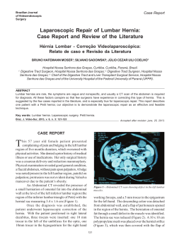
SPINE Volume 30, Number 16S, pp S68 –S72
©2005, Lippincott Williams & Wilkins, Inc.
Dynamic Stabilization in the Surgical Management
of Painful Lumbar Spinal Disorders
Russ P. Nockels, MD
Study Design. A literature review.
Objective. To evaluate the mechanisms of action and
effectiveness of posterior dynamic stabilization devices in
the management of painful spinal disorders.
Summary of Background Data. Dynamic stabilization
may provide pain relief by altering the transmission of
abnormal loads across the degenerated disc space.
Methods. A Medline search was conducted.
Results. Articles supporting abnormal load transmission
across the disc space and clinical reviews of currently available posterior dynamic systems were included.
Conclusions. Posterior dynamic stabilization systems
may provide benefit comparable to fusion techniques, but
without the elimination of movement. Further study is required to determine optimal design and clinical indications.
Key words: dynamic stabilization, degenerative disc
disease, lumbar spinal disorders, low back pain, motion
preservation, lumbar fusion. Spine 2005;30:S68 –S72
Current surgical management of the painful lumbar motion segment is imperfect. Improvements are necessary in
the predictability of pain relief, the reduction of treatment related morbidities, and an overall improvement in
the clinical success rates of pain reduction and functional
improvement. Recent advances in fusion techniques have
elevated arthrodesis rates, without an equivalent improvement in relief of pain.1 Fusion is intended to alleviate pain secondary to abnormal motion, or instability.2
Recent reports, however, have demonstrated relative
success with implants that permit movement rather than
eliminate it. These observations have promoted a more
thorough understanding of alternate mechanisms that
cause low back pain, particularly those centered on patterns of load transmission in the disc space.
Abnormal patterns of load transmission are recognized as a principal cause of osteoarthritic changes in
other joints.3 Spinal osteoarthritic changes may be due to
similar forces across the lumbar disc. Dynamic stabilization, or “soft stabilization,” systems seek to alter the
mechanical loading of the motion segment by unloading
the disc, without the loss of motion required by fusion
From the Departments of Neurological Surgery, Loyola University
Medical Center, Maywood, IL.
Acknowledgment date: April 25, 2005. Acceptance date: May 24,
2005.
The device(s)/drug(s) that is/are the subject of this manuscript is/are not
FDA-approved for this indication and is/are not commercially available in the United States.
No funds were received in support of this work. No benefits in any
form have been or will be received from a commercial party related
directly or indirectly to the subject of this manuscript.
Address correspondence and reprint requests to Russ P. Nockels, MD,
c/o Department of Neurological Surgery, 2160 South First Avenue,
Maywood, IL 60153; E-mail: [email protected]
S68
surgery. The concept is particularly appealing with
greater recognition of the negative effects of fusion on
adjacent spinal segments and global functioning of the
spine following surgery. Additionally, the concept may
help explain the common clinical scenario in which back
pain is primarily related to position or posture, rather
than movement of the lumbar spine. The clinical application of this concept will be explored, along with the
international experience to date, and potential future
directions.
Rationale for Dynamic Stabilization
For two decades, the dominant surgical justification for
fusing the painful motion segment has been the concept
of instability.4,5 Yet, instability is difficult to define.
When abnormal motion is present on flexion-extension
radiographs, especially in the setting of spondylolisthesis, fusion seems an effective option.6 By this standard,
however, relatively few patients with low back pain have
overt subjective or objective evidence of instability.
Low back symptoms often implicate abnormal loading rather than motion as the primary source of pain.
Many patients complain of postural or positional pain as
a prevailing symptom.7 Radiographs of these patients
often fail to demonstrate motion on dynamic studies.
Furthermore, many patients with low back pain fail to
improve following a successful lumbar fusion.1,8 These
observations suggest that low back pain may have etiologies related to load, and successful treatments may exist
beyond fusion.
Pain at a symptomatic motion segment may originate
from the vertebral endplates, the disc anulus, vertebral periosteum, facet joints, and/or surrounding supportive soft tissue structures.9 As the lumbar spine ages, these structures
undergo well-described degenerative changes, such as disc
space dehydration and collapse, and corresponding facet
arthropathy. The increased stiffness that accompanies these
changes may further aggravate global spinal function, by
diminishing sagittal balance and disrupting coronal and
sagittal contour.10,11
The pathologic changes within the disc space may result in abnormal transmission of load across the endplates. It has been well established that, in other weightbearing joints, abnormal load transmissions result in
degenerative changes, and the resultant pain may be diminished by a properly placed osteotomy.3 For example,
in younger patients, alteration of the load transmission
across a painful hip joint is often treated in this way. By
comparing the role of the normal and degenerated disc
on load transmission, we may see the same principles
applied to the painful lumbar motion segment.
Dynamic Stabilization • Nockels S69
The normal disc consists of a homogeneous gel of
collagen and proteoglycan.12 The normal disc is therefore isotropic, like a fluid-filled bag, a property that allows it to transmit load uniformly across the vertebral
endplates.13,14 Importantly, this asset allows the disc to
distribute equal loads across its surface regardless of position. In this way, flexion, extension, and lateral bending are all accommodated.
Disc degeneration alters the isotropic properties of the
disc.14,15 The disc becomes nonhomogeneous, with areas
of fragmented and condensed collagen, fluid and gas.16
Load transmission over the endplates, therefore, becomes uneven. Focal loading of the endplate cartilage
and subchondral bony trabeculae can occur with certain
positions, leading to endplate thinning and destruction.17,18 Since a clear-cut association exists between
posture and load on the disc, it may follow that certain
upright postures may cause pain via the inability of the
degenerative disc to distribute the load evenly.19,20 At
the same time, the corresponding loss of disc space height
reduces tension of the anulus, leading to infolding and
fractures of the anular structures.21
The loads across the disc space are now highly dependent on position. This “stone-in-the-shoe” phenomenon
helps explain the association some patients may have
between their individual pattern of disc composition and
postural pain. 22 A pressure profilometry study by
McNally and Adams revealed the anisotropic properties
of the degenerated disc.14 This study demonstrated that
the pattern of loading, rather than the absolute levels of
loading, was related to pain generation in the degenerated spine.23 The concept may also help explain the lack
of correlation between degrees of disc degeneration and
back pain, since individual anatomic and consequential load transmission changes vary highly form one
person to another. In addition, the continued progression of these changes results in the disc becoming more
collagenized and homogeneous. Therefore, the very
aged disc may once again distribute loads more evenly,
resulting in a degree of spontaneous relief of pain with
time.24
Altering the load transmission across the degenerated
disc may therefore be beneficial. Furthermore, such benefit could be accomplished without the elimination of
movement. Dynamic stabilization devices place the posterior structures under tension and create a focal increase
in lordosis. This process may shift load transmission so
that certain positions are more tolerable, and may limit
motion so that painful positions are not experienced.22
Dynamic stabilization has several theoretical advantages over fusion. By allowing limited motion, dynamic
stabilization may negate the deleterious effects of fusion
on adjacent levels and on overall sagittal balance.25 Fusion has been implicated in accelerated disease of adjacent motion segments, and in the case of surgical posterior distracting procedures, major deformities such as
flat back syndrome.26 Even well-performed fusions impose considerable postural stress on levels above the fu-
sion. Fusions form L4 to S1 place considerable rotatory
stress on the sacroiliac joints during sitting.27 Dynamic
systems may allow the motion segment to be altered in an
anatomic fashion when subjected to postural changes.
Furthermore, solid posterolateral fusions do not stop
loading of the disc. Although the pattern of load transmission may be altered, fusion may also prevent the spine
form, taking up a position where normal loading occurs.
If the transmission of load across the disc space is a
source of pain, the results of anterior fusion should be
superior to posterior fusion, and in some studies, they
are.2,28 It is interesting to note that the most successful
interbody fusions are associated with development of
bone around the cage, increasing the surface area for
load transmission.29,30 Without such bone, interbody
devices may transmit enormous loads to the vertebral
endplate.31 It is possible that the success of large interbody fusions is due to a favorable alteration of load
transmission. Posterior stabilization systems may provide similar benefit by altering disc loads without the loss
of movement. By creating a more normal loading pattern, dynamic stabilization may replicate the success of
large volume interbody fusions, without the loss of
movement.
Dynamic systems, therefore, may work by limiting
movement to a zone or range where normal or near normal loading may occur or by preventing a spinal position
where abnormal loading may occur.
Device History
Outside of North America, lumbar dynamic stabilization
devices have been used for over a decade. Typically, these
systems provide dynamic, or “soft” stabilization by providing a posterior tension band. This places the motion
segment in extension while allowing limited movements
in other planes. The Graf ligament system was one of the
first such devices used. It consists of a posterior nonelastic band that serves as a ligament between two pediclebased screws32 (Figure 1). The inventor, Henri Graf,
thought that abnormal rotatory motion was responsible
for pain generation; therefore, the device was primarily
designed to control rotatory movement by locking the
lumbar facets in the extended position. Limited flexion
was allowed within the range of normal movement. By
providing posterior tensioning, the system probably unloads the anterior disc and may redistribute the load
transmission of the painful disc. Although widely used,
the clinical effects of the Graf system have not been rigorously studied. Some analyses, however, have demonstrated clinical success of the Graf ligamentoplasty similar to fusion procedures.33–36 In two separate studies,
clinical rates of excellent to good outcomes were in the
75% range with 2-year follow-up.33,37 It is recommended by the authors that the device be used in younger
patients with adequate lumbar musculature, and in
whom facet arthropathy is minimal. A prospective study
of 88 patients by Konno and Kikuchi with low-grade
spondylolisthesis, application of the Graf system seemed
S70 Spine • Volume 30 • Number 16S • 2005
Figure 1. Graf ligamentoplasty: The implant is shown disassembled and in situ. The components include a nonelastic band, which
is secured around two pedicle screw heads by a metal band.
to diminish low back pain complaints as compared to
decompression alone at 4 years.38 The device has also
been associated with an increase in lateral recess stenosis
due to abutment of the facets in extension, infolding of
the ligamentum flavum, and reduction of the neural foramen.35 This effect was primarily due to the segmental
lordosis associated with its application and resulted in
some early surgical complications.37
The Dynesys system (Zimmer Spine, Warsaw, IN) includes a design that provides both controlled flexion and
extension by combining a tension band with a plastic
tube, which resides between pedicle screws (Figure 2). In
flexion, motion is controlled by tension on the band,
while during extension the plastic cylindrical tubes act as
Figure 2. Dynesys System: Pedicle screws are connected by a
central cord surrounded by a cylindrical plastic bumper.
a partially compressible spacer, thereby allowing limited
extension.39,40 Indeed, these plastic cylinders can be partially weight bearing in extension. In order to function
properly, application of the Dynesys device must follow
careful technical guidelines. Inappropriately long plastic
spacers, for example, may cause a focal kyphosis, a scenario that has been associated with poor outcomes.41
The Dynesys system may have some advantage over pure
band-like devices in that it provides some protection
against compression of the posterior anulus, a structure
known to contribute to painful load bearing. Biomechanical testing of in vitro reconstructed human cadaveric spines indicates that Dynesys provides 1° to 3° of
greater movement at L3–L4 than rigid fixation in both
flexion and extension. Compared with the intact spine,
the implant permits similar degrees of extension but restricts flexion by 30%.40 Stoll et al reported on a multicenter experience with 83 consecutive patients, indicating an overall improvement in the Oswestry scores from
54 to 23.41 Grob et al recently reported a retrospective
series of 31 patients followed over 2 years implanted
with the Dynesys device.42 Although 67% of patients
reported improvement in back pain, overall improvement in quality of life was only 50%, and 19% required
reoperation in the follow-up period.42 The authors note
that these rates compare favorably with fusion, although
randomization was not performed. The device is currently undergoing evaluation by the Food and Drug Administration in the United States.
Other devices have sought to improve on these early
designs, including the Leeds-Keio ligamentoplasty for
spondylolisthesis,43,44 and the FASS (fulcrum-assisted
soft stabilization) system.22,45 The latter uses a slightly
anteriorly placed flexible fulcrum to maintain distraction
of the posterior anulus during extension. This allows unloading of the disc space while still providing motion.46
The Leeds-Keoi device was studied by Muchida et al,
who reported a reduction of back and leg pain with use
of the device comparable to fusion in spondylolisthesis.43
Despite these generally favorable results, however, it
should be noted that these studies are not of sufficient
size or caliber to provide convincing data regarding the
benefit of dynamic stabilization in the management of
low back pain. Furthermore, no motion device is cleared
for use in the United States at present.
Dynamic Stabilization • Nockels S71
Clinical Application of Dynamic Devices
Posterior dynamic lumbar spinal implants may have advantages over rigid systems in both fusion and nonfusion
applications. By allowing bone graft in the interbody
space to experience increased compressive loads during
flexion, these devices may increase the likelihood of bony
fusion across the interbody space (Wolfe’s Law). It is also
likely that these devices would be eventually used as
stand alone devices without arthrodesis. Under these circumstances, the device would provide stabilization
across the motion segment without complete elimination
of movement.
To function properly, these devices need to work in
harmony with intact soft tissue supporting structures of
the motion segment. Ideally, therefore, they would be
implanted with minimal damage to the muscular and
ligamentous structures that participate in normal spinal
motion. At present, surgical implantation of dynamic
stabilization devices is very invasive, with resulting disruption of the muscle and ligamentous structures. It is
well recognized that these soft tissue structures play an
important in normal spinal movement, balance, and load
transmission. Over time, these devices would ideally
need the natural support of muscles and ligaments to
function. Fortunately, advances in the surgical implantation of fusion-related devices, such as interbody cages,
rods, and screws, have provided minimally invasive techniques. These techniques are far less disruptive of these
supporting structures and are associated with faster recovery times. Optimal performance of dynamic systems
would seem likely to result from a coupling of these minimally invasive techniques with the implant itself. Indeed, minimally invasive surgical techniques may be
much more beneficial when used with motion-sparing
devices than with fusion in long-term results.
Special challenges exist with regard to the long-term
tolerance of dynamic implants with regard to the screwbone interface, as well as the fatigability of the composite
materials. Cyclical loading and unloading of the device
as a consequence of daily physical activities may cause
screw loosening, or implant breakage. Reports of screw
loosening in the Dynesys system experience suggest that
progress needs to be made in this area.41
Other motion-preserving devices, discussed elsewhere, may be placed between the spinous processes.47
Posterior pedicle-based devices, such as those described
here, will probably be positioned along procedures involving larger decompressions, or those involving multilevel mild degrees of spondylolisthesis and/or anterior
loss of height.
Conclusion
Dynamic stabilization systems have theoretical advantages over rigid spinal implants. They may allow similar
or improved patient outcomes compared with fusions in
patients in whom disc load characteristics represent a
modifiable solution over the sagittal plane of the verte-
bral endplates. Some design features must be addressed,
as well as placement of the devices with preservation of
the surrounding spinal structures. Ultimately, a welldesigned system would need to prove its clinical effectiveness in a well-designed prospective randomized clinical trial.
Key Points
Dynamic stabilization systems have reported
rates of clinical benefit comparable to fusion in a
few small clinical series.
● Abnormal load transmission across the degenerated disc space may be a source of postural low
back pain.
● Dynamic stabilization may alter the load transmission across the disc space into a zone of diminished pain generation while allowing limited
motion.
●
References
1. Boos N, Webb JK. Pedicle screw fixation in spinal disorders: a European
view. Eur Spine J 1997;6:2–18.
2. Zdeblick TA. A prospective, randomized study of lumbar fusion: preliminary
results. Spine 1993;18:983–91.
3. Troum OM, Crues JV 3rd. The young adult with hip pain: diagnosis and
medical treatment, circa 2004. Clin Orthop 2004;418:9 –17.
4. Dvorak J, Panjabi MM, Chang DG, et al. Functional radiographic diagnosis
of the lumbar spine: flexion-extension and lateral bending. Spine 1991;16:
562–71.
5. Kwon BK, Vaccaro AR, Grauer JN, et al. Indications, techniques, and outcomes of posterior surgery for chronic low back pain. Orthop Clin North
Am 2003;34:297–308.
6. Detwiler PW, Marciano FF, Porter RW, et al. Lumbar stenosis: indications
for fusion with and without instrumentation. Neurosurg Focus 1997;3:e4;
discussion 1 p following e4.
7. Smith D, McMurray N, Disler P. Early intervention for acute back injury:
can we finally develop an evidence-based approach? Clin Rehabil 2002;
16:1–11.
8. Gibson JN, Grant IC, Waddell G. The Cochrane review of surgery for lumbar disc prolapse and degenerative lumbar spondylosis. Spine 1999;24:
1820 –32.
9. Bogduk N. The innervation of the lumbar spine. Spine 1983;8:286 –93.
10. Fujiwara A, Lim TH, An HS, et al. The effect of disc degeneration and facet
joint osteoarthritis on the segmental flexibility of the lumbar spine. Spine
2000;25:3036 – 44.
11. Fujiwara A, Tamai K, An HS, et al. The relationship between disc degeneration, facet joint osteoarthritis, and stability of the degenerative lumbar
spine. J Spinal Disord 2000;13:444 –50.
12. Hukins DW. A simple model for the function of proteoglycans and collagen
in the response to compression of the intervertebral disc. Proc R Soc Lond B
Biol Sci 1992;249:281–5.
13. McMillan DW, McNally DS, Garbutt G, et al. Stress distributions inside
intervertebral discs: the validity of experimental ‘stress profilometry.’ Proc
Inst Mech Eng [H] 1996;210:81–7.
14. McNally DS, Adams MA. Internal intervertebral disc mechanics as revealed
by stress profilometry. Spine 1992;17:66 –73.
15. Krag MH, Seroussi RE, Wilder DG, et al. Internal displacement distribution
from in vitro loading of human thoracic and lumbar spinal motion segments:
experimental results and theoretical predictions. Spine 1987;12:1001–7.
16. Moore RJ, Vernon-Roberts B, Fraser RD, et al. The origin and fate of herniated lumbar intervertebral disc tissue. Spine 1996;21:2149 –55.
17. Ariga K, Miyamoto S, Nakase T, et al. The relationship between apoptosis of
endplate chondrocytes and aging and degeneration of the intervertebral disc.
Spine 2001;26:2414 –20.
18. Simpson EK, Parkinson IH, Manthey B, et al. Intervertebral disc disorganization is related to trabecular bone architecture in the lumbar spine. J Bone
Miner Res 2001;16:681–7.
S72 Spine • Volume 30 • Number 16S • 2005
19. Nachemson A. Towards a better understanding of low-back pain: a review
of the mechanics of the lumbar disc. Rheumatol Rehabil 1975;14:129 – 43.
20. Schultz A, Andersson G, Ortengren R, et al. Loads on the lumbar spine:
validation of a biomechanical analysis by measurements of intradiscal pressures and myoelectric signals. J Bone Joint Surg Am 1982;64:713–20.
21. Jinkins JR. Acquired degenerative changes of the intervertebral segments at
and suprajacent to the lumbosacral junction: a radioanatomic analysis of the
nondiscal structures of the spinal column and perispinal soft tissues. Eur J
Radiol 2004;50:134 –58.
22. Mulholland RC, Sengupta DK. Rationale, principles and experimental evaluation of the concept of soft stabilization. Eur Spine J 2002;11(suppl 2):
198 –205.
23. McNally DS, Shackleford IM, Goodship AE, et al. In vivo stress measurement can predict pain on discography. Spine 1996;21:2580 –7.
24. Modic MT. Degenerative disc disease and back pain. Magn Reson Imaging
Clin North Am 1999;7:481–91, viii.
25. Korovessis P, Papazisis Z, Koureas G, et al. Rigid, semirigid versus dynamic
instrumentation for degenerative lumbar spinal stenosis: a correlative radiological and clinical analysis of short-term results. Spine 2004;29:735– 42.
26. Okuda S, Iwasaki M, Miyauchi A, et al. Risk factors for adjacent segment
degeneration after PLIF. Spine 2004;29:1535– 40.
27. Lazennec JY, Ramare S, Arafati N, et al. Sagittal alignment in lumbosacral
fusion: relations between radiological parameters and pain. Eur Spine J
2000;9:47–55.
28. Schofferman J, Slosar P, Reynolds J, et al. A prospective randomized comparison of 270 degrees fusions to 360 degrees fusions (circumferential fusions). Spine 2001;26:E207–12.
29. McAfee PC. Interbody fusion cages in reconstructive operations on the spine.
J Bone Joint Surg Am 1999;81:859 – 80.
30. Whitecloud TS 3rd, Castro FP Jr, Brinker MR, et al. Degenerative conditions
of the lumbar spine treated with intervertebral titanium cages and posterior
instrumentation for circumferential fusion. J Spinal Disord 1998;11:
479 – 86.
31. Polikeit A, Ferguson SJ, Nolte LP, et al. The importance of the endplate for
interbody cages in the lumbar spine. Eur Spine J 2003;12:556 – 61.
32. Graf H. Lumbar instability: surgical treatment without fusion. Rachis 1992;
412:123–37.
33. Grevitt MP, Gardner AD, Spilsbury J, et al. The Graf stabilisation system:
early results in 50 patients. Eur Spine J 1995;4:169 –75; discussion 35.
34. Gardner A, Pande KC. Graf ligamentoplasty: a 7-year follow-up. Eur
Spine J 2002;11(suppl 2):157– 63.
35. Hadlow SV, Fagan AB, Hillier TM, et al. The Graf ligamentoplasty procedure: comparison with posterolateral fusion in the management of low back
pain. Spine 1998;23:1172–9.
36. Brechbuhler D, Markwalder TM, Braun M. Surgical results after soft system
stabilization of the lumbar spine in degenerative disc disease–long-term results. Acta Neurochir (Wien) 1998;140:521–5.
37. Markwalder TM, Wenger M. Dynamic stabilization of lumbar motion segments by use of Graf’s ligaments: results with an average follow-up of 7.4
years in 39 highly selected, consecutive patients. Acta Neurochir (Wien)
2003;145:209 –14; discussion 14.
38. Konno S, Kikuchi S. Prospective study of surgical treatment of degenerative
spondylolisthesis: comparison between decompression alone and decompression with Graf system stabilization. Spine 2000;25:1533–7.
39. Rajaratnam SS, Shepperd JAN, Mulholland RC. Dynesis stabilization of the
lumbo-sacral spine. Second Combined Meeting of the BSS BASS BCSS SBSR,
Birmingham, AL, 2002.
40. Schmoelz W, Huber JF, Nydegger T, et al. Dynamic stabilization of the
lumbar spine and its effects on adjacent segments: an in vitro experiment.
J Spinal Disord Tech 2003;16:418 –23.
41. Stoll TM, Dubois G, Schwarzenbach O. The dynamic neutralization system
for the spine: a multi-center study of a novel non-fusion system. Eur Spine J
2002;11(suppl 2):170 – 8.
42. Grob D, Benini A, Junge A, et al. Clinical experience with the Dynesys
semirigid fixation system for the lumbar spine: surgical and patient-oriented
outcome in 50 cases after an average of 2 years. Spine 2005;30:324 –31.
43. Mochida J, Suzuki K, Chiba M. How to stabilize a single level lesion of
degenerative lumbar spondylolisthesis. Clin Orthop 1999:126 –34.
44. Mochida J, Toh E, Suzuki K, et al. An innovative method using the LeedsKeio artificial ligament in the unstable spine. Orthopedics 1997;20:17–23.
45. Sengupta DK. Dynamic stabilization devices in the treatment of low back
pain. Orthop Clin North Am 2004;35:43–56.
46. Sengupta DK, Mulholland RC. Fulcrum assisted soft stabilization system: a
new concept in the surgical treatment of degenerative low back pain. Spine
2005;30:1019 –29.
47. Caserta S, La Maida GA, Misaggi B, et al. Elastic stabilization alone or
combined with rigid fusion in spinal surgery: a biomechanical study and
clinical experience based on 82 cases. Eur Spine J 2002;11(suppl 2):192–7.
Baixar