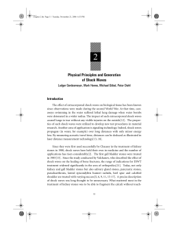
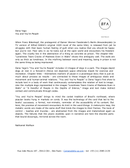
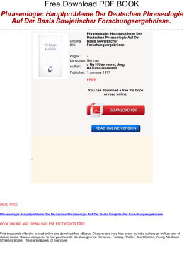
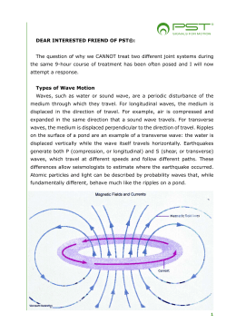
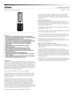
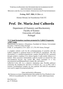
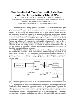
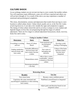
Chapter 18.fm Page 193 Tuesday, November 21, 2006 6:41 PM
18
rESWT in Heel Spur (Fasciitis Plantaris)
Gerald Hupt1
Rupert Diesch,2 Thomas Straub,3 Emil Penninger,3 Thomas Frölich,4
Jakob Schöll,5 Heinz Lohrer,5 Dr. Theodor Senge6
17
Introduction
The overall prevalence of fasciitis plantaris is about 15%. Lateral x-ray exams
on caucasians revealed plantar and/or dorsal heel spurs in 15.7% whereby both
legs were affected in 11%[32]. The incidence rises with age and is comparable for
different continents such as Europe, Africa and America[2].
The primary symptom is pain, frequently associated with restrictions in
range of motion (ROM)[3, 9, 14]. A multitude of conservative treatments have
been described[30, 40, 41]. These include ultrasound [10, 37], lontopheresis [16]
and low-energy laser [12] in addition to physical therapy, steroid injections and
non-steroid antiphlogistics, however without evidence brought today of their
efficacy. Operative therapy is recommended only following failure of conservative
action[1].
1
Urological clinic and policlinic - University of Cologne
2
Private practice, Friedrichshafen
3
Private practice, Dingolfing
4
Private practice, Stuttgart
5
Sportsmedicine Institute of Frankfurt
6
Urological clinic – Uniersity of Bochum, Herne
193
Chapter 18.fm Page 194 Tuesday, November 21, 2006 6:41 PM
194
EXTRACORPOREAL SHOCK WAVE THERAPY
The introduction of extracorporeal shock waves for the treatment of urolithiasis has revolutionized renal calculi therapy. Additional applications focus on
other calcull such as gallstones, pancreatic stones and salivary duct stones[28, 34,
35]. Since 1986 we have tested the effect of shock waves on the healing of wound
and bone fractures in experimental models and were first to demonstrate the
osteogenetic potential of shock waves[21-23]. This led to the therapy of pseudarthroses with shock waves. Finally, soft tissue diseases such as calcific tendonitis of
the shoulder, epicondylitis lateralis and medialis and fasciitis plantaris have also
been treated increasingly in recent years[7, 15, 31].
Apart from the conventional generation of shock waves, these can also be
produced ballistically (Lithoclast) and were first used in urology (for endoscopic
stone fragmentation). These processes are significantly more economical. Our
own studies on soft tissue and bones of rabbits and monkeys showed that the
results of treatment with ballistically produced radial shock waves coincided with
those obtained following therapy with focused extracorporeal shock waves.
Accordingly, one can state that the shock waves produced by either method are
comparable in terms of their biological effect. This study examines the effect of
radial shock waves on fasciitis plantaris.
Materials and Methods
All shock wave treatments were performed with the Swiss DolorClast® (Figure 1), a modified device compared to the Swiss LithoClast®, which is a unit for
Figure 1.
Therapy of the Fasciitis plantaris.
Chapter 18.fm Page 195 Tuesday, November 21, 2006 6:41 PM
rESWT IN HEEL SPUR (FASCIITIS PLANTARIS)
195
Figure 2. Pain at night.
the endoscopic treatment of calculi[27, 42]. The modifications were based on animal studies conducted on rabbits and rhesus monkeys[19]. In plantar heel pain,
the tip of the applicator was positioned at the point where the greatest pain was
reported via patient feedback (Figure 2). Shock waves were delivered via a customary medium (ultrasound gel).
Patients
One hundred and three consecutive patients with chronic fasciitis plantaris
were examined as part of a multicentric, prospective, randomized and placebocontrolled study. Patients were enrolled if they reported a pain history of at least 6
months with at least 2 different failed attempts at a conservative treatment indicating the need for surgery. Exclusion criteria were defined as poor health condition (Karnofsky index < 70), a specific intervention within the last two weeks,
pregnancy, disturbance in coagulation, tumors in the area to be treated and systemic diseases which could be interpreted as possible causes of pain in differential
diagnostics (e.g. collagenosis, rheumatic diseases).
Patients were randomized at a 1:1 ratio into a verum or sham group. While
both groups obtained identical treatments, the test set-up was modified in the
control group so that no shock waves would be transmitted. Up to 3 treatments
were conducted without local anesthesia. Local anesthesia was administered only
in exceptional cases when pain was no longer tolerable. Follow-up exams were
conducted after 1, 4, 12 and 52 weeks.
Chapter 18.fm Page 196 Tuesday, November 21, 2006 6:41 PM
196
EXTRACORPOREAL SHOCK WAVE THERAPY
Total
Verum group
Control group
50.4 ± 11.7
50.4 ± 11.3
50.6 ± 12.3
Female
77
39
38
Male
26
16
10
Right position
49
27
22
Left position
54
28
26
Pain duration
(months)
24.0 ± 27.5
23.7 ± 27.4
24.6 ± 28.1
Age (years)
Table 1.
Demographic data.
If pain persisted after 4 weeks in patients from the control group, blinding
was terminated and admission to the treatment group was authorized.
Questionnaires were filled out by the treating orthopedist or surgeon and
forwarded anonymously to the study center for statistical evaluation (SPSS).
Results
One hundred and three patients were admitted to the study. Fifty five
patients were randomized into the verum group, 48 into the control group.
Demographic data (Table 1) as well as symptoms and initial findings (Table 2)
show homogeneity at baseline.
Treatments were performed at an initial pressure of 4 bar with 2000 shock
waves. Local anesthesia was required for 5 patients (9%) in the verum group and
3 patients (6%) in the control group. Local symptoms could be observed immediately following treatment (Table 3), but they disappeared after 1 week.
Eighty-four patients were examined up to 52 weeks after the last rESWT.
Night pain, pain at rest and walking improved significantly in the treatment
group compared to placebo (Figures 3-5). In fact, an increasing improvement
throughout the entire follow-up period could be observed. In the control group
no significant change between initial findings and follow-up could be found.
Patients who dropped out of the control group 4 weeks after rESWT due to persistent pain received an unblinded active treatment. They finally achieved results
similar to the primary treatment group.
Chapter 18.fm Page 197 Tuesday, November 21, 2006 6:41 PM
rESWT IN HEEL SPUR (FASCIITIS PLANTARIS)
Total
Verum group
Control group
Night pain
32.0
36.4
27.1
Restriction in daily life
95.1
92.7
97.9
Restriction in sports
66.0
74.5
56.3
Restriction in occupation
52.4
58.2
45.8
0.0
0.0
0.0
Restricted
57.3
49.1
66.7
Not restricted
14.6
16.4
12.5
Start-up pain
23.3
27.3
18.8
Redness
1.0
0.0
2.1
Overheating
1.9
0.0
4.2
Swelling
6.8
3.6
10.4
Scars
1.0
1.8
0.0
Injection sites
1.0
1.8
0.0
Skewfoot
21.4
20.0
22.9
Splayfoot
35.9
30.9
41.7
Flatfoot
39.8
34.5
45.8
2.9
0.0
6.3
Maximum walking time
Pes equinocavus
Table 2. Pathology and findings giving as percent (%).
Total
Verum group
Control group
48.5
76.4
37.5
Petechial bleedings
9.7
18.2
0.0
Hematoma
2.9
5.5
0.0
Swelling
20.4
34.5
4.2
Pain
18.4
32.7
2.1
Irritation
Table 3. Postoperative adverse events (%).
197
Chapter 18.fm Page 198 Tuesday, November 21, 2006 6:41 PM
198
EXTRACORPOREAL SHOCK WAVE THERAPY
Figure 3.
Rest pain.
Limitations in walking time and daily activities in the verum group were persistent in 36% and 34% respectively. Limitations in sports and profession, were
persistent in 52% and 50%, even though the extent of the limitation had
decreased significantly. Comparative values for the control group are above 70%.
The majority of patients would repeat the treatment after 1 week. This
remained unchanged in the verum group. In the control group this value drops
down after 4 weeks and once again after 12 weeks (Figure 5). This observation
Figure 4.
Walking pain.
Chapter 18.fm Page 199 Tuesday, November 21, 2006 6:41 PM
rESWT IN HEEL SPUR (FASCIITIS PLANTARIS)
199
Figure 5. Acceptance of reapplication.
correlates with the patients’ satisfaction. After 12 weeks, over 90% of patients
report an improvement and more than 60% are even completely satisfied. In the
control group, this is documented as 10% (Figure 6).
No clinically relevant side effects were found, but minor petechial bleeding
and swelling were reported.
Discussion
In the past 30 years, the effects of many physical factors on the healing processes of bone and soft tissue have been studied. With the consideration of
Figure 6. Patient satisfaction.
Chapter 18.fm Page 200 Tuesday, November 21, 2006 6:41 PM
200
EXTRACORPOREAL SHOCK WAVE THERAPY
extracorporeal shock waves for the treatment of urolithiasis, a new physical
medium was introduced to medicine[4, 5]. Shock waves are able to generate
effects without any surgery. These shock wave were used in urolithiasis. As of
1985 gall stones and pancreatic and salivary duct calculi were treated with shock
waves also[28, 34, 35]. Common to all these therapies is a fragmentation of the
calculi by the shock waves.
Shock waves were used for the first time in 1986 for the purpose of stimulating healing processes rather than fragmenting stones. Low energetic shock waves
were known to stimulate wound healing but high energetic shock waves did not.
These findings were observed in superficial skin wounds in pigs[20]. In addition,
shock waves were shown to have an osteogenetic effect, which led to the use of
shock waves in the treatment of pseudarthrosis[11, 15, 17, 18, 22, 24, 25, 36, 38,
39]. The positive effects on wound healing were no longer used until recent studies have shown again these effects. The wound healing effect is discussed further
within the chapter written by Wolfgang Schaden and Richard Thiele.
In the case of fasciitis plantaris, for a long time almost no multicentric, controlled studies have examined conservative or operative processes. In the meantime, several excellent prospective randomized placebo-controlled studies on the
effect of focused ESWT have been published, some with partially contradictory
results[43, 44, 45]. Therefore, an evaluation of the efficacy of focused ESWT is
not conclusively possible but it is certain that a conservative therapy of ESWT is
indicated prior to surgery. The recent reviews have shown the evidence of shock
waves on chronic plantar heel pain.
This study reports for the first time on the effect of radial ESWT (rESWT) for
the treatment of heel spur. The patients enrolled in this trial had at least two
unsuccessful conservative therapies and the pain duration was longer than 6
months. This corresponds to a negative selection.
The subjective success rates of conventional extracorporeal shock wave therapy are shown as 50 to 75% painfree or significant pain reduction[6, 8, 29, 33].
Radial shock wave therapy obtains comparable success rates. The well known placebo effect, which is device associated, was controlled by a sham rESWT group.
The essential difference and advantage compared to conventional focused shock
wave therapy is: rESWT is easy to administer, no imaging is required, no local
anesthesics are required and the cost is significantly reduced. In case of failed
rESWT, surgical options remain possible.
Chapter 18.fm Page 201 Tuesday, November 21, 2006 6:41 PM
rESWT IN HEEL SPUR (FASCIITIS PLANTARIS)
201
The side effects of radial therapy are similar to those of focused ESWT such
as transitory pain, petechial bleeding or subcutaneous hematomata in up to 4%
of the patients[26]. However, local skin symptoms are clearly more frequent in
radial therapy which is easy to explain because of the high energy close to the tip
of the applicator. After 1 week, no more side effects were found and no neurological disorders occurred. Local irritation therefore does not appear to reach clinical
relevance.
The use of shock waves in orthopedics remains controversial and continues
to be documented by insufficient studies[13].
A multitude of shock wave studies have been published but only few prospective randomized controlled studies are available. Nevertheless, this therapy for
fasciitis plantaris has been found to be effective and is not to be classified as “lifestyle” therapy. The high recommendation by patients and physicians supports the
clinical evidence that ESWT is effective, much more than any surgical or injection
technique.
The radial shock wave therapy examined in our study appears to be clinically
effective without side effects. Therefore it is reasonable to use the rESWT for the
treatment of chronic fasciitis. In addition, rESWT provides for a very economical
treatment when compared to conventional shock wave processes.
Literature
1.
2.
3.
4.
5.
6.
Anderson, R.B., M.D. Foster: Operative treatment of subcalcaneal pain. Foote Ankle
(US) 9(1989) 317-323
Banadda, B.M., O. Gona, D.M. Ndlovu, R. Vaz: Calcaneal spurs in a black African
population. Foot Ankle 13(1992) 352-354
Baxter, D.E., C.M. Thigpen: Heel pain: operative results. Foot Ankle 5(1984) 16-25
Chaussy, C., Extracorporeal shock wave lithotripsy. 1982, Basel: Karger.
Chaussy, C., F. Eisenberger, K. Wanner, F. Forssmann, W. Hepp, E. Schmiedt, W.
Brendel: The use of shock waves for the destruction of renal calculi without direct
contact. Urol. Res. 4(1976) 175
Dahmen, G.P., R. Franke, V. Gonchars, K. Poppe, S. Lentrodt, S. Lichtenberger, S.
Jost, J. Montigel, V.C. Nam, G. Dahmen: Die Behandlung knochennaher
Weichteilschmerzen mit Extrakorporaler Stosswellentherapie (ESWT), Indikation,
Technik und bisherige Ergebnisse. In: Chaussy, C., F. Eisenberger, D. JochamD.
Wilbert (Hrsg.): Die Stosswelle - Forschung und Klinik, Attempto Verlag, Tübingen
(1995) 175-186.
Chapter 18.fm Page 202 Tuesday, November 21, 2006 6:41 PM
202
7.
8.
9.
10.
11.
12.
13.
14.
15.
16.
17.
18.
19.
20.
21.
22.
23.
24.
25.
EXTRACORPOREAL SHOCK WAVE THERAPY
Dahmen, G.P., L. Meiss, V.C. Nam, B. Skruodies: Extrakorporale
Stosswellentherapie (ESWT) im knochennahen Weichteilbereich an der Schulter.
Extracta Orthopaedica 11(1992) 25-27
Diesch, R., G. Haupt: Extracorporeal shock waves in the treatment of
pseudarthrosis, calcific tendinitis of the shoulder and calcaneal spur. In: Siebert,
W.M. Buch (Hrsg.): Springer Verlag, Berlin Heidelberg New York (1997) 131-135.
DuVries, H.L.: Heel spur (calcaneal spur). A.M.A. Arch. Surg. 74(1957) 536
Ebenbichler, G., K.L. Resch: Kritische Überprüfung des therapeutischen
Ultraschalls. Wien. Med. Wochenschr. 144(1994) 51-53
Ekkernkamp, A., A. Bosse, G. Haupt, A. Pommer: Der Einfluß der extrakorporalen
Stosswellen auf die standardisierte Tibiafraktur am Schaf. In: Ittel, T., G. SieberthH.
Matthiass (Hrsg.): Aktuelle Aspekte der Osteologie, Springer, Berlin Heidelberg
New York (1992) 307-310.
Ernst, E., V. Fialka: Low-dose Lasertherapie: eine kritische Prüfung der klinischen
Wirksamkeit. Schweiz. Med. Wochenschr. 123(1993) 949-954
Fritze, J.: Extrakorporale Stosswellentherapie (ESWT) in orthopädischer
Indikation: Eine ausgewählte Übersicht. Versicherungsmedizin 50(5) (1998) 180185
Furey, J.G.: Plantar fasciitis: the painful heel syndrome. J. Bone Joint Surg. 75A(1975) 672-673
Graff, J., Die Wirkung hochenergetischer Stosswellen auf Knochen und
Weichteilgewebe. 1989, Bochum: Habilitationsschrift, Ruhr-Universität Bochum.
Grossi, E., G.C. Monza, S. Pollavini, L. Bona: NSAID ionisation inthe management
of soft tissue rheumatism: role played by the drug, electrical stimulation and
suggestion. Clin. Exp. Rheumatol. 4 (1986) 265-267
Haist, J.: Die Osteorestauration via Stosswellenanwendung. Eine neue Möglichkeit
zur Therapie der gestörten knöchernen Konsolidierung. In: Chaussy, C., F.
Eisenberger, D. JochamD. Wilbert (Hrsg.): Die Stosswelle - Forschung und Klinik,
Attempto Verlag, Tübingen (1995) 157-161.
Haist, J., D. Steeger, U. Witzsch, R.A. Bürger, U. Haist: The extracorporeal
shockwave therapy in the treatment of disturbed bone union. 7th Int. Conference
on Biomedical Engineering, Singapore (1992) 222-224
Haupt, G., Behandlung von Harnsteinen, Knochen und Weichgewebe mit
ballistischer Energie: Leistungsfähigkeit und Systemerweiterung einer neuen
minimal-invasiven Therapietechnik. 1997: Ruhr-Universität Bochum.
Haupt, G., M. Chvapil: Effect of shock waves on the healing of partial-thickness
wounds in piglets. J. Surg. Res. 49(1990) 45-48
Haupt, G., A. Ekkernkamp, M. Chvapil, A. Haupt, B. Gerety: Der Einfluß
extrakorporaler Stosswellen auf die Knochenbruchheilung. Hefte zur
Unfallheilkunde 220(1991) 524
Haupt, G., A. Haupt, A. Ekkernkamp, B. Gerety, M. Chvapil: Influence of shock
waves on fracture healing. Urology 39(1992) 529-532
Haupt, G., A. Haupt, B. Gerety, M. Chvapil: Enhancement of fracture healing with
extracorporeal shock waves. J. Urol. 143(1990) 230A
Haupt, G., A. Haupt, T. Senge: Die Behandlung von Knochen mit extrakorporalen
Stosswellen - Entwicklung einer neuen Therapie. In: Chaussy, C., F. Eisenberger, D.
JochamD. Wilbert (Hrsg.): Stosswellenlithotripse, Aspekte und Prognosen,
Attempto Verlag, Tübingen (1993) 120-126.
Haupt, G., P. Katzmeier: Anwendung der hochenergetischen extrakorporalen
Stosswellentherapie bei Pseudarthrosen, Calcific tendinitis der Schulter und
Ansatztendinosen (Fersensporn, Epicondylitis). In: Chaussy, C., F. Eisenberger, D.
Chapter 18.fm Page 203 Tuesday, November 21, 2006 6:41 PM
rESWT IN HEEL SPUR (FASCIITIS PLANTARIS)
26.
27.
28.
29.
30.
31.
32.
33.
34.
35.
36.
37.
38.
39.
40.
41.
42.
203
JochamD. Wilbert (Hrsg.): Die Stosswelle - Forschung und Klinik, Attempto Verlag,
Tübingen (1995) 143-146.
Haupt, G., A. Menne, M. Schulz: Medizinisches Instrument zum Erzeugen und
Weiterleiten von extrakorporalen nicht fokussierten Druckwellen auf biologisches
Gewebe. Patentanmeldung (1997)
Haupt, G., R. Olschewski, S. Hartung, T. Senge: Comparison of laser and ballistic
systems by in vitro lithotripsy with a standardized stone model. J. Endourol.
7(1993) S62
Iro, H., H. Schneider, C. Födra, G. Waitz, N. Nitsche, H.H. Heinritz, J. Benninger,
C. Ell: Shockwave lithotripsy of salivary duct stones. Lancet 339(1992) 1333-1336
Krischek, O., J.-D. Rompe, B. Herbsthofer, B. Nafe: Symptomatische niedrigenergetische Stosswellentherapie bei Fersenschmerzen und radiologisch
nachweisbarem plantaren Fersensporn. Z. Orthop. 136(1998) 169-174
Labelle, H., R. Guibert, N. Newman, M. Fallaha, C.-H. Rivard: Lack of scientific
evidence for the treatment of lateral epicondylitis of the elbow. J. Bone. Joint. Surg.
74-B(1992) 646-651
Loew, M., W. Jurgowski: Erste Erfahrungen mit der Extrakorporalen StosswellenLithotripsie (ESWL) ind der Behandlung der Tendinosis calcarea der Schulter. Z.
Orthop. 131(1993) 470-473
Riepert, T., T. Drechsler, R. Urban, H. Schild, R. Matern: Häufigkeit,
Altersabhängigkeit und Geschlechtsverteilung des Fersensporns. Analyse der
Röntgenmorphologie bei 1027 Patienten der mitteleuropäischen Population. Rofo
Fortschr. Geb. Röntgenstr. Neuen Bildgeb. Verfahr. 162(1995) 502-505
Rompe, J.D., K. Küllmer, P. Eysel, H.M. Riehle, R. Bürger, B. Nafe:
Niedrigenergetische extrakorporale Stosswellentherapie ESWT beim plantaren
Fersensporn. Orthop. Praxis 32(1996) 271-275
Sauerbruch, T., M. Delius, G. Paumgartner, J. Holl, O. Wess, W. Weber, W. Hepp, W.
Brendel: Fragmentation of gallstones by extracorporeal shock waves. New Engl. J.
Med. 314(1986) 818-822
Sauerbruch, T., J. Holl, M. Sackmann, R. Werner, R. Wotzka, G. Paumgartner:
Disintegration of a pancreatic duct stone with extracorporeal shock waves in a
patient with chronic pancreatitis. Endoscopy 19(1987) 207-208
Schleberger, R.: Anwendung der extrakorporalen Stosswelle am Stütz- und
Bewegungsapparat im mittelenergetischen Bereich. In: Chaussy, C., F. Eisenberger,
D. JochamD. Wilbert (Hrsg.): Die Stosswelle - Forschung und Klinik, Attempto
Verlag, Tübingen (1995) 166-174.
Stratford, P.W., D.R. Levy, S. Gauldie, D. Miseferi, K. Levy: The evaluation of
phonophoresis and friction message as treatments for extensor carpi radialis
tendinitis: a randomized controlled trial. Physiotherapy Canada 41(1989) 93-99
Sukul, K., E.J. Johannes, E. Pierik, G. van Eijck, M. Kristelijn: The effect of high
energy shock waves focused on cortical bone: an in vitro study. J. Surg. Res.
54(1993) 46-51
Valchanou, V.D., P. Michailov: High energy shock waves in the treatment of delayed
and nonunion fractures. Internat. Orthopaedics (SICOT) 15(1991) 181-184
Verhaar, J., G. Walenkamp, A. Kester, H. van Mameren, T. van der Linden: Lateral
extensor release for tennis elbow. A prospective long-term follow-up study. J. Bone
Joint Surg. 75-A(1993) 1034-1043
Wittenberg, R., S. Schaal, G. Muhr: Surgical treatment of persistent elbow
epicondylitis. Clin. Orthop. 278(1992) 73-80
Zhong, P., G.M. Preminger: In vitro comparison of different modes of
intracorporeal shock wave lithotripsy. J. Urol. 153(1995) S47
Chapter 18.fm Page 204 Tuesday, November 21, 2006 6:41 PM
204
EXTRACORPOREAL SHOCK WAVE THERAPY
43. Rompe JD, et al. Shock wave application for chronic plantar fasciitis in running
athletes – a prospective, randomized, placebo- controlled trial. Am J Sports Med
2003; 31:268-275
44. Buchbinder R, Ptasznik R, Gordon J, Buchanan J, Prabaharan V, Forbes A:
Ultrasound-guided extracorporeal shock wave therapy for plantar fasciitis. A
randomized controlled trial. JAMA 288: 1364-1372, 2002
45. Haake M, Buch M, Schoellner C, Goebel F: Extracorporeal shock wave therapy for
plantar fasciitis: randomised controlled multicentre trial. British Medical Journal
327: 1-5, 2003
Baixar