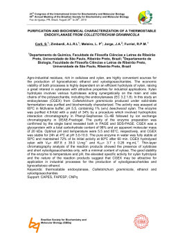
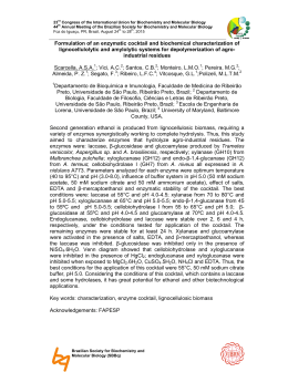
1. RETROSPECTIVE:1. RETROSPECTIVE
29/4/09
14:19
Página 28
medigraphic
Localizador
06-074
Artemisa
en línea
Originales
Retrospective analysis of leg ulcers cases
at the university hospital, Faculty of
Medicine of Ribeirão Preto, University
of São Paulo (1991-2001)
Análise retrospectiva dos casos de úlceras de perna do hospital das clínicas
da Faculdade de Medicina de Ribeirão Preto da Universidade de São Paulo
(1991-2001)
FM Barbetta, EL Mazzucato, AM Salathiel, NT Foss, MAC Frade
From the Dermatology Division of the Department of Internal Medicine. Faculty of Medicine of Ribeirão Preto.
University of São Paulo. Ribeirão Preto. SP. Brazil.
Correspondence:
Marco Andrey Cipriani Frade
Divisão de Dermatologia
Av. Bandeirantes, 3900 - Monte Alegre - Ribeirão Preto - SP
CEP: 14.049.900
Fax/Fone: 55-16-36330236
e-mail: [email protected]
Summary
Introduction: Leg ulcers are a syndrome characterized by loss of tegument that affect the lower extremities usually caused by vascular system
dysfunction, representing a high morbidity chronic disease especially among the aged. This study determined the epidemiological characteristics of
leg ulcer patients seen at Clinical Hospital Ribeirão Preto from 1991 to 2001, and their main etiologies and the resolving nature of the treatments
applied were investigated.
Method: This was a descriptive retrospective study (case series) analyzing 199 medical records of leg ulcer patients, coded by the Medical Records
from 1991 to 2001.
Results: 51% of the patients were women and 49% men, with respective median ages of 65 and 66 years, and 81% were white. Etiologically, 49%
were classified as venous ulcers, 15% arterial, 9% neuropathic, 2% anemic, and 20% were not classified. Approximately 40% of the patients had
been wounded for more than 1 year, 13% died, 40% discharged, and 45% evolved with chronic disease. Among the discharged patients, 64% of
those with arterial ulcers and 80% of those with neurotrophic ulcers underwent amputation of the segment involved. Regarding venous ulcers, only
37% of the patients were discharged, after local dressing in 56% of cases and after skin graft in only one.
Comment: Leg ulcer disease was found to be an important chronic disease among the patients seen at the Clinical Hospital Ribeirão Preto from 1991
to 2001, with severe consequences regarding the arterial and neurotrophic ulcers, although the etiologic diagnosis was unknown or neglected in
20.1% of the cases.
(FM Barbetta, EL Mazzucato, AM Salathiel, NT Foss, MAC Frade. Retrospective analysis of leg ulcers cases at the university hospital, Faculty of Medicine of Ribeirão Preto, University of São Paulo (1991-2001). Med Cutan Iber Lat Am 2009;37(1):28-32)
Key words: leg ulcers, retrospective study.
Resumo
Introdução: Úlcera de perna é uma síndrome caracterizada por perda circunscrita ou irregular do tegumento, que acomete extremidades dos membros inferiores cuja causa geralmente está na disfunção do sistema vascular. Doença crônica de alta morbidade principalmente na população idosa. O
estudo busca características epidemiológicas dos pacientes com úlceras de perna atendidos no Hospital das Clínicas de Ribeirão Preto, de 1991 a
2001, conhecer suas principais etiologias e a resolubilidade dos tratamentos realizados.
Metodologia: Estudo descritivo e retrospectivo (série de casos), da análise de 199 prontuários médicos dos pacientes com úlceras de perna, codificados pelo Arquivo Médico, de 1991 a 2001.
28
Med Cutan Iber Lat Am 2009;37(1):28-32
1. RETROSPECTIVE:1. RETROSPECTIVE
29/4/09
14:19
Página 29
FM Barbetta et al. Retrospective analysis of leg ulcers cases at the university hospital, Faculty of Medicine of Ribeirão Preto, University of São Paulo (1991-2001)
Resultados: 51% dos pacientes eram do sexo feminino, 49% masculino, medianas de idade de 65 e 66 anos respectivamente, e 81% brancos. Etiologicamente, 49% foram úlceras classificadas como venosas, 15% arteriais, 9% neuropáticas, 2% anêmicas e 20% sem classificação. Aproximadamente 40% apresentavam a doença por mais de 1 ano, 13% evoluíram para óbito, 40% para alta ambulatorial e 45% para cronicidade da doença.
Dentre os pacientes com alta, 64% dos casos de úlceras arteriais e 80% das neurotróficas sofreram amputação do segmento acometido. Quanto às
venosas, apenas 37% dos pacientes obtiveram alta, sendo 56% por curativos locais e somente 1 caso por enxertia cutânea.
Conclusões: A doença úlcera de perna foi uma enfermidade crônica importante, principalmente as venosas, dentre os pacientes atendidos no Hospital das Clínicas de Ribeirão Preto de 1991 a 2001, com repercussões graves como as arteriais e neurotróficas, apesar do diagnóstico etiológico negligenciado/desconhecido em 20,1% dos casos.
Palavras chave: úlcera de perna, estudo retrospectivo.
Leg ulcers are a syndrome characterized by the circumscribed or irregular loss of tegument (dermis or epidermis)
that may reach subcutaneous tissue and underlying tissues. The disease involves the lower extremities and the
cause usually is related to some dysfunction of the arterial
or venous vascular system.[1] The classification may be
based on etiology and therefore leg ulcers are divided into
those due to venous insufficiency (venous ulcers), to arterial insufficiency (arterial ulcers), to neuropathy (neuropathic ulcer/diabetic foot), to red blood cell abnormalities
(anemic ulcers), to obliterating thromboangiitis, as well as
those due to causes such as trauma, neoplasia, infections,
panniculitis, and pyoderma gangrenosum.[1-4] Among the
main predisposing factors are age (elderly individuals), sex
(females), obesity, profession (prolonged standing up), vulnerability of the legs to traumas and infections, increased
venous pressure, reduced arterial flow, and a family history.[1,6]
Venous ulcers present characteristic signs such as evening ankle edema and red-brown spots due to pigmentation
generated by red blood cell extravasation and later transformation of hemoglobin to hemosiderin (hypostatic purpura).
Eczema, cellulitis and streptococcal infection may be associated with the signs and symptoms. Ulceration frequently
arises after an initial trauma. The habitual localization is in
the lower third and inner surface of the leg, in the supramalleolar region. Usually there is a single ulcer that progresses
slowly, representing an ulcer of variable shape and size. At
first it presents irregular borders and a hemorrhagic or purulent fundus, but with time the borders become callous and
adhere to underlying tissues. Leishmaniasis, sporotrichosis,
neoplasias, syphilis and tuberculosis should be excluded in
the differential diagnosis. The possibility of stasis should be
considered as a factor contributing to the picture involved in
these affections.
Leg or foot ulcers occurring in elderly individuals, often
diabetic and/or hypertensive, may be fundamentally triggered by cutaneous ischemia depending on truncular
arterial lesions. This usually occurs after a traumatic
injury. The ulcers are irregular, with pallor, absence of sta-
sis, delayed color after elevation of the limb, reduction or
absence of pulsation in the foot arteries, and pain of variable intensity. Some diabetics have degenerative arteriocapillary disease, which leads to the formation of ischemic
ulcers in the skin of the lower limbs. These ulcers are
deep, painful and non-edematous, and are preferentially
located in the lower third of the leg, on the lateral surface
or on the malleoli.[7]
Ulcers of neuropathic origin are chronic and occur in an
area of anesthesia due to trauma or pressure. Anemic ulcers
usually occur in young Black women, are located in the
lower third of the leg, present nonspecific characteristics
and are diagnosed on the basis of the absence of signs of
stasis and of blood count findings (sickle-cell erythrocytes).[7]
The University Hospital of the Faculty of Medicine of
Ribeirão Preto, University of São Paulo, is a tertiary reference
hospital serving the population of the macro region of Ribeirão Preto, which consists of 80 municipalities with approximately 3 million inhabitants according to the 2000 IBGE
Census.
The objective of the present study was to describe the clinical and epidemiological characteristics of patients with leg
ulcers seen at HCFMRP-USP during the period from 1991 to
2001, and to determine the main etiologies, the possibility of
resolution and the treatments performed.
Material and methods
The authors analyzed 199 medical records of the patients
with lower limb ulcers as the main diagnosis or associated
with other major diseases coded in the list of problems of the
Medical Records Service (SAME) of HCFMRP-USP, attended during the period from 1991 to 2001. The following data
were collected: first and last year of treatment, etiologic diagnosis of the leg ulcer, demographic data (sex, color, marital
status, origin), age, discharge condition (death, ulcer resolution or active ulcer requiring ambulatory follow-up), and treatment (amputation of the limb, skin graft, dressings or vascular surgery).
Med Cutan Iber Lat Am 2009;37(1):28-32
29
1. RETROSPECTIVE:1. RETROSPECTIVE
29/4/09
14:19
Página 30
FM Barbetta et al. Retrospective analysis of leg ulcers cases at the university hospital, Faculty of Medicine of Ribeirão Preto, University of São Paulo (1991-2001)
60
Number of patients
50
Male
40
Female
30
20
10
0
0-25
26-50
51-75
76-100
Figure 1. Patient age range (years),
HCFMRP-USP, 1991-2001.
Age (years)
Results
Comment
The median age of the 199 patients whose records were evaluated was 65 years for women and 66 years for men (range:
12-92 years). Distribution by age range and sex is presented
in Figure 1. Other demographic data for the sample studied
are listed in Table 1. The etiologies of the ulcers are illustrated in Figure 2.
Data regarding time of ulcer evolution and the possibility
of resolution are listed in Table 2, and the treatments used
related to etiologic classification are listed in Table 3.
Little information is currently available in the literature about
leg ulcers, including countries like Brazil, especially regarding prevalence and epidemiology.[5] Callam et al.[4] estimated that approximately 1% of the world population presents chronic leg ulceration in some phase of life. However,
this, as well as other information about the epidemiology of
leg ulcers is probably underestimated when the general
population is considered, since many cases are not reported
or are not correctly followed and documented by a single
integrated service, but are handled by different specialties in
almost all sectors of health services.[5,8]
Table 1. Data of the patients with leg ulcers seen at HCFMRP-USP
during the period from 1991 to 2001.
Demographic data
%
Sex
Male
Female
98
101
49
51
Color
White
Non-white
163
36
82
18
Married
Single
Divorced
Widowed
121
40
6
32
61
20
3
16
Ribeirão Preto
Other towns in the RP region
Other States
111
82
6
56
41
3
Marital status
Origin
Total
n = number of patients.
30
n
Med Cutan Iber Lat Am 2009;37(1):28-32
199
20%
5%
49%
2%
9%
15%
Venous
Arterial
Neurotrophic
Anemic
Mixed
Not classified
100
Figure 1. Etiologic classification of leg ulcers, HCFMRP-USP, 19912001.
1. RETROSPECTIVE:1. RETROSPECTIVE
29/4/09
14:19
Página 31
FM Barbetta et al. Retrospective analysis of leg ulcers cases at the university hospital, Faculty of Medicine of Ribeirão Preto, University of São Paulo (1991-2001)
Table 2. Resolution and follow-up time of patients with leg ulcers seen at HCFMRP-USP during the period from 1991 to 2001.
Clinical course
<1m
1m-1y
1a5y
>5y
n
%
n
%
n
%
n
1. Death
11
42
10
38
3
12
2
2. Discharge from the service
15
19
34
44
27
34
3. Active ulcer
10
16
31
35
29
32
4. Only one visit
–
Total
36
–
21
–
75
39
Total
%
n
%
8
26
13
2
3
78
39
20
16
90
45
5
3
199
100
–
59
30
24
10
n = number of patients; m = month; y = year.
A longitudinal study carried out in the United States in
2001 reported that leg ulcers due to phlebopathy occur in
approximately 18/100,000 Americans each year.[7,9] With
the increased life expectancy of the population, this problem
has become even more relevant[7-9] because of its chronic
nature, cost of treatment and severe complications such as
amputations ans malignancies.
In the present study, venous ulcers were the most frequent (49%) although their frequency was lower than that
reported in the literature (80-90%), arterial ulcers affected 5
to 10% of patients, and the remaining cases were of neuropathic or mixed origin.[5] However, recent studies have indicated a slight increase in the frequency of arterial or associated ulcers, perhaps due to the current changes in age
distribution in the population and to the improved diagnostic
techniques for the detection of arterial disease,[5] with a
consequent decrease in the frequency of venous ulcers. In
addition, the high index of unclassified ulcers should be
pointed out, a fact that also corroborates the low index of
venous ulcers. Neurotrophic ulcers (9%) were also frequent
in the present study. The high frequency of patients coming
from other towns in the Ribeirão Preto region may be explained by the fact that these were more serious cases inadequately treated in the municipalities of origin. Regarding the
age ranges, there was a predominance of elderly patients,
with 55% of the subjects being older than 61 years.
Among the patients who died, 42% died within the first
month of follow-up at the hospital, while 80% died within one
year of follow-up. These were mainly patients in serious condition whose had been admitted due to another cause, with a
secondary diagnosis of leg ulcers and with a consequent
inadequate investigation and etiologic definition of the condition.
Of all the patients with venous ulcers studied here, only
37% were discharged from the service, with 56% of them
being treated only with local dressings. Only one patient was
Table 3. Treatments applied according to etiologic type of ulcer to the patients who were discharged from HCFMRP-USP during the period from
1991 to 2001.
Etiology
AM
n
SG
HD
%
n
%
n
VS
%
Discharge
Total
n
%
n
%
n
%
Venous
2
6
1
3
20
56
13
36
36
37
97
49
Arterial
13
64
3
14
5
23
–
–
21
73
29
15
4
80
1
20
–
–
–
–
5
28
18
9
1
20
5
2
Neurotrophic
Anemic
1
100
Mixed
1
25
–
–
3
75
–
–
4
40
10
5
Not classified
4
33
4
33
3
33
–
–
11
28
40
20
24
31
9
11
32
41
13
17
78
39
199
100
Total No. of patients
n = number of patients.
AM = amputation; SG = skin graft; HD = healing with dressings; VS = vascular surgery; Discharge = number of patients who were discharged from the Hospital.
Med Cutan Iber Lat Am 2009;37(1):28-32
31
1. RETROSPECTIVE:1. RETROSPECTIVE
29/4/09
14:19
Página 32
FM Barbetta et al. Retrospective analysis of leg ulcers cases at the university hospital, Faculty of Medicine of Ribeirão Preto, University of São Paulo (1991-2001)
submitted to a skin graft, a treatment performed in only
4.5% of the present cases of leg ulcers although it could
accelerate the discharge of these patients, thus increasing
the resolution of these cases in a tertiary level hospital.
A large number of amputations was observed among the
patients with ulcers of arterial and neurotrophic etiology
(64% and 80%, respectively), with respective discharge
rates of 73% and 28%. These data show a reserved prognosis for the patients with arterial ulcers admitted to HCFMRPUSP who required amputations followed by discharge, whereas the neurotrophic ulcers presented a more chronic
course, as shown by the higher percentage of amputations
and the low rate of discharge.
The present results show that lower limb ulcers, especially venous ones, represented an important chronic disease among the patients seen at HCFMRP-USP during the
period from 1991 to 2001, involving a population of elderly
individuals and often causing serious consequences such
as arterial and neurotrophic ones, although their etiologic
diagnosis had been neglected or was unknown in 20.1% of
cases. Finally, it is clear that there is the need for a better
interaction among medical specialties for the care of
patients with leg ulcers which should be of a multidisciplinary type, with a significant improvement in care regarding
the diagnosis, treatment and re-adaptation of these
patients.
Bibliography
1. Frade MAC, Cursi IB, Andrade FF, Soares SC,
Ribeiro WS, Foss NT et al. Estudo clínico-epidemiológico de úlceras de perna em Juiz de
Fora e região. 9º Simpósio Internacional de Iniciação Científica da Universidade de São
Paulo (SIICUSP), Ribeirão Preto, 2001.
2. Frade MAC, Cursi IB, Andrade FF, Soares SC,
Ribeiro WS, Santos AV, Foss NT. Úlceras de
perna: um estudo de casos em Juiz de ForaMG (Brasil) e região. An Bras Dermatol
2005;80:41-6.
3. Grinell F, Ho CH, Wysocki A. Degradation of
fibronectin and vitronectin in chronic wound
32
Med Cutan Iber Lat Am 2009;37(1):28-32
fluid: Analysis by cell blotting, immunoblotting
and cell adhesion assays. J Invest Dermatol
1992;98:410-6.
4. Callam MJ, Harper DR, Dale JJ, Ruckley CV.
Chronic ulcer of the leg: clinical history. BMJ
1987;294:1389-91.
5. Phillips TJ. Chronic cutaneous ulcers: etiology
and epidemiology. J Invest Dermatol 1994;
102:38S-41S.
6. Cordts PR, Hanrahan LM, Rodriguez AA, Woodson J, LaMorte WW, Menzoian JO. A prospective,
randomized trial of Unna’s boot versus Duoderm
CGF hydroactive dressing plus compression in
the management of venous leg ulcers. J Vasc
Surg 1992;15:480-6.
7. Fivenson D, Scherschum L. Clinical and economic impact of Apligrat for the treatment of
nonhealing venous leg ulcers. Int J Dermatol
2003;42: 960-65.
8. Gourdin FW, Smith JG Jr. Etiology of venous ulceration. Southern Medical Journal 1993;86:1142-5.
9. Heit JA, Rooke TW, Silverstein MD, Mohr DN,
Lohse CM, Petterson TM et al. Trends in the
incidence of venous stasis syndrome and
venous ulcer: a 25 year population-based
study. J Vasc Surg 2001;33:1022-7.
Baixar