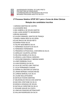
Perez et al
Chest films in the diagnosis of LV dysfunction in Chagas' disease
Arq Bras
Cardiol
Artigo
Original
2003; 80: 208-13.
Value of the Radiological Study of the Thorax for Diagnosing
Left Ventricular Dysfunction in Chagas' Disease
Amanda Arantes Perez, Antonio Luiz Pinho Ribeiro, Márcio Vinícius Lins Barros,
Marcos Roberto de Sousa, Roberto José Bittencourt, Fernando Santana Machado,
Manoel Otávio Costa Rocha
Belo Horizonte, MG - Brazil
Objective - To determine the value of the radiological
study of the thorax for diagnosing left ventricular dilation
and left ventricular systolic dysfunction in patients with
Chagas' disease.
Methods - A cross-sectional study of 166 consecutive
patients with Chagas’ disease and no other associated diseases. The patients underwent cardiac assessment with
chest radiography and Doppler echocardiography. Sensitivity, specificity, and positive and negative predictive values
of chest radiography were calculated to detect left ventricular dysfunction and the accuracy of the cardiothoracic
ratio in the diagnosis of left ventricular dysfunction with the
area below the ROC curve. The cardiothoracic ratio was
correlated with the left ventricular ejection fraction and the
left ventricular diastolic diameter.
Results - The abnormal chest radiogram had a sensitivity of 50%, specificity of 80.5%, and positive and negative predictive values of 51.2% and 79.8%, respectively, in
the diagnosis of left ventricular dysfunction. The cardiothoracic ratio showed a weak correlation with left ventricular ejection fraction (r=-0.23) and left ventricular diastolic diameter (r=0.30). The area calculated under the
ROC curve was 0.734.
Conclusion - The radiological study of the thorax is
not an accurate indicator of left ventricular dysfunction;
its use as a screening method to initially approach the
patient with Chagas’ disease should be reevaluated.
Key words:
chest radiography, left ventricular dysfunction, Chagas’ disease
Hospital das Clínicas da Faculdade de Medicina da UFMG and Ecoar - Medicina
Diagnóstica
Mailing address: Antonio Luiz Pinho Ribeiro – Rua Campanha, 98/101 – 30310770 - Belo Horizonte, MG, Brazil – E-mail: [email protected]
English version by Stela Maris C. e Gandour
Chagas’ disease currently affects 20 million individuals in Latin America 1. In Brazil, approximately 5 million
chagasic patients exist 1,2. Chagas’ disease has a wide
variety of clinical presentations, ranging from completely
asymptomatic individuals, who represent the great majority
of the patients, to others with severe, sometimes fatal,
cardiac alterations. Of the indicators of a poor prognosis,
left ventricular dysfunction stands out as the major predictor of morbidity and mortality 3-7.
The radiological study of the thorax is a routine investigative method to initially assess patients with Chagas’
disease, aiming at detecting cardiac impairment and at characterizing the clinical form of the disease.8 In other cardiac
diseases, however, conventional radiography of the thorax
is known not to allow an accurate assessment of the degree
of ventricular dysfunction 9,10.
We determined the value of the radiological study of
the thorax in the diagnosis of left ventricular dilation and left
ventricular systolic dysfunction on Doppler echocardiography of patients with Chagas’ disease. The prevalence of
echocardiographic and electrocardiographic alterations
predictive of a worse prognosis in patients with a normal
and enlarged cardiac silhouette was also determined.
Methods
To determine the value of chest radiography for detecting left ventricular dilation and left ventricular systolic dysfunction in patients with Chagas’ disease, we carried out a
cross-sectional study on 166 consecutive patients with
Chagas’ disease, whose ages ranged from 21 to 70 years.
The patients were selected from the Referral Outpatient Care
Unit for Chagas’ Disease of the Hospital das Clínicas of the
Universidade Federal de Minas Gerais and in the “Orestes
Diniz” Training and Referral Center for Infectious and Parasitic Diseases according to inclusion and exclusion criteria
from January 1998 to July 1999.
Arq Bras Cardiol, volume 80 (nº 2), 208-13, 2003
208
Arq Bras Cardiol
2003; 80: 208-13.
Perez et al
Chest films in the diagnosis of LV dysfunction in Chagas' disease
Individuals with positive serology for Trypanossoma
cruzi according to 2 different techniques or more, including
indirect immunofluorescence, indirect hemagglutination,
and ELISA were considered chagasic.
The exclusion criteria were as follows: 1) impossibility
or no availability to perform the examinations; 2) systemic
arterial hypertension, operationally defined as blood
pressure ≥ 160/95 mmHg, or use of hypotensive drugs; 3)
history compatible with coronary heart disease, according
to clinical evaluation; 4) previous episode suggestive of
acute rheumatic disease; 5) diabetes mellitus or reduced
tolerance to glucose, according to the definition of the National Diabetes Data Group; 6) thyroid dysfunction manifested as abnormal levels of thyroid stimulating hormone
(TSH) and free thyroxin (T4F); 7) renal failure, defined as an
increase in the serum levels of creatinine (> 1.2 mg/dL for
females and > 1.5 mg/dL for males) and urea (> 36 mg/dL for
females and > 42 mg/dL for males); 8) chronic obstructive
pulmonary disease, according to the presence of suggestive history, physical examination, electrocardiogram, and
radiological alterations; 9) electrolytic and fluid disorders
(serum levels of sodium > 145 mg/dL and serum levels of
potassium > 5.5 mg/dL); 10) significant anemia defined as
hemoglobin < 10 g/dL; 11) alcoholism, defined as a mean
week consumption above 420 g of ethanol (daily mean > 60g
of ethanol); 12) any other acute or chronic significant systemic disease; 13) pregnancy defined by the laboratory
finding of plasma levels of chorionic gonadotropin > 5
mUImL; and 14) presence of atrial fibrillation or implanted
pacemaker.
All individuals selected underwent complementary investigations, which comprised chest radiography in posteroanterior and lateral views and Doppler echocardiography.
The examinations proposed were performed with the
written informed consent of all patients taking part in the
study, which was approved by the Committee on Ethics in
Research of the Federal University of Minas Gerais (UFMG).
The well being and rights of all patients studied were
considered.
Chest radiography was performed according to the conventional technique at the Radiological Unit of the Hospital
das Clínicas of the UFMG and analyzed by one observer who
ignored the echocardiographic findings. The cardiac silhouette and the cardiothoracic ratio were assessed. The cardiothoracic ratio was obtained by the relation between the transverse diameter of the heart and the transverse diameter of the
thorax at the level of the right diaphragmatic cupula. The
transverse diameter of the heart was calculated by adding
the greatest segment obtained in the cardiac area to the right
of the central axis (T1) and the greatest segment obtained in
the cardiac area to the left of the central axis (T2) (fig. 1).
The echocardiographic parameters were obtained by an
experienced echocardiographer, who ignored the findings of
the chest radiography, and used HDI 5000 equipment. The
measurements of the M mode and of the contractility of the
regional wall were performed according to the recommendations of the American Society of Echocardiography 11. The
Fig. 1 – Conventional chest radiography. The cardiothoracic ratio was obtained dividing the transverse diameter of the heart by the transverse diameter of the thorax at
the level of the right diaphragmatic cupula. The transverse diameter of the heart was
calculated by adding the greatest segment obtained in the cardiac area to the right of
the central axis (T1) and the greatest segment obtained in the cardiac area to the left of
the central axis (T2).
ejection fraction was obtained with the Simpson method (fig.
2), and the score of motility of the left ventricular wall was
obtained by using the model of 16 segments.
Left ventricular dysfunction was defined by the
presence of left ventricular dilation (left ventricular diastolic
diameter > 55 mm) or depressed ejection fraction (ejection
fraction < 0.50), or both, obtained on Doppler echocardiography.
The cardiac silhouette was classified as normal or enlarged based on the subjective analysis of the cardiac area
observed on chest radiography in posteroanterior and lateral views. Chest radiography with a cardiothoracic ratio
greater than 0.50 or an enlarged cardiac silhouette, or both,
was considered abnormal.
The quantitative variables were described by the
mean and standard deviation or the median and interquartile
range; the qualitative variables were described by frequency. The correlation between them was obtained with
the Spearman correlation coefficient. The patients with a significant discordance between the chest radiographic
findings and those on Doppler echocardiography were
identified and their cases were carefully reviewed. The normality test for quantitative variables (Ryan – Anderson) and
the Bartlett test for homogeneity of variance were performed. In all tests, p<0.05 was used for rejecting the null hypothesis.
The sensitivity, specificity, and positive and negative
predictive values of the abnormal chest radiography on the
diagnosis of left ventricular dysfunction were calculated.
The performance of the different values of the cardiothoracic ratio on the detection of left ventricular dysfunction in
patients with Chagas’ disease was assessed with the ROC
(receiver-operator-characteristic) curve. The area below the
209
Perez et al
Chest films in the diagnosis of LV dysfunction in Chagas' disease
curve varies from zero to 1 and measures the accuracy of a
diagnostic test. Considering 0.5 as the cut point established
in the literature 12,13, the sensitivity, specificity, and positive
and negative predictive values of the cardiothoracic ratio
on the diagnosis of left ventricular dysfunction were calculated.
The patients were divided into 2 groups according to
the finding of normal or enlarged cardiac silhouette. In each
group, the characteristics of the indices obtained on
Doppler echocardiography (left ventricular diastolic diameter, ejection fraction, regional contractility, presence of
aneurysm, and left ventricular diastolic function) and on
electrocardiography (duration of the QRS complex, heart
rate, presence of ventricular extrasystoles, and intraventricular blocks) were assessed. The cardiac silhouette was
confronted with the cardiothoracic ratio. The comparisons
were performed with ANOVA, the Kruskal-Wallis test or the
Pearson chi-square test, depending on the type and distribution of the variable studied.
Results
The study comprised 166 patients with Chagas’
disease, 55% of whom were males, and their ages ranged
from 21 to 70 (mean = 42.6) years. Forty-eight (29%) patients
had left ventricular dysfunction.
The abnormal chest radiogram had a sensitivity of
50%, specificity of 80.5%, and positive and negative predictive values of 51.2% and 79.8%, respectively, in diagnosing
left ventricular dysfunction.
The cardiothoracic ratio correlated weakly with the
ejection fraction (r = -0,23; 95% CI = -0.37 to -0.08; p<0.05)
and with the left ventricular diastolic diameter (r = 0.30; 95%
Table I - General, echocardiographic, electrocardiographic, and
radiological characteristics of chagasic patients with normal and
enlarged cardiac silhouette
Normal silhouette
Absolute number
Age (years)#
Males
LVd (mm)*
LVd > 55 mm
EF (%)*
EF < .50
LVD
Aneurysm
Score *
Diastolic dysfunction
HR #
QRS (ms)#
VES
RBBB
CTR*
Enlarged silhouette
119
41.9 ± 9.0
57
50 (29-74)
19
62 (26-69)
11
20
18
1.00 (1-2.61)
8
64.1 ± 11.7
0.095 ± 0.024
7
19
0.44 (0.34-0.49)
47
45.9 ± 10.6
50
55 (46-77)
49
57 (23-72)
43
51
28
1.31 (1-2.81)
33
62.5 ± 10.7
0.119 ± 0.030
19
55
0.52 (0.42-0.52)
P
0.02
0.54
<0.01
<0.01
<0.01
<0.01
<0.01
0.27
<0.01
<0.01
0.74
<0.01
0.01
<0.01
<0.01
Data in percentages, except # mean ± SD, and * median (minimum and
maximum). LVd- left ventricular diastolic diameter; EF- ejection fraction;
LVD- left ventricular dysfunction; aneurysm = aneurysm of the left ventricular
apex; score = score of the motility of the left ventricular wall; HR- heart rate;
QRS- duration of the QRS complex; VES- ventricular extrasystoles; RBBBright bundle-branch block; CTR = cardiothoracic ratio.
210
Arq Bras Cardiol
2003; 80: 208-13.
Fig. 2 – The ejection fraction was obtained with the Simpson’s method.
CI = 0.15 to 0.43; p<0.05) (fig. 3). The cardiothoracic ratio
greater than 0.5 showed sensitivity of 41.7%, specificity of
88.1%, and positive and negative predictive values of 58.9%
and 78.7%, respectively, in detecting left ventricular dysfunction. The area calculated under the ROC curve was
0.734±0.04 (fig. 4).
The comparisons between the echocardiographic and
electrocardiographic parameters of patients with normal
and enlarged cardiac silhouette are shown in table I. The
patients with Chagas’ disease and an enlarged silhouette
were significantly older than those with normal silhouette.
The presence of left ventricular dysfunction was significantly more frequent in patients with an enlarged cardiac silhouette, and a similar pattern was observed for the presence of diastolic dysfunction and alterations in the segmentary contractility. A significant increase in the duration of
the QRS complex was evident in patients with an enlarged
cardiac area. The occurrence of ventricular extrasystoles
and complete right bundle-branch block was significantly
greater in the group of patients with an enlarged cardiac silhouette. On Doppler echocardiography, no significant difference was found between the 2 groups in regard to the
presence of left ventricular aneurysms.
Discussion
The value of measuring cardiac size based on radiological film has decreased as compared with that based on
Doppler echocardiography, which provides a more precise
analysis of the diameter of the cardiac chambers and of the
left ventricular function, and also other important functional
data, such as evaluation of the diastolic function and segmentary contractility. Although cardiac size may be determined by chest radiography, many cardiac and extracardiac
factors influence this measure, such as the examination
technique, the patient’s biotype, the patient’s physiological
status, thoracic alterations (scoliosis, pectus excavatum),
the size of the lungs, the breathing phase, the cardiac cycle
phase, and heart rate at the time of examination 12,13. Therefore, chest radiography may only provide a raw and sub-
Arq Bras Cardiol
2003; 80: 208-13.
Perez et al
Chest films in the diagnosis of LV dysfunction in Chagas' disease
75
70
65
LVd
EF
60
50
55
45
40
35
30
25
20
0.3
0.4
0.5
0.6
0.7
0.3
CTR
0.4
0.5
0.6
0.7
CTR
Fig. 3 – Correlation between the cardiothoracic ratio, the ejection fraction, and the left ventricular diastolic diameter.
1
0.9
0.8
Sensitivity
0.7
0.6
0.5
0.4
0.3
0.2
0.1
0
0
0.2
0.4
0.6
0.8
1
1 - Specificity
Fig. 4 – Performance of the cardiothoracic ratio in the diagnosis of left ventricular
dysfunction in chagasic patients. Area under the ROC curve = 0.734.
jective estimation of the cardiac size through the classification of the cardiac silhouette as normal or enlarged. More
objective numerical parameters provided by noninvasive
examinations, such as Doppler echocardiography, are required for an accurate estimation of the diameter of the
cardiac chambers.
In this study, in accordance with the results obtained in
patients with heart failure 9,10, chest radiography alone was
not an accurate indicator of the degree of left ventricular
dysfunction in patients with Chagas’ disease, although a
weak and significant correlation has been observed between the cardiothoracic ratio and the left ventricular diastolic diameter (r = 0.30; p<0.05) and between the cardiothoracic ratio and the ejection fraction (r = -0.23; p<0.05). These
values are different from those of a previous study 14, in
which a clear correlation between the radiological and echocardiographic measurements in patients with Chagas’
disease was observed. In that study 14, a small sample of 22
patients was divided into 2 different groups according to the
cardiac size obtained on radiological imaging. Samples
representing extreme values of cardiothoracic ratio and
cardiac diameter were compared; patients with cardiothoracic ratio near the maximum limit, which is considered normal in the literature 12,13, were not assessed. In addition, the
authors 14 considered a cardiothoracic ratio of 0.5 as abnormal, contrary to the maximum limit value adopted for the cardiothoracic ratio in our study 12,13.
Conventional chest radiography and electrocardiography are the initial investigation techniques used in a
patient with Chagas’ disease to assess cardiac impairment
and to characterize the clinical form of the disease 15.
Absence of alterations on chest radiography is required to
characterize the chronic undetermined form of Chagas’
disease 8, even knowing that, if assessed on more refined
methods, these patients may have significant cardiac alterations 16. However, in our study, chest radiography had a
low sensitivity (50%) for detecting left ventricular dilation
and left ventricular systolic dysfunction, in addition to a
negative predictive value lower than 80%, having, therefore, limited value as a screening test. The overall performance of the cardiothoracic ratio was also limited, with an
area under the ROC curve of 0.734.
The presence of cardiac enlargement on radiography
is a reliable indicator of left ventricular dysfunction as
follows: a cardiothoracic ratio greater than 0.5 has a specificity of 88.1% and a positive predictive value of 58.9%. In
addition, an enlarged cardiac silhouette is associated with
electrocardiographic and echocardiographic markers of
cardiac impairment, such as the presence of right bundlebranch block and ventricular extrasystoles, although it
does not relate to other significant abnormalities, such as
the presence of ventricular aneurysm. Therefore, although
inadequate for screening left ventricular dysfunction, chest
radiological study plays an important role in the clinical
assessment of patients with Chagas’ disease and defined
211
Perez et al
Chest films in the diagnosis of LV dysfunction in Chagas' disease
heart disease. In addition to providing elements about the
pulmonary circulation in patients with heart failure, a significantly enlarged cardiac silhouette is an indicator of the severity of the disease and of a worse prognosis 5.
Left ventricular dysfunction is the major prognostic
factor in Chagas’ disease 3-7. Identification of the group of
individuals with left ventricular dysfunction among the numerous populations of patients with Chagas’ disease is a
challenge in clinical practice. These individuals are candidates for preventive treatment with angiotensin-converting
enzyme inhibitors, which can delay the appearance of symptoms and reduce mortality in the presence of left ventricular
dysfunction 17. Although the beneficial effects of these
drugs on the survival of patients with Chagas’ heart disease
have not been formally tested, their beneficial clinical and
hemodynamic effects on patients with Chagas’ heart disease are evident 18,19. In addition, these drugs are indicated
in all patients with left ventricular dysfunction, symptomatic
or not, independent of its etiology 20.
The electrocardiogram is particularly useful for identifying the low-risk population, because a normal examination
is associated with an excellent prognosis in the midterm 21-23.
The additional value of chest radiography in the initial
assessment of patients with Chagas’ disease and a normal
electrocardiogram is questionable, because the examination
has little sensitivity for detecting left ventricular dysfunction, the major indicator of risk in Chagas’ disease. Currently, even echocardiography, although able to show abnormalities in segmentary contractility 24 or more subtle alterations on Doppler tissue imaging 25, seems to have its routine
use questionable in this population with an excellent prognosis. On the other hand, because individuals with typical
electrocardiographic alterations may have either depressed
or within the normal range left ventricular systolic function,
additional examinations are indicated to identify the presen-
Arq Bras Cardiol
2003; 80: 208-13.
ce of left ventricular dysfunction. Considering the insufficient diagnostic performance of chest radiography, echocardiography appears to be one method of choice for the
subsequent evaluation of cardiac impairment in patients
with abnormal electrocardiographic findings, allowing the
stratification of patients into high- and low-risk groups and
the selection of candidates for therapy with angiotensinconverting enzyme inhibitors 23. However, some limitations
of echocardiography should be considered, especially the
difficulty in performing the examination in rural areas where
the disease is endemic, the existence of patients without an
adequate window for the examination, and the need for an
experienced examiner. Measurement of the type B natriuretic peptide is an important marker of left ventricular dysfunction in nonchagasic patients 26, and it may also be a useful
tool in the initial evaluation of patients with Chagas’ disease. Data obtained in our study indicate that the method has
an excellent diagnostic performance in detecting left ventricular dysfunction in chagasic patients 27. Because of the
high prevalence of Chagas’ disease among us, cost-benefit
studies could quantify the medical, social, and economic
impact of the application of different approaches in the
initial evaluation of these patients.
In conclusion, although specific, chest radiography has
little sensitivity in diagnosing left ventricular systolic dysfunction; its use as a screening method in the initial approach
to patients with Chagas’ disease should be reevaluated.
Acknowledgements
We thank the National Council on Scientific and Technological Development (Conselho Nacional de Desenvolvimento Científico e Tecnológico - CNPq) and the Dean of
Research at the UFMG (Pró-Reitoria de Pesquisa da UFMG).
References
1.
2.
3.
4.
5.
6.
7.
8.
9.
WHO Expert Committee on the Control of Chagas Disease. Control of Chagas
Disease 1991: 1-95.
Dias JCP. Situación actual de la enfermedad de Chagas en las Américas. In: Madoery R, Madoery C, Cámera ML, editors. Actualizaciones en la Enfermedad de
Chagas. Buenos Aires: Congresso Nacional de Medicina, 1993: 1-29.
Hagar JM, Rahimtoola SH. Chagas’ heart disease in the United States. N Engl J
Med 1991; 325: 763-8.
Espinosa RA, Pericchi LR, Carrasco HA, Escalante A, Martinez O, González R.
Prognostic indicators of chronic chagasic cardiopaphy. Int J Cardiol 1991; 30:
195-202.
Carrasco HA, Parada H, Guerrero L, Duque M, Durán D, Molina C. Prognostic
implications of clinical, eletrocardiographic and hemodynamic findings in
chronic Chagas’disease. Int J Cardiol 1994; 43: 27-38.
Mady C, Cardoso RHA, Barreto ACP, Luz PL, Bellotti G, Pileggi F. Survival and
predictors of survival in patients with congestive heart failure due to Chagas’
cardiomypathy. Circulation 1994; 90: 3098-102.
Bestetti RB, Dalbo CMR, Freitas OC, Teno LAC, Castilho OT, Oliveira JSMO.
Noninvasive predictors of mortality for patients with Chagas’ heart disease. A
multivariate stepwise logistic regression study. Cardiology 1994; 84: 261-7.
Reunião de Pesquisa Aplicada em Doença de Chagas. Validade do conceito de forma
crônica indeterminada de doença de Chagas. Rev Soc Bras Med Trop 1985; 18: 46.
Clark AL, Coats AJ. Unreliability of cardiothoracic ratio as a marker of left ventri-
212
10.
11.
12.
13.
14.
15.
16.
17.
cular impairment: comparison with radionuclide ventriculography and echocardiography. Postgrad Med J 2000; 76: 289-91.
Philbin EF, Garg R, Danisa K, et al. The relationship between cardiothoracic
ratio and left ventricular ejection fraction in congestive heart failure. Arch Intern
Med 1998; 158: 501-6.
Schiller NB, Shah PM, Crawford M, et al. Recommendations for quantitation of the
left ventricle by two-dimensional echocardiography. American Society of Echocardiography Committee on Standards, Subcommittee on Quantitation of TwoDimensional Echocardiograms. J Am Soc Echocardiography 1989; 2: 358-67.
Steiner RM, Levin DC. Radiology of the heart. In: Braunwald E, editor. Heart
disease. 4th ed. Philadelphia: WB Saunders Co., 1992: 204-34.
The Criteria Committee of the New York Heart Association. Nomenclature and
criteria for diagnosis of diseases of the heart and great vessels. 9th ed. New York:
Copyright, 1994: 259-63.
Pereira-Barreto AC, Mady C, Arteaga-Fernandez E, et al. Valor do índice cardiotorácico na avaliação do comprometimento miocárdico. Correlação com avaliação ecocardiográfica. Rev Hosp Clin Fac Med S Paulo 1983; 38: 40-5.
Rassi Jr A, Marin-Neto JA. Estado da Arte – Cardiopatia chagásica crônica. Rev
Soc Cardiol Estado de São Paulo 2000; 10: 6-32.
Ribeiro ALP, Rocha MOC. Forma indeterminada da doença de Chagas: considerações acerca do diagnóstico e do prognóstico. Rev Soc Bras Med Trop 1998;
31: 301-14.
Effect of enalapril on mortality and the development of heart failure in asympto-
Arq Bras Cardiol
2003; 80: 208-13.
18.
19.
20.
21.
22.
Perez et al
Chest films in the diagnosis of LV dysfunction in Chagas' disease
matic patients with reduced left ventricular ejection fractions. The SOLVD
Investigattors N Engl J Med 1992; 327: 685-91.
Roberti RR, Martinez EE, Andrade JL, et al. Chagas cardiomyopathy and captopril. Eur Heart J 1992; 13: 966-70.
Khoury AM, Davila DF, Bellabarba G, et al. Acute effects of digitalis and enalapril on the neurohormonal profile of chagasic patients with severe congestive
heart failure. Int J Cardiol. 1996; 57: 21-9.
Hunt SA, Baker DW, Chin MH, et al. ACC/AHA guidelines for the evaluation
and management of chronic heart failure in the adult: a report of the American
College of Cardiology/American Heart Association Task Force on Practice Guidelines (Committee to Revise the 1995 Guidelines for the Evaluation and Management of Heart Failure). Am Coll Cardiol, 2001. Web site disponível em http://
www.acc.org
Forichon E. Contribuition aux estimation de morbiditê et de mortalité dans la maladie de Chagas (Trypanosomose américaine). Rev Patol Trop 1975; 1: 57-78.
Maguire JH, Hoff R, Sherlock I, et al. Cardiac morbidity and mortality due to
23.
24.
25.
26.
27.
Chagas’ disease: prospective electrocardiographic study of a Brazilian community. Circulation 1987; 75: 1140-5.
Xavier SS, Sousa AS, Borges-Pereira J, Hasslocher-Moreno A. Nova proposta de
classificação do acometimento cardíaco na fase crônica da doença de Chagas. Rev
Soc Bras Med Trop 1999; 32: 121.
Ortiz J, Barretto AC, Matsumoto AY, et al. Alteração contrátil segmentar na forma
indeterminada da Doença de Chagas. Estudo ecocardiográfico. Arq Bras Cardiol
1987; 49: 217-20.
Barros MV, Rocha MO, Ribeiro AL, Machado FS. Doppler tissue imaging to evaluate early myocardium damage in patients with undetermined form of Chagas’ disease and normal echocardiogram. Echocardiography 2001; 18: 131-6.
McDonagh TA, Robb SD, Murdoch DR, et al. Biochemical detection of left-ventricular systolic dysfunction. Lancet 1998; 351: 9-13.
Ribeiro ALP, Reis AM, Barros MVL, et al. Brain natriuretic peptide in the diagnosis of systolic left ventricular dysfunction in Chagas disease. Lancet 2002;
360: 461-2.
Bula resumida – MICARDIS® Telmisartam - Uso adulto - Forma farmacêutica e apresentações: Comprimidos de 40 mg: embalagens com 14 e 28 comprimidos. Comprimidos de 80 mg: embalagens com 14 e 28
comprimidos. Composição: Cada comprimido contém 40 mg ou 80 mg de telmisartam. Excipientes q.s.p. 1 comprimido. Indicações: Tratamento da hipertensão arterial, como monoterapia ou em associação com
outros agentes anti-hipertensivos. Contra-indicações: Hipersensibilidade ao ingrediente ativo ou aos excipientes. Gravidez e lactação. Obstrução biliar. Disfunção hepática ou renal grave. Intolerância hereditária
à frutose. Precauções: Hipertensão renovascular: pacientes com estenose arterial renal bilateral ou estenose da artéria com um único rim funcionando: risco aumentado de hipotensão grave e insuficiência renal.
Disfunção renal ou transplante hepático: monitoração periódica dos níveis séricos de potássio e creatinina. Não há experiência em pacientes com transplante renal recente. Desidratação: hipotensão
sintomática, especialmente após a primeira dose, pode ocorrer em pacientes que têm volemia e/ou sódio depletado, o que deve ser corrigido antes do início da terapêutica com MICARDIS. Outras condições de
estimulação do SRAA e condições dependentes da atividade SRAA (insuficiência cardíaca congestiva grave): hipotensão aguda, hiperazotemia, oligúria ou, raramente, insuficiência renal aguda.
Hiperaldosteronismo primário: não se recomenda o uso de MICARDIS. Estenose valvar aórtica e mitral e cardiomiopatia hipertrófica obstrutiva: Recomenda-se precaução especial. Hipercalemia: recomendase monitoração adequada dos níveis séricos de potássio em pacientes de risco. Diuréticos poupadores de potássio, suplementos de potássio, sais de potássio ou outros medicamentos que podem aumentar
os níveis de potássio, como a heparina: podem levar a um aumento da potassemia. Portanto, nestas situações MICARDIS deve ser administrado com cautela. Distúrbios hepatobiliares: pode-se esperar redução
da depuração em pacientes com disfunções obstrutivas do sistema biliar ou insuficiência hepática, pois a eliminação da droga é principalmente biliar. Intolerância à frutose: os comprimidos de MICARDIS contém
sorbitol; portanto, é inadequado para pacientes com intolerância hereditária à frutose. Outros: menor eficácia na redução da pressão arterial na população negra do que na população não-negra. Cardiopatia
isquêmica ou doença cardiovascular isquêmica pode resultar em infarto do miocárdio. Interações medicamentosas: MICARDIS pode aumentar o efeito hipotensor de outros agentes anti-hipertensivos. Observouse um aumento de 20% da concentração plasmática média de digoxina. Relataram-se aumentos reversíveis das concentrações séricas de lítio e de toxicidade; portanto, recomenda-se cuidadosa monitoração
do uso concomitante com lítio. Gravidez e lactação: Contra-indicado. Reações adversas: As reações adversas à droga obtidas a partir de todos os estudos clínicos com telmisartam foram: Infecções do trato
urinário, infecções do trato respiratório superior, ansiedade, visão anormal, vertigem, dor abdominal, diarréia, boca seca, dispepsia, flatulência, dor de estômago, eczema, aumento de suor, artralgia, dor nas
costas, cãibras nas pernas ou dores nas pernas, mialgia, sintomas de tendinite, dor no peito, sintomas de gripe. Além disso, desde a introdução de telmisartam no mercado, relataram-se casos raros de eritema,
prurido, desmaio, insônia, depressão, vômito, hipotensão, bradicardia, taquicardia, dispnéia, eosinofilia, trombocitopenia, fraqueza e perda de eficácia. Relataram-se casos isolados de angioedema, urticária
e outros eventos relacionados. Investigações: Raramente, observaram-se diminuição na hemoglobina ou aumento no ácido úrico. Observaram-se aumentos na creatinina ou nas enzimas hepáticas. Efeitos
na habilidade de dirigir e utilizar máquinas: Ainda não se realizaram estudos específicos. Contudo, ao dirigir ou operar máquinas, pode ocasionalmente ocorrer tontura ou sonolência. Posologia: A dose
recomendada é de 40 mg uma vez ao dia. Alguns pacientes podem apresentar benefício com dose diária de 20 mg. Em casos em que a pressão arterial pretendida não seja atingida, a dose de MICARDIS pode
ser aumentada para no máximo 80 mg uma vez ao dia. Alternativamente, MICARDIS pode ser usado em combinação com diuréticos tiazídicos, como a hidroclorotiazida, para se obter uma redução maior da
pressão arterial. Quando se considerar um aumento da dose, deve-se levar em conta que o máximo efeito anti-hipertensivo é geralmente atingido quatro a oito semanas após o início do tratamento. MICARDIS
pode ser administrado com ou sem alimento. Insuficiência renal: Não há necessidade de ajustes de dose em pacientes com insuficiência renal leve a moderada. Telmisartam não é removido do sangue por
hemofiltração. Insuficiência hepática: Nos pacientes portadores de insuficiência hepática leve a moderada, não se deve exceder a dose diária de 40 mg. Pacientes idosos: Não são necessários ajustes de doses.
Crianças e adolescentes: Não há dados de segurança e eficácia de MICARDIS em crianças e adolescentes. VENDA SOB PRESCRIÇÃO MÉDICA - MS - 1.0367.0110 - Boehringer Ingelheim do Brasil Química
e Farmacêutica Ltda.
Bula resumida – MICARDIS® HCT – Telmisartam/Hidroclorotiazida - Uso adulto - Composição: Cada comprimido de MICARDIS HCT contém 40 mg/12,5 mg ou 80 mg/12,5 mg de telmisartam/hidroclorotiazida.
Embalagens com 14 e 28 comprimidos. Excipientes q.s.p. 1 comprimido. Indicações: Tratamento da hipertensão arterial. MICARDIS HCT, como associação de dose fixa, é indicado em pacientes cuja pressão
arterial não é adequadamente controlada com telmisartam ou hidroclorotiazida isoladamente. Posologia: MICARDIS HCT deve ser administrado uma vez ao dia. A dose de MICARDIS pode ser aumentada
gradativamente antes de substituí-lo pelo MICARDIS HCT. A substituição direta da monoterapia pelas combinações fixas pode ser considerada. MICARDIS HCT 40/12,5 mg pode ser administrado em pacientes
cujas pressões sangüíneas não sejam adequadamente controladas por MICARDIS 40 mg ou hidroclorotiazida. MICARDIS HCT 80/12,5 mg pode ser administrado em pacientes cujas pressões arteriais não sejam
adequadamente controladas por MICARDIS 80 mg ou por MICARDIS HCT 40/12,5 mg. O máximo efeito anti-hipertensivo é obtido após 4 a 8 semanas de tratamento. Quando necessário, MICARDIS HCT pode ser
administrado com outros anti-hipertensivos. MICARDIS HCT pode ser administrado com ou sem alimento. Devido ao componente hidroclorotiazida, MICARDIS HCT não deve ser usado em pacientes com
disfunção renal grave. Nos casos leves a moderados, não se observaram efeitos adversos renais e não é necessário ajuste de dose. Recomenda-se monitoração periódica da função renal. Nos casos de
insuficiência hepática leve a moderada, não se deve exceder a dose de 40/12,5 mg uma vez ao dia. MICARDIS HCT não é indicado em pacientes com insuficiência hepática grave. Não são necessários ajustes
de dose conforme a idade. Ainda não se estabeleceram a segurança e a eficácia de MICARDIS HCT em pacientes menores de 18 anos. Contra-indicações: Hipersensibilidade aos componentes ativos,
excipientes ou substâncias derivadas de sulfonamidas (como HCT). Gravidez e lactação. Colestase e distúrbios obstrutivos biliares. Insuficiência hepática ou renal grave (depuração de creatinina <30 ml/min).
Hipopotassemia refratária e hipercalcemia. Precauções: Não utilizar nos casos de colestase, distúrbios biliares obstrutivos ou insuficiência hepática grave. Usar com cautela nos casos de função hepática
alterada ou doença hepática progressiva, pois pode ocorrer coma hepático. Há maior risco em pacientes com estenose arterial renal bilateral ou estenose com um único rim funcionando. Micardis HCT não deve
ser administrado em pacientes portadores de disfunção renal grave. Não há experiência quanto à administração de MICARDIS HCT em pacientes com grave insuficiência renal ou com um rim transplantado
recentemente. Recomenda-se a monitoração periódica dos níveis séricos de potássio, creatinina e ácido úrico. A azotemia associada a diuréticos tiazídicos pode ocorrer em pacientes com função renal alterada.
Pode ocorrer hipotensão sintomática em pacientes que têm volemia e/ou sódio depletado. Insuficiência cardíaca congestiva grave ou doença renal, estenose da artéria renal tratada concomitantemente com
drogas que afetem o sistema renina-angiotensina-aldosterona foram associados com hipotensão aguda, hiperazotemia, oligúria ou, raramente, insuficiência renal aguda. Aldosteronismo primário: não se
recomenda o uso de telmisartam, precaução especial em estenose valvar aórtica ou mitral e cardiomiopatia hipertrófica obstrutiva. Como tiazidas podem prejudicar a tolerância à glicose, pode ser necessário
ajuste no tratamento em diabéticos e pode ocorrer manifestação de diabetes latente. A hidroclorotiazida aumenta os níveis de colesterol e triglicérides; contudo, nenhum ou poucos efeitos adversos foram
relatados na dose de 12,5 mg contida em MICARDIS HCT. Pode ocorrer hiperuricemia ou precipitação de gota devido a hidroclorotiazida. Pode ser necessária monitoração periódica dos níveis séricos de
eletrólitos, uma vez que a hidroclorotiazida pode causar desequilíbrio eletrolítico ou de fluidos (hipopotassemia, hiponatremia e alcalose hipoclorêmica). O tratamento concomitante com telmisartam pode
reduzir a hipopotassemia induzida por diuréticos, devido aos efeitos antagônicos. Diuréticos poupadores de potássio, suplementos de potássio ou substitutos do sal contendo potássio devem ser utilizados
concomitantemente com cautela. Não há evidências de que MICARDIS HCT reduza ou previna hiponatremia induzida por diuréticos. A deficiência de cloreto é geralmente leve. Tiazidas podem causar elevação
dos níveis séricos de cálcio e magnésio. Interromper o uso antes dos testes de função da paratireóide. Não utilizar em intolerância hereditária à frutose. A redução excessiva da pressão arterial no caso de doença
cardiovascular isquêmica pode ser prejudicial. Relatou-se exacerbação ou ativação do lupus eritematoso sistêmico com o uso de hidroclorotiazida. Recomenda-se cautela no uso de anti-hipertensivos ao dirigir
e operar máquinas. Interações medicamentosas: Relataram-se aumentos reversíveis das concentrações séricas de lítio durante administração concomitante com inibidores da enzima conversora de
angiotensina. Relataram-se casos de interação com antagonistas dos receptores da angiotensina II. O risco de toxicidade por lítio pode ser aumentado com o uso de MICARDIS HCT. Recomenda-se a monitoração
dos níveis séricos de lítio durante o uso concomitante. O efeito de depleção de potássio da hidroclorotiazida é atenuado pelo efeito poupador de potássio do telmisartam, mas é potencializado por outros diuréticos
caliuréticos, laxantes, corticosteróides, ACTH, anfotericina, carbenoxolona, penicilina G sódica, ácido salicílico e derivados. Por outro lado, o uso concomitante de diuréticos poupadores de potássio,
suplementos/sais de potássio ou outras drogas podem aumentar os níveis séricos de potássio. Nesses casos, recomenda-se a monitoração dos níveis plasmáticos de potássio. Recomenda-se monitoração
periódica de potássio na administração de MICARDIS HCT com digitálicos, antiarrítmicos e drogas que são conhecidamente indutoras de torsades de pointes. Telmisartam pode aumentar o efeito hipotensor de
outros agentes anti-hipertensivos. Observou-se um aumento de 20% da concentração plasmática média de digoxina. Não se identificaram outras interações de importância clínica. Interações com a
hidroclorotiazida: álcool, barbitúricos ou narcóticos (potencialização da hipotensão ortostática); drogas antidiabéticas (ajustes de dose podem ser necessários); metformina (risco de acidose láctica);
colestiramina e resina colestipol (absorção de hidroclorotiazida é prejudicada); corticosteróides, ACTH (depleção eletrolítica, principalmente aumento de hipopotassemia); glicosídeos digitálicos
(hipopotassemia ou hipomagnesemia induzida por tiazídicos favorece o aparecimento de arritmias cardíacas induzidas por digitálicos); drogas antiinflamatórias não-esteroidais (a administração de droga
antiinflamatória não-esteroidal pode reduzir o efeito diurético, natriurético e anti-hipertensivo dos diuréticos tiazídicos em alguns pacientes). Os efeitos das aminas hipertensoras podem ser levemente
diminuídos. Os relaxantes musculares esqueléticos não-despolarizantes são potencializados pela hidroclorotiazida. Medicamentos uricosúricos podem requerer ajustes. Verificou-se aumento da incidência
de reações de hipersensibilidade ao alopurinol. Os efeitos hiperglicêmicos dos beta-bloqueadores e diazóxido podem ser aumentados pelas tiazidas. Os agentes anticolinérgicos (por exemplo, atropina,
biperideno) podem aumentar a biodisponibilidade das tiazidas. As tiazidas podem aumentar os eventos adversos da amantadina. As tiazidas podem reduzir a excreção renal de drogas citotóxicas (por exemplo,
ciclofosfamida, metotrexato). Gravidez e lactação: É contra-indicado. Se ocorrer gravidez, telmisartam deve ser descontinuado o mais breve possível. As tiazidas atravessam a barreira placentária e podem
causar distúrbios eletrolíticos no feto e é possível que ocorram outras reações. Relataram-se casos de trombocitopenia ou icterícia neonatal com o tratamento tiazídico materno. Tiazidas são excretadas no leite
humano e podem inibir a lactação. Reações adversas: Foram relatadas as seguintes reações adversas: Bronquite, faringite, sinusite, infecções do trato respiratório superior, infecções do trato urinário,
sialadenite, eosinofilia, anemia aplástica, anemia hemolítica, depressão da medula óssea, leucopenia, neutropenia/agranulocitose, trombocitopenia, alergia, reações anafiláticas, perda do controle da diabete,
hipercolesterolemia, hiperuricemia, hipopotassemia, causa ou aumento da depleção de volume, desequilíbrio eletrolítico, hiponatremia, anorexia, perda de apetite, hiperglicemia, ansiedade, depressão,
inquietação, tontura, desmaio, insônia, escotomas, parestesia, distúrbios do sono, visão alterada, visão borrada temporariamente, xantopsia, vertigem, bradicardia, taquicardia, arritmias cardíacas, hipotensão,
hipotensão postural, angiite necrotizante (vasculite), dispnéia, sofrimento respiratório (incluindo pneumonite e edema pulmonar), dor abdominal, diarréia, dispepsia, gastrite, dor de estômago, boca seca,
flatulência, vômito, constipação, pancreatite, icterícia (icterícia hepatocelular ou colestática), eczema, aumento do suor, eritema, prurido, reações cutâneas do tipo lupus eritematoso, vasculite cutânea, reações
de fotossensibilidade, erupções cutâneas, reativação do lupus eritematoso cutâneo, necrólise epidérmica tóxica, atralgia, artrose, dor nas costas, dor nas pernas, mialgia, cãibras nas pernas, sintomas de
tendinite, fraqueza, espasmo muscular, nefrite intersticial, disfunção renal, glicosúria, impotência, sintomas de gripe, dor, dor no peito, perda da eficácia, febre. Investigações: Diminuição da hemoglobina, aumento
do ácido úrico, aumento na creatinina, aumento das enzimas hepáticas, aumento dos triglicérides. Assim como ocorre com outros antagonistas da angiotensina II, relataram-se casos isolados de angioedema,
urticária e outras reações relacionadas. VENDA SOB PRESCRIÇÃO MÉDICA. - MS 1.0367.0134 Boehringer Ingelheim do Brasil Química e Farmacêutica Ltda.
213
Baixar