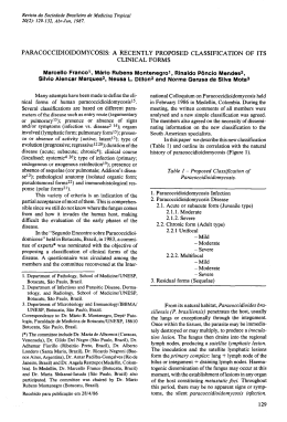
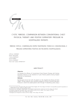
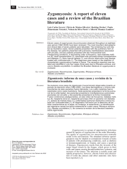
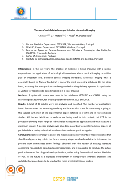
Three-Dimensional Contrast-Enhanced
Magnetic Resonance Pulmonary Angiography in
the Study of Pulmonary Vascular Pathology [114]
BOBAN THOMAS, PEDRO FARTO E ABREU, JOSÉ ROQUETTE, RAFAEL FERREIRA
Cardiology Service, Hospital Fernando Fonseca, Amadora, Portugal
Cardiothoracic Surgical Department, Hospital Santa Marta, Lisbon, Portugal
Rev Port Cardiol 2003; 22 (12) : 1471-1476
ABSTRACT
Objectives: To assess the feasibility of
performing pulmonary angiography using
MRI with contrast enhancement in patients
with pulmonary vascular disease.
Methods: We present our experience in ten
individuals, two controls and eight patients
who underwent the exam after injection of a
gadolinium-based contrast agent on a 1 Tesla
MR scanner using a time-of-flight sequence
and breath-holding during injection of
contrast.
Results: Pathology in the main pulmonary
artery and its major branches was detected
easily while resolution at the segmental and
subsegmental levels was inadequate.
Conclusion: Contrast-enhanced magnetic
resonance pulmonary angiography is feasible
on a 1 Tesla MR scanner for the study of
pathology of the main pulmonary artery and
its major branches, like massive pulmonary
embolism. However its ability to detect and
define distal vessel pathology as found in
chronic thromboembolic pulmonary
hypertension and small pulmonary emboli is
limited.
Key words
Pulmonary angiography; MRI;
Thromboembolism
RESUMO
Angiografia Pulmonar por Ressonância
Magnética Tridimensional optimizada
por Contraste, no Estudo da patologia
Vascular pulmonar
Objectivos: Avaliar a possibilidade de
realização de angiografia pulmonar utilizando
imagem por ressonância magnética
optimizada por contraste, em doentes com
patologia vascular pulmonar.
Métodos: É apresentada a nossa experiência
em dez indivíduos, dois controlos e oito
doentes que foram submetidos ao exame
após injecção de um agente de contraste
baseado em Gadolinium com recurso a um
equipamento de ressonância magnética de 1
Tesla, utilizando uma sequência com time-offlight e interrupção da respiração durante a
injecção de contraste.
Resultados: A patologia do tronco da artéria
pulmonar e dos seus ramos principais foi
facilmente detectada enquanto que os níveis
de resolução segmentar e subsegmentar
foram inadequadas.
Conclusão: A angiografia pulmonar por
ressonância magnética de 1 Tesla optimizada
por contraste, é possível para o estudo da
patologia do tronco da artéria pulmonar e dos
seus ramos principais, nomeadamente
embolismo pulmonar. No entanto, a sua
capacidade de detectar e definir patologia
dos vasos distais como a habitualmente
encontrada na hipertensão pulmonar
associada a tromboembolismo crónico, é
limitada.
Palavras-Chave
Angiografia pulmonar; Imagem por ressonância magnética;
Tromboembolismo
Recebido para publicação: Fevereiro de 2003 • Aceite para publicação: Novembro de 2003
Received for publication: February 2003 • Accepted for publication: November 2003
1471
INTRODUCTION
T
he high incidence of pulmonary embolism
has led to the development of a plethora of
diagnostic techniques and algorithms, each
with its own advantages, disadvantages, advocates and dissidents. Among the various imaging techniques, pulmonary angiography using
MRI is relatively new and not widely used (1).
However, MRI is the only technique that detects lower-limb deep venous thrombosis and
pelvic vein thrombosis by direct thrombus imaging (DTI) without contrast (2, 3). The ability to
assess cardiac function in the same exam motivated some investigators to use MRI to study
pulmonary emboli (4, 5). Initially, MRI used timeof-flight (TOF) sequences without contrast. Because these techniques were inadequate, gadolinium-based contrast agents, which decreased
imaging times and improved resolution, were
developed (6-9). However, imaging the pulmonary
vasculature using MRI with contrast presents
unique challenges. We investigated the possibility of using this technique in the imaging of
pulmonary embolism.
METHODS
1472
MRI used three-dimensional angiography
techniques that we have described in the study
of aortic pathology (10). Briefly, a three-dimensional time-of-flight related sequence was used
on a 1.0 Tesla GE Signa scanner with a prescan preparation time of 29 msec, time to repetition (TR) of 8.7 msec and a time-to-echo (TE)
of 2.9 msec with a slice thickness of approximately three millimeters, a field of view of 48
x 48 cm, a flip angle of 45 degrees, a matrix of
256 x 160 and a bandwidth of 31.2 kHz, using
a standard body coil applied to the thorax with
no flow compensation. The imaging slab was
placed coronally on a previously acquired sagittal localizing scan. Image acquisition was
done in the anterior to posterior direction.
Forty milliliters of a commercially available
gadolinium-based contrast agent (Magnevist®,
Schering Lusitana) was injected during the
scan to maintain opacification for the entire
duration of the scan, which usually lasted between 18 and 24 seconds with breath-holding.
Injection of the contrast agent commenced during the initiation of the breath-holding maneuver, which lasted about five seconds before the
initiation of the sequence. Reconstruction in a
three-dimensional format was completed after
all the individual slices were acquired with the
number of excitations (NEX) kept constant at
one. Image interpretation was performed by
post-processing of the 3D data with multi-planar
reformatting (MPR), providing tomographic
images in virtually any plane and maximum
intensity projection (MIP) providing images
analogous to conventional contrast angiography
along with 3D reconstruction images that can
be rotated. Inspection of the lumen for filling
defects was performed in reconstructions in the
axial, coronal and sagittal planes.
Conventional pulmonary angiography (CPA)
was also performed in some patients with a low
osmolar contrast (Ultravist, Schering Lusitana)
using a power injector and 25 ml of contrast
(at 10 ml/sec) in each major branch of the
main pulmonary artery. Scanning was performed in the AP and oblique (right and left at
30 degrees) directions, using either a 7F multipurpose catheter or an 8F pulmonary artery
flotation catheter with side-holes for injection.
Patients: Two controls and eight patients
with the following pulmonary artery pathology
underwent MR studies: one with an aorto-pulmonary window, one with stenosis of the pulmonary valve with post-stenotic aneurysm of
the pulmonary artery, three with massive pulmonary embolism (two of whom underwent
thrombolytic therapy and one embolectomy),
one with a pulmonary artery sarcoma, and two
with chronic thromboembolic pulmonary
hypertension (CTEPH) considered ineligible
for thromboendarterectomy. Six of the eight patients also underwent CPA.
RESULTS
In the three patients with massive pulmonary embolism (two of whom had hemodynamic
compromise) the embolus was clearly visible in
the right branch of the main pulmonary artery
(MPA) and disappeared after thrombolysis in
two patients (Figs. 1-2), and the third patient
underwent surgical embolectomy after CPA
(Figs. 3-4). The patient with pulmonary artery
tumor underwent both exams and the tumor
was clearly visible in the left branch of the
main pulmonary artery along with extension to
the lobar artery of the left lower lobe (Figs. 5-6).
This space-filling lesion was diagnosed as a
large thrombus initially but did not resolve
with anticoagulant therapy and was not associated with right ventricular dysfunction or
Fg. 1 A single slice showing a massive embolus in the right
branch of the main pulmonary artery.
Fig. 3 Conventional pulmonary angiography showing truncation
of the major branch to the right upper lobe.
pulmonary hypertension. The MR angiography
also confirmed the presence of the aorto-pulmonary window (and aneurysm of the MPA)
detected during right heart catheterization and
CPA. The distal obliterative disease in the patients with CTEPH was visible in CPA but resolution on contrast-enhanced magnetic reso-
Fig. 2 After thrombolytic therapy, the right branch of the MPA
is free of thrombus. Opacification in the left atrium is seen immediately below the MPA branches showing that this slice has
captured images of both the arterial and venous phases of contrast injection.
Fig. 4 A single slice of CMRPA shows the obstructing thrombus.
nance pulmonary angiography (CMRPA) was
inadequate at the segmental and subsegmental
levels. In some individual slices it was easy to
mistake a branch of the PA for a tributary of
the pulmonary vein, which could confound interpretation.
1473
1474
Fig. 5 Gradient echo sequence showing a mass in the left
branch of the main pulmonary artery.
Fig. 6 CMRPA shows the mass in a coronal slice.
DISCUSSION
A few anatomical and technical details deserve comment. The pulmonary vasculature divides in three directions – from medial to lateral, anterior to posterior and superior to
inferior – within the lung. Hence some slices
will image some branches that may be coursing
the lung in the superior-to-inferior direction
while others may be doing so in the anteriorto-posterior direction. This means that some
vessels will be cut along their short axis while
others along their long axis. This anatomical
feature of the pulmonary vasculature can compromise resolution with some vessels in a slice
being clearly visible while others are blurred.
The data acquired in MRI (called the individual k-lines that add to what is technically
called the k-space which eventually forms the
image) has to coincide with maximal contrast
presence in the MPA and its branches (11) .
However, the time between the arterial phase
enhancement and the initiation of the venous
phase in the pulmonary vasculature is extremely short. If image acquisition is commenced
too late contrast may not be present in the
MPA or branches, while imaging too early may
show the contrast in the superior vena cava
and the right heart chambers with very little
enhancement of the MPA. Some commercially
We used MRI to image space-occupying lesions (presumed to be emboli) in the MPA and
its major branches up to the lobar level on a 1
Tesla machine. Resolution was inadequate to
interpret images at the segmental and subsegmental levels, even in the controls. Some authorities have discounted the importance of
emboli at the subsegmental levels. However,
the subsegmental vasculature is important in
those with marginal cardiopulmonary reserve,
and therefore a vascular imaging technique
should preferably have adequate resolution at
this level. Imaging on scanners with higher
gradient rise times (slew rates) provides higher
resolution and image quality and shorter durations, permitting a better evaluation at the segmental level, while subsegmental images may
still be inadequate. In patients with CTEPH
with laminated organized thrombus at the segmental level, thromboendarterectomy performed by experienced surgeons has resulted in
considerable clinical improvement (10). Prior to
this procedure, it is important to delineate the
extent of thrombus clearly to define the surgical approach. In our patients only CPA could
demonstrate this.
available programs (SMARTPREP) are available that claim better coordination of contrast
injection and image acquisition. Our experience with this program, though limited, has
not been encouraging. Recent improvements
with software resulting in fluoro-triggered
MRA sequences provide better coordination
between bolus arrival and image acquisition
and may improve the quality of images. Image
analysis by MPR and MIP is also time-consuming, tedious and unreliable due to the overlap of vessels mentioned above. Therefore even
many experts do not recommend MRI to rule
out PE in an acute situation.
The ability to image the leg veins and the
pelvic veins is unique to MR (although spiral
CT can perform leg venography, albeit with
contrast). If further technical improvements
can be made in sequences with better bolusimage acquisition coordination, this technique
may provide a comprehensive approach to imaging the venous and pulmonary vasculature in
embolic disease. In venous thromboembolic disease vascular imaging confirms the clinical
consequence of a thrombophilic state that is
treated by anticoagulation. Therefore imaging
techniques have their inherent limitations in
this clinical context and do not stand alone.
Optimal management of PE should involve an
optimal clinical assessment for hemodynamic
instability, laboratory measurement of D-dimer
levels using a sensitive assay, echocardiography to assess RV dysfunction and an imaging modality that can detect thrombi, as
shown in a recent study (12). One study even
provided evidence for the safety of managing
patients for suspected PE based on the pretest
probability and the D-dimer result alone, avoiding any type of diagnostic imaging (13).
The emergence of spiral CT, which is also
available in our institution, as a robust technique has further diminished the interest of
some groups in MRI. Single-detector helical
CT performed extremely well as the primary
diagnostic test to rule out PE, with a total mortality of 4.1 % in patients with a normal helical
CT, none dying of fatal PE, only one among
246 patients developing a nonfatal PE and
another patient developing venous thromboembolism (14). Multi-detector CT can provide higher
sensitivity for small subsegmental emboli, thinner imaging sections (up to 1 mm), shorter
imaging times and more extensive coverage of
the thorax (15). However at this time and to the
best of our knowledge, although spiral CT
techniques do exist for the venous system in
the lower extremity (16), the pelvic veins and
cardiac function cannot be assessed. Right
ventricular dysfunction is a powerful predictor
of outcome in patients with pulmonary embolism (17, 18). Although this information can be obtained by transthoracic echocardiography, not
even transesophageal echocardiography can
image the left branch of the MPA completely
because of the interposed trachea. MRI with
contrast provides information on the systemic
venous and pulmonary vasculature and cardiac
function, although significant limitations exist.
The development of multidetector CT technology along with the limitations of MRI will probably relegate the latter technique to a secondary role in the diagnostic imaging of PE, unless further technical developments overcome
the current limitations.
LIMITATIONS
It needs to be stated clearly that this study
involved only MR, precluding us from launching into a discussion on any relative merits
of the technique compared to spiral CT other
than those mentioned in the preceding paragraphs. Scanner strengths up to 3 Tesla are
currently being applied in MR angiography
studies.
The number of patients is very small and
we were unable to gather an adequate number
of patients in each group due to logistical and
financial constraints, in order to make conclusive statements on each pathological state. The
diagnosis of pulmonary artery tumor was a serendipitous finding and we do not attempt to
state that this technique is confirmatory in the
diagnosis of these conditions, as only a spacefilling defect will be seen and other sequences
like spin echo with better definition of tissue
characteristics based on T1 or T2 weighting is
more useful.
REFERENCES
1. Gupta A, Frazer CK, Ferguson JM, et al. Acute pulmonary
embolism: Diagnosis with MR angiography. Radiology 1999;
210:353-9.
2. Stern JB, Abehsera M, Grenet D, et al. Detection of pelvic
vein thrombosis by magnetic resonance angiography in patients with acute pulmonary embolism and normal lower limb
compression ultrasonography. Chest 2002;122:115-21.
3. Fraser DGW, Moody AR, Morgan PS, Martel AL, Davidson. Diagnosis of lower-limb deep venous thrombosis: A
prospective study of magnetic resonance direct thrombus
imaging. Ann Intern Med 2002;136:89-98.
4. Meaney JFM, Weg JG, Chenevert TL, Stafford-Johnson D,
Hamilton BH, Prince MR. Diagnosis of pulmonary embolism
1475
with magnetic resonance angiography. N Engl J Med 1997;
336:1422-7.
5. Ruehm SG, Goyen M, Barkhausen J, Kroger K, Bosk S,
Ladd ME, Debatin JF. Rapid magnetic resonance angiography for detection of atherosclerosis. Lancet 2001;357:
1086-91.
6. Vrachiolitis TG, Bis KG, Shetty AN, Ravikrishan KP.
Contrast-enhanced three-dimensional MR angiography of the
pulmonary vascular tree. Int J Cardiovasc Imaging. 2002;18:
283-93.
7. Oudkerk M, van Beek EJ, Wielopolski P, et al. Comparison of contrast-enhanced magnetic resonance angiography
and conventional pulmonary angiography for the diagnosis of
pulmonary embolism: a prospective study. Lancet 2002; 359:
1643-7.
8. Bloomgarden DC, Rosen MP. Newer diagnostic modalities
for pulmonary embolism. Pulmonary angiography using CT
and MR imaging compared with conventional angiography.
Emerg Med Clin North Am 2001;19:975-94.
9. Thomas B, Roquette J, Ferreira R. Three-dimensional
contrast enhanced magnetic resonance angiography in aortic
diseases. Rev Port Cardiol 2002;21:839-47.
10. Jamieson SW. Pulmonary thromboendarterectomy. Heart
1998;79:118-20.
11. Bogaert J, Duerinckx AJ, Rademakers FE. Magnetic Resonance of the Heart and Great Vessels. Clinical Applications. pp 24-25 Springer Eds. 2002.
12. Kucher N, Luder CM, Dornhofer T et al. Novel management strategy for patients with suspected pulmonary embolism. Eur Heart J 2003;24:366-76.
13. Wells PS, Anderson DR, Rodger M et al. Excluding pulmonary embolism at the bedside without diagnostic imaging:
management of patients with suspected pulmonary embolism
presenting to the emergency department by using a simple
clinical model and D-Dimer. Ann Intern Med. 2001;135: 98107.
14. van Strijen MJL, de Monye W, Schiereck J et al. Singledetector helical computed tomography as the primary diagnostic test in suspected pulmonary embolism: a multicenter
clinical management study of 510 patients. Ann Intern Med.
2003;138:307-14.
15. Ghaye B, Szapiro D, Mastora I et al. Peripheral pulmonary arteries: how far in the lung does multi-detector row
spiral CT allow analysis? Radiology. 2001:219:629-36.
16. Yoshida S, Akiba H, Tamakawa M, et al. Spiral CT venography of the lower extremities by injection via an arm
vein in patients with leg swelling. Br. J Radiol 2001;74:
1013-6.
17. Ribeiro A, Lindmarker P, Juhlin-Dannfelt A, Johnson H,
Jorfeldt L. Echocardiography Doppler in pulmonary embolism: right ventricular dysfunction as a predictor of mortality
rate. Am Heart J. 1997;134:479-87.
18. Grifoni S, Olivotto I, Cecchini P, et al. Short-term clinical outcome of patients with pulmonary embolism, normal
blood pressure and echocardiographic right ventricular
dysfunction. Circulation 2000;101:2817-22.
Address for reprints:
Pedido de separatas para:
1476
BOBAN THOMAS, MD
Cardiology Service
Hospital Fernando Fonseca, IC-19
2790-276 AMADORA
Baixar