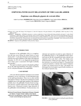
ARTIGO / ARTICLE
SPONTANEOUS COMBINED INTERNAL AND EXTERNAL
BILIARY FISTULAE IN ASSOCIATION WITH GALLSTONES
AND GLIOMATOSIS OF THE GALLBLADDER*
Fístulas Biliares em Combinação Espontânea Interna e
Externa Associadas a Cálculos e Glimatose de Vesícula Biliar
Cícero de Andrade Urban1, Linei Augusta Brolini Dellê Urban2,
Rubens Silveira de Lima1 and Luiz Fernando Bleggi-Torres3
)*564)+6
Spontaneous combined internal and external biliary fistula is a very rare complication of biliary tract disease. Only
two previous cases have been reported so far, and all of them with previous episodes of acute cholecystitis, treated without
surgery. We describe a 27-year-old female patient (the younger reported to date) who presented with a long history of
neglected gallbladder inflammation complicated by neuroglial implants on the peritoneum and on the gallbladder wall due
to an ovarian teratoma. She had spontaneous cholecystocutaneous fistula and cholecystocolic fistula that were successfully
treated by cholecystectomy and excision of the fistulous tracts.
Key words: fistula; gliomatosis peritonei; cholecystitis; gallstones.
RESUMO
As fístulas combinadas internas e externas são complicações muito raras das patologias biliares. Apenas dois
casos foram previamente reportados, ambos com episódios prévios de colecistite aguda, tratada clinicamente, sem
cirurgia. Os autores descrevem, neste estudo, uma paciente de 27 anos de idade (a mais jovem relatada na
literatura), que se apresentou com uma longa história de colecistite calculosa, complicada por implantes neurogliais
no peritônio e na parede da vesícula biliar, originários de um teratoma ovariano maduro. Ela apresentou fístula
colecistocutânea e colecistocólica, que foi tratada com sucesso pela colecistectomia e excisão do trajeto fistuloso.
Palavras
e: fistula; gliomatose peritonial; colicistite; vesícula biliar.
alavras--chav
chave:
*Realizado no Serviço de Anatomia Patológica da Universidade Federal do Paraná e no Serviço de Cirurgia Oncológica do Hospital Nossa Senhora
das Graças, Curitiba
1
Médicos do Serviço de Cirurgia Oncológica, Hospital Nossa Senhora das Graças, Curitiba, PR. Enviar correspondência para C.A.U. Rua Marechal
Hermes, 550 apto.12; 80530-230 Curitiba, PR, Brasil. Fax: (41) 335-5898.
2
Médica Residente do Serviço de Radiologia, Hospital de Clínicas, Universidade Federal do Paraná.
3
Chefe do Serviço de Anatomia Patológica, Hospital de Clínicas, Universidade Federal do Paraná.
Revista Brasileira de Cancerologia, 2001, 47(3): 273-76
273
Urban, C.A., Urban, L.A.B.D., Lima,R.S. and Bleggi-Torres, L.F.
INTRODUCTION
Spontaneous biliary fistula is a rare
complication of gallstones, occurring in 3 per
cent of all patients undergoing biliary surgery.1
Such fistulae usually occur between the biliary
tree and the gastrointestinal tract (internal
fistulae). Spontaneous external biliary fistula
was common in the last century, but became
rare because of early diagnosis of biliary tract
disease. 2 We report the third case of
simultaneous external and internal biliary
fistulae in a 27-year-old female patient. This
is the younger patient reported to date, and
the first with the association of long time biliary
tract disease, gallstones and gliomatosis
peritonei with deposits on the gallbladder wall
due to an ovarian teratoma.
CASE REPORT
A 27-year-old woman was admitted for the
first time six years ago with an episode of
acute cholecystitis and an ultrassound scan
confirmed the presence of gallstones. At that
time the patient refused surgical treatment.
After that first admission she had 3 more
episodes of biliary pain. The second previous
admission was two years ago when she was
submitted to an exploratory laparotomy due
to a 30 cm partially cystic mass replacing the
left ovary. Ascites and multiple peritoneal
deposits were also present. A left salpingooophorectomy and omental biopsy were
performed and histological examination
showed, at that time, a well differentiated solid
teratoma with omental deposits of benign
neuroglial tissue. No evidence of extraabdominal disease was found at that time. On
her third and last admission she presented
with a one year history of discharging sinus
affecting her right subcostal region. A
sinogram revealed cholecystocutaneous
fistulae, multiple large gallstones,
cholecystocolic fistulae; the remaining of
biliary system appeared within normal limits
(Figure 1). Another laparotomy was then
performed. A thick walled contracted
gallbladder containing several large stones was
274 Revista Brasileira de Cancerologia, 2001, 47(3): 273-76
Figura 1 - Contrast sinogram showing: the catheter
introduced in cutaneous fistulae (arrow), gallbladder (g),
colon (c) and duodenun (d).
adherent to the anterior portion of the
abdominal wall, and there was a further
communication with the transverse colon. The
fistulae were divided, and the defect in the
transverse colon was then surgically closed.
There was no mass, but the peritoneum
persisted with multiple deposits. Postoperative
recovery was uneventful. Histological
examination of the gallbladder showed
extensive fibrosis of the wall with deposits of
well differentiated circumscribed neuroglial
tissue (Figure 2). In the fistula there was only
fibrous tissue. At present, 28 months after
her third admission, she is doing well, and
the wound and fistulae healed completely.
DISCUSSION
Spontaneous external biliary fistulae was
a common complication of biliary tract
disease in the last century. The first
description is attributed to Thilesus in 1670
while Courvoisier in 1890 described further
169 cases. After these initial reports, Naunyn
Spontaneous Combined Internal and External Biliary Fistulae in Association with Gallstones and Gliomatosis of the Gallbladder
in 1896 reported 184 cases, and Bonnet
(1897) 122 cases. However only 16 cases
have been described in the last 50 years, and
only two cases of spontaneous combined
internal and external biliary fistulae have been
reported to date.3,4
Spontaneous biliary fistulae are usually a
complication of acute suppurative
cholecystitis associated with cholelithiasis, and
are uncommon in biliary disease without
gallstones (0,6%). Perforation in such cases
occurs probably because of bacterial
dissemination, steroids, polyarteritis nodosa,
typhoid, and trauma.2 The necrosis of the
gallbladder wall and perforation leads to drain
throughout the abdominal wall to the surface
of the skin (external fistulae), or to the
abdominal viscera such as the duodenum
(77%), colon (15%), or rarely into the
bronchial tree, stomach, and even urinary tract
(internal fistulae).4
The gliomatosis peritonei in association
with ovarian teratoma is also a rare condition,
with few cases reported in the literature.
Robboy and Scully reviewed 12 cases of
gliomatosis peritonei in association with
ovarian teratoma and showed good prognosis
of patients with this kind of secondary
deposits due to the benign behavior of mature
implanted tissues. The implants of the ectopic
glial tissue are probably due to capsular defects
in the primary ovarian tumor, although lymph
node spread has also been described.4,5 The
behavior of ectopic neural tissue is not
completly understood.
Cytogenetic analysis of benign ovarian
teratomas usually reveals normal karyotypes
(46,XX). There are very few data reporting
chromosome abnormalities in this type of
tumor. Surti in 1990 found 7% of the numerical
alterations (trysomy, triploidy and mosaicism)
in 102 ovarian teratomas analysed.6
So far this seems to be the first case in the
literature describing the association of
gliomatosis peritonei in the wall of the
gallbladder with cholelithiasis and further
complicated by spontaneous combined
internal (colon) and external biliary fistulae.
The two previous cases affected old women
(81 and 84-year-old) with previous episodes
Figura 2 - Histological section of gallbladder showing serosal adipose tissue (*) incontinuity
with nests of glial cells (G) (HEx100).
of acute choleystitis treated without surgery
because of their poor health conditions.3,4 In
the present report the development of the
fistulae could be due to the long history of
neglected gallbladder inflammation
complicated by the association with the
presence of ectopic neural tissue implants on
the wall of gallbladder, which contributed to
a poor contraction and stasis.
The treatment for such fistulae is
cholecystectomy with the excision of the
fistulous tract. However, in high risk patients,
percutaneous removal of the stones should be
also considered, so that the cholecystocutaneous
fistula might close spontaneously. 3 The
treatment of choice to gliomatosis peritonei is
the simple excision of any symptomatic
masses.7,8
REFERENCES
1. Andley M, Biswas RS, Ashok S, Somshekar G,
Gulati SM. Spontaneous cholecystocutaneous
fistula secondary to calculous cholecystitis. AJG
1996;91:1646.
2. Birch BRP, Cox SJ. Spontaneous external biliary
fistula uncomplicated by gallstones. Postgrad
Med J 1991;67:391-2.
3. Davies MG, Tadros E, Gaine S, McEntee GP,
Gorey TF, Hennessy TPJ. Combined internal
and external biliary fistulae treated by
percutaneous cholecystlithotomy. Br J Surg
1989;76:1258.
4. Reed MWR, Tweedie JH. Spontaneous
simultaneous internal and external biliary
fistulae. Br J Surg 1985;72:538.
Revista Brasileira de Cancerologia, 2001, 47(3): 273-76
275
Urban, C.A., Urban, L.A.B.D., Lima,R.S. and Bleggi-Torres, L.F.
5. Luesley DM, Monypenny IJ, Fielding JW,
Chan KK. Gliomatosis peritonei associated
with ovarian teratomas. Br J Obstet Gynaecol
1983; 90:668-70.
6. Surti U, Hoffner L, Chakravarti A, Ferrel RE.
Genetics and biology of human ovarian
teratomas. I Cytogenetic analysis and mechanism
276 Revista Brasileira de Cancerologia, 2001, 47(3): 273-76
of origin. Am J Hum Genet 1990;47:635-43.
7. Favara BE, Franciosi RA. Ovarian teratoma and
neuroglial implants on the peritoneum. Cancer
1973;31:678-81.
8. Gatti L, Franchini R, Di Lorenzo B, Gullotti
G, Messina G. La fistola colecisto-cutanea
spontanea. Minerva Chir 1989; 44:2263-5.
Baixar